


30
Annals of Cancer Research and Therapy Vol. 29 No. 1, 2021
Ann. Cancer Res. Ther. Vol. 29, No. 1, pp. 30-33, 2021
Introduction
Thyroglossal cysts (TGC) and fistulas (TGF) are
formed as a result of the violation of the thyroid duct
lining (TDL), a path of ontogenetic migration of the
thyroid gland from the place of its foundation. From the
root of the tongue to the site of its usual location on the
front surface of the neck. TGC forms from days 16-24
of embryonic development at the bottom of the primitive
pharynx in the zone that developed from days 12–18 of
embryogenesis of the pharyngeal membrane separating
the mouth fossa and the primary intestinal cavity. At this
site, thickening of the entoderm occurs, and by day 28,
it begins to sink to the subject mesenchyme and forms
a thyroid diverticulum (diverticulum thyroideum). As
the bookmark of the thyroid sinks into the tongue tissue
and moves along the primary intestine, the diverticulum
deepens and transforms into TDL. In week 5 of embryo-
genesis, the developing hyoid bone splits from the TGP,
creating two unequal parts: the shorter tongue (duсtus
lingual) and the longer thyroid (ductus thyroids). In 5%–
7% of cases, TDL is associated with the horn of the hy-
oid bone, while it is associated with the div of the hyoid
bone in the remaining cases. Thus, the hyoid bone is the
most important reference point among the topographic
characteristics of the TGC
1-6)
.
By days 40–50 of embryogenesis, the TGC reaches its
usual position at the front surface of the neck. By week 8
of embryonic development, the TGP is obliterated, trans-
forming into fibrous epithelium that is gradually reduced
with age. On evaluating HR data, Harnsberger et al.
detected residual elements of the reduced TGP in more
than 7% of autopsies
4)
. Bogdanov et al. revealed induced
TGF residues in 25% of patients with TGC
5)
.
Anatomical evidence of the existence and ontoge-
netic migration of TGC is the blind tongue hole (natural
boundary between the div and root of the tongue and
site of the original location of the bookmark of the TGC
and sometimes found a pyramidal fraction of the TGH,
ULTRASOUND IMAGING OF THYROGLOSSAL CYSTS OF THE NECK
TO THE HYOID BONE
Lalita Yunusova
1)
, Toru Aoyama
2)
, Gayrat Ikramov
1)
, Bakhodir Halmanov
1)
, Junichi Sakamoto
3)
,
Hurriyat Kurbanbaeva
1)
1)
Tashkent State Dental Institute, Republic of Uzbekistan
2)
Department of Surgery, Yokohama City University, Japan
3)
Tokai Central Hospital, Japan
Abstract
Background: The present study attempted to clarify the typical anatomical variants of Thyroglossal cysts (TGC).
Patients and methods: Clinically and epidemiologically 67 previously non-experienced patients with TGC 1.5 to 73.0 years
old were examined.
Results: Based on clinical and ultrasound examinations of 121 patients with 67 thyroglossal cysts, the most typical cyst of
anatomical variations was specified. It was noted that, concerning the hyoid bone, thyroglossal cysts may be suprahyoid
(located at the root of the tongue), parahyoid (broadly adjoining the hyoid), prelingual (located in the front of the hyoid in
the hypo lingual region), postlingual (located behind the hyoid bone in the prenatal and peri-laryngeal spaces), or sublingual
(located the book from the hyoid bone). An ultrasound examination facilitated the identification of thyroglossal cysts with-
out clinical manifestations (23 sublingual cysts among 37 [62.2%] were incidentally revealed by the ultrasound examina-
tion), which is important when selecting the most appropriate surgical treatment.
Conclusion: Ultrasound studies facilitate the identification of TGCs located at the root of the tongue without any clinical
manifestations, which is important when determining the degree of surgical treatment to perform.
Keywords: thyroglossal cysts, ultrasound, topographic, anatomical variants
(Received January 14, 2021; Accepted February 1, 2021)
Corresponding author
: Toru Aoyama, Department of Surgery, Yokohama City
University, 3-9 Fukuura, Kanazawa-ku, Yokohama 236-0004, Japan. TEL: 81-45-
787-2800, E-mail: t-aoyama@lilac.plala.or.jp

31
XXXXXXXXXXXXXXXXXXXXXXXXXX
departing upwards from the isthmus of the TGC. In in-
ternational medical practice, cysts and fistulae arising
from the induced remnants of the TGP are considered
thyroglossal, while in domestic practice the term “me-
dian cysts and fistulae of the neck” is used more often. In
our opinion, these terms are not interchangeable, as the
very definition of TGC reflects not only the etiology but
also the probability of pathological changes throughout
the thyroid duct, from the root of the tongue to the usual
site of the thyroid, anddoes not correlate with the term
“middle cysts and fistulae of the neck”. At the same time,
the understanding that in the presence of a “median cyst
of the neck” is very likely the existence of changes in
the root of the tongue (in the form of a cyst or a fistula),
is otherwise organized the diagnostic process and, most
importantly, determines the treatment tactics.
In the existing topographic and anatomical classifica-
tions of TGC, the location of the cyst relative to the hyoid
bone is emphasized. Authors have classified cysts located
above the hyoid bone as being located in front of the hy-
oid bone, in the hypoplastic region of the front surface of
the neck
1)
. This definition is easy for operating surgeons
to comprehend but is not quite accurate with regard to
the topographic anatomy. It should be noted that current-
ly available classifications do not reflect the full range of
anatomical variants of TGC, which may result in misun-
derstandings between diagnosticians and surgeons.
The present study attempted to clarify the typical ana-
tomical variants of TGC.
Materials and Method
Clinically and epidemiologically 67 previously non-
experienced patients with TGC 1.5 to 73.0 years old were
examined. Echography was performed using ultrasound
scanners (an SLE-501; Lithuania; and an Affiniti-70;
Philips, Holland) with linear sensors with frequencies of
7.5-12 MHz.
If necessary, the differential diagnosis of cystic and
solid formations (mainly lymph nodes) was performed by
visualizing the blood flow (e.g. via color Doppler map-
ping or energy Doppler).
Results
The distribution of patients by age is presented in
Table 1. As can be seen, the peak of detection of TGC
not complicated by the presence of external cyst falls on
the age of 3 to 12 years old (50.8%). In general, potential
patients in children’s specialized departments (under 17
years old) account for 67.2% of patients. A total of 90
cysts were detected among the 67 patients: 14 patients
had 2 cysts each, 3 had 3 cysts each, and 1 had 4 cysts.
Therefore, 18 (26.9%) patients with TGC had multiple
cysts. In all cases, additional cysts (23 cysts in 18 pa-
tients) were located at the root of the tongue (Fig. 1). The
following variants of TGC to the hyoid bone were re-
vealed: suprahyoid cysts (located at the tongue root) (Figs.
2–4), parahyoid (widely adjoining to hyoid bone) (Fig.
5), sublingual (located in front of the hyoid bone in the
sublingual region) (Fig. 6), located behind the hyoid bone
in the prenatal and peri-laryngeal space (Fig. 7), and lo-
cated behind the hyoid bone (Figs. 8, 9). All prelingual
and sublingual cysts were associated with the hyoid bone
either by a fibrous sore throat or by their spurs.
Table 1 Patients background
Total number of patients
Age ranges
67
up to 3 years
3–7 years
8–13 years
14–17 years
between 18
and 25 years 26–45 years 46–60 years over 60 years
Absolute
6
15
19
5
9
4
6
3
Relative, %
8.9
22.4
28.4
7.5
13.4
6.0
8.9
4.5
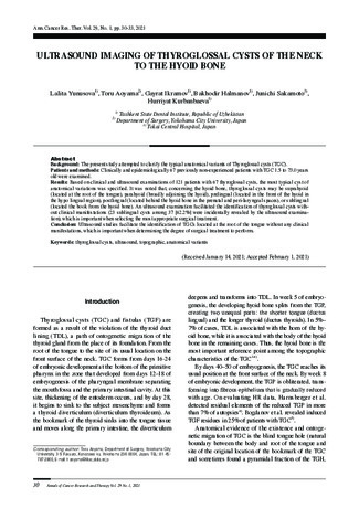
Fig. 1 THC. multiple cysts in the root of
the language (arrow)

32
Annals of Cancer Research and Therapy Vol. 29 No. 1, 2021
Fig. 3 THC. variant with rear
position of cyst in the
root of tongue. The cyst
is located behind the
line connecting the hyoid
bone and the blind hole
in the tongue, and has a
sauce with oropharynx
through the mucous root
of the tongue (arrows).
Fig. 4 THC. variant with a cyst
in the front position at
the root of the tongue.
The cyst is located at the
front of the line connect-
ing the hyoid bone and
the blind tongue open-
ing, spreading over the
mouth diaphragm from
the hyoid bone in the di-
rection of the chin austle
(arrows).
Fig. 2 THC. variant with central
location of cyst in the
root of language. The
cyst is located in the line
connec ting the hyoid
bone and the blind hole
of the tongue (arrows).
Fig. 9 De e p lo c ation of the
cyst. The cyst reaches
the cartilage or mucous
membrane of the larynx
(arrows).
Fig. 8 THC. The upper position
of the cyst between the
muscles of the sublin-
gual group (arrows).
Fig. 7 THC. The cyst is located
behind the hyoid bone
in the prenatal glandular
space (arrows).
Fig. 5 THC. The cyst is widely
adjacent to the hyoid
bone (arrows).
Fig. 6 The cyst is located at the
front of the hyoid bone in
the hypodlingual region
(arrows).

33
XXXXXXXXXXXXXXXXXXXXXXXXXX
Discussion
According to clinical examination data, the most
frequently identified types were sublingual (40.3%) and
circumlingual (25.4%) cysts, while the most frequently
found type was supra-lingual (root) cysts (20.9%). An
ultrasound investigation enabled the identification of
cysts at the root of the tongue without clinical manifes-
tations, which changed the idea about the frequency of
TGC of different localization. The most common TGC
variant was supernodalinguous cysts (41.1% of observa-
tions). Twenty-three of 37 sublingual cysts (62.2%) were
randomly detected on ultrasound and had no clinical
manifestations. Of the 37 sublingual TGCs, 17 (45.9%)
had a central position at the root of the tongue (along the
line connecting the hyoid bone and the blind hole of the
tongue, the conditional course of the tongue portion of
the thyroid duct), not adjoining the mucous membrane of
the tongue root; 14 (37.9%) were located behind this line,
widely adjoining the mucous membrane of the tongue
root or with a source in the oropharynx through the mu-
cous membrane of the tongue root; and 6 (16.2%) were in
front of this line, spreading over the jaw-lingual muscles
(mouth diaphragm) from the hyoid bone in the direction
of the chin. Of the 27 TGCs with hyoid localization, 19
(70.3%) were superficially located (between the breast
hyoid and shield hyoid muscles or in front of them), and
8 (29.7%) were deeply located, reaching the cartilage or
laryngeal mucosa.
Thus, the existing TGC classifications can be refined
with the detailed elaboration of the anatomical cyst lay-
out topography while still meeting the needs of surgeons.
To the hyoid bone, TGC can be defined as suprahyoid
(located at the root of the tongue), sublingual (located in
the root of the tongue), above sublingual (located at the
front of the hyoid bone - above the hyoid region), behind
the hyoid (located behind the hyoid bone at the epiglot-
tis), and behind sublingual (located behind the hyoid
bone in the epiglottis and collateral regions). Sublingual
TGCs may be centrally located at the root of the tongue,
along a line connecting the hyoid bone and the blind hole
of the tongue. A third type of tongue root cysts according
to Bezrukov
1)
; rear position relative to this line, broadly
adjoining the mucous membrane of the root of the tongue
or originating in the oropharynx through the mucous
root of the tongue (second type of cysts of the root of the
tongue according to Bezrukov
1)
) and the front position
relative to this line, spreading over the mouth diaphragm
from the hyoid bone in the board of the chin muscle
(first type of cysts of the root of the tongue according to
Bezrukov)
1)
.
Ultrasound studies facilitate the identification of
TGCs located at the root of the tongue without any clini-
cal manifestations, which is important when determining
the degree of surgical treatment to perform. Sublingual
TGCs may be located superficially (between the muscles
under the sublingual group) and deeply (near the throat,
reaching the cartilage or laryngeal mucosa). Of note, the
proposed classification of TGC describes only the most
frequently occurring (and therefore typical) variants and
does not reflect the diversity of manifestations of this
malformation. However, a thorough understanding of
the anatomical variants of TGC will help improve the
reliability of the diagnosis of this type of pathology, and
detailing the peculiarities of TGC location is important
for planning the scope of surgery.
Acknowledgments:
This study is supported, in part, by the nonprofit organization
Epidemiological and Clinical Research Information Network
(ECRIN).
References
1) Moshe Yehuda, Melissa E. Schechter, Nora Abu Ghanem,
Gilad Golan, Gilad Horowitz. European Archives of Oto-
Rhinolaryngology. 2017;16:15–19.
2) Wei S., Livolsi V.A., Baloch Z.W. Pathology of thyroglossal duct:
an institutional experience. Endocr. Pathol. 2015;26:75–79.
3) Hilary Pitner. Diagnostic Accuracy of Midline pediatric neck
masses. American Academy of Otolaryngology–Head and Neck
Surgery Foundation, 2019;46:55–69.
4) Erikci V., Hosgr M. Management of congenital neck lesions in chil-
dren. J. Plast. Reconstr. Aesthet. Surg. 2014;67:e217–22.
5) Hill S., Maddalozzo J. Congenital lesions of epithelial origin.
Otolaryngol Clin. N. Am. 2015;48:209–223.
6) Lim-Dunham J.E., Toslak E., Alsabban K., Aziz A., Okur G.,
Longo K.C. Ultrasound risk stratification for malignancy using the
2015 American Thyroid Association management guidelines for
children with thyroid nodules and differentiated thyroid cancer.
Pediatr Radiol. 2017;47(4):429–436.