


JNROnline Journal
ISSN: 2320-3358 (e)
ISSN: 0972-5547(p)
Journal of Natural Remedies
Vol. 21, No. 9(1), (2021)
55
EVALUATION OF THE EFFECTIVENESS OF ANTI-INFLAMMATORY
THERAPY OF CONJUNCTIVITIS AND BLEPHARITIS BASED ON
CYTOLOGICAL ANALYSIS
Djamalova Shirin Abdumuratovna
1
, Yangieva Nodira Rahimovna
1
,
Kuryazova Zebiniso Xushnudovna
1
, Mirbabayeva Feruza Abdusamadovna
1
,
Axrarov Abdusamad Aristanovich
2
1
Tashkent State Dental Institute,
2
Tashkent Medical Academy
Tashkent, Uzbekistan
ABSTRACT
Introduction.
In the complex treatment of inflammatory eye diseases, anti-inflammatory therapy
takes the leading place. The use of glucocortico steroids, which have powerful anti-inflammatory
and antiallergic effects, carries the risk of serious side effects. Therefore, it was quite logical to
develop and introduce into ophthalmological practice non-steroidal anti-inflammatory drugs,
which are only slightly inferior to them in their anti-inflammatory activity. On the basis of a
cytological analysis of the state of the conjunctiva, to assess the effectiveness of a new domestic
ophthalmic drug "0.5% benzketozone ointment" in the treatment of conjunctivitis and blepharitis
of infectious etiology.
Materials and methods.
To assess the effectiveness of 0.5% benzketozone ophthalmic ointment
(Registration certificate No. 06-07.), A study was carried out on 134 (218 eyes) patients. In the
control group, patients received traditional treatment, in the main group, 0.5% benzketozone
ointment was additionally prescribed to the traditional treatment. Cytological examination was
carried out by the method of modified impression cytology.
Results.
The data of modified impression cytology showed that the inclusion of benzketozone
ointment in the traditional treatment significantly reduces the phenomena of exudation and
proliferation, which at the subcellular level is manifested by a decrease in the number of basophils
and eosinophils, restoration of the structure of epithelial cells and normalization of the nuclear-
cytoplasmic ratio at an earlier time than in control groups, a faster decrease in the preparations of
protein and tissue detritus.
Conclusion.
The inclusion of benzketozone eye ointment in the complex of traditional therapy
increases regenerative activity, improves metabolic processes and alleviates the symptoms of
inflammatory eye lesions
KEY WORDS:
non-steroidal anti-inflammatory drugs, conjunctivitis, blepharitis.
INTRODUCTION
Inflammatory lesions of the organ of vision occupy a leading place among eye diseases. Causing
the so-called "red eye" syndrome, these diseases of an infectious and, less often, non-infectious
nature are among the most common diseases of the organ of vision. [1, 2, 3, 4]. According to the
calculated data [5, 6, 7], among the total number of patients with inflammatory eye diseases, the
majority are patients with conjunctivitis (66.7%) and blepharitis (23.3%), inflammatory lesions of
the cornea are less common (4.2% ) and the inner membranes of the eye - uveitis, chorioretinitis,
neuritis (5.8%), but it is these diseases that are a common cause of decreased vision and blindness.
In the Republic of Uzbekistan, according to data for 2007 - 2011, 20% of all registered cases were
patients with inflammatory lesions. Of these, 42% were patients with conjunctivitis, 40% - with
blepharoconjunctivitis, 5% - blepharitis, 8% were patients with barley and chalazion, and 5% -
with keratitis, episcleritis, uveitis and chorioretinitis [8].
In the complex treatment of inflammatory eye diseases, anti-inflammatory therapy takes the
leading place [9, 10, 11, 12]. Medicinal components in this area are mainly aimed at preventing or
reducing the release of inflammatory mediators. Glucocorticosteroids and non-steroidal anti-
inflammatory drugs have an effect on this process [13].

Journal of Natural Remedies
Vol. 21, No.9(1), (2021)
56
The use of glucocorticosteroids, which have a powerful anti-inflammatory and antiallergic effect,
is associated at the same time with the risk of developing necrotic changes and gross scarring of
the cornea, impaired transparency of the lens with the formation of posterior subcapsular cataract,
a possible increase in intraocular pressure with the likely subsequent development of glaucoma.
Therefore, it was quite logical to develop and introduce into ophthalmological practice non-
steroidal anti-inflammatory drugs, which are only slightly inferior in their anti-inflammatory
activity to glucocorticosteroids and have no side effects inherent to them. However, the use of
non-steroidal anti-inflammatory drugs is limited by the fact that until recently, only a few of them
were produced in the form of ophthalmic dosage forms [14].
Purpose of the research is to study the basis of cytological analysis of the state of the conjunctiva,
to assess the effectiveness of a new domestic ophthalmic drug "0.5% benzketozone ointment" in
the treatment of conjunctivitis and blepharitis of infectious etiology.
MATERIALS AND METHODS
To assess the effectiveness of 0.5% benzketozone ophthalmic ointment (Registration certificate
No. 06-07.), The study was conducted on 134 (218 eyes) patients with inflammatory eye diseases.
The age of the observed patients ranged from 16 to 82 years. Of these, 64 are men and 67 are
women. The study included patients with conjunctivitis and blepharitis with reliably established
bacterial etiology on the basis of culture.
In the control group, patients received traditional treatment: washing the eyes with disinfectant
solutions (furacilin solution 1: 5000), instillation of antibacterial drops (30% sodium sulfacyl,
0.25% chloramphenicol solution); the patients of the main group were additionally prescribed
0.5% benzketozone ointment to the traditional treatment.
Cytological examination was carried out by the method of modified impression cytology [15]. In
this study, a milipore filter (CELLULOSE ACETATE FILTER, Pore size - 0.8 µm, Sartorius AG,
Germany) was inserted into the conjunctival cavity, retracting the lower eyelid, and then the lower
eyelid was brought into contact with the surface of the eyeball with low pressure. At the next
retraction of the lower eyelid, a filter strip with "imprinted" epithelial cells of both bulbar and tarsal
conjunctiva, as well as the cornea, "printed" on both its surfaces was removed. The prints were
immediately transferred onto a specially prepared degreased glass slide. Then the smears were dried
and fixed with the Mein-Grunveld fixative for 1-3 minutes. After fixation, the prints were washed
with distilled water, then stained with Romanovsky-Giemsa paint for 20-30 min, washed with
distilled water and then dried. The study and photographic registration of cytological preparations
were carried out on the "Fotomikroscope - III" ("Opton", Germany) with a magnification -
eyepiece 10; lens 90.
Biomaterials were taken from patients upon admission, on the 3rd, 7th and 10th days from the
start of treatment.
Statistical processing of digital data was carried out using a Pentium IV computer, the method of
multiple analysis using Microsoft Excel 7.0 application programs. The reliability of differences
between the groups in terms of the studied characteristics was carried out using the Student's test,
the differences were considered significant if the probability of coincidence was less than P <0.05.
RESULTS
In the cytological preparations of the control and main groups of patients with conjunctivitis and
blepharitis, in the first days of the study, there was a predominance of morphological signs of
inflammation. Often, the preparations contained a large amount of mucous protein mass and
fibrinous filaments, tissue detritus (Fig. 1).

Journal of Natural Remedies
Vol. 21, No.9(1), (2021)
57
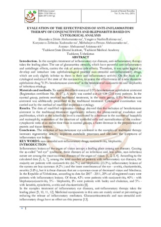
Figure: 1. Condition upon admission. A dense inflammatory infiltrate consisting of fibrin
filaments, tissue detritus and leukocytes. The epithelium is in a state of dystrophy and
destruction. Giemsa staining. Magnification: eyepiece 10, objective 90.
The inflammatory infiltrate was dominated by fibrin filaments and lumpy protein masses, among
which were located epithelial cells in a state of wrinkling and destruction. Polynuclear leukocytes,
which contained basophils and eosinophils with signs of active degranulation, densely surrounded
both epithelial cells and tissue detritus with microorganisms, which also indicated the
predominance of alteration and exudation. On the part of the epithelial cells of the conjunctiva,
polymorphic changes were noted.
An enlargement of the nucleus with hyperchromasia and the appearance of a nucleolus was
observed. In the cytoplasm, degenerative and dystrophic changes were noted in the form of the
appearance of eosinophilic inclusions, vacuolization of the peripheral part of the cytoplasm. The
appearance of multicore symplasts was noted. At this time of the study, the indicators of the
nuclear-cytoplasmic ratio averaged 0.069, which is significantly lower than the norm - 0.2 (table).
Table 1. Changes in the nuclear-cytoplasmic ratio during treatment in patients with
conjunctivitis and blepharitis
Patient groups Upon
enrolment
3
rd
day
7
th
day
10
th
day
Control
0,069±0,0007
0,062±0,0005
0,074±0,0009
0,087±0,002
Main
0,075±0,0067
0,225±0,003*
0,291±0,005*
0,333±0,003*
Note: * - significant difference from control: P <0.001.
Further dynamics in the groups with traditional treatment and with the inclusion of benzketozone
ointment differed significantly.
In the control group, on the 3rd day of the study, the morphological signs of inflammation
persisted. The amount of tissue detritus, protein mass, and fibrinous filaments did not significantly
decrease. Epithelial cells were mainly in a state of destruction and shrinkage. Eosinophilic
inclusions and vacuolization of the cytoplasm persisted in the cytoplasm. The nuclei retained
hyperchromasia and the nucleolus. The indicators of the nuclear-cytoplasmic ratio are still low and
amounted to 0.062 (Fig. 2.).
Figure: 2. 3rd day of traditional treatment. The inflammatory infiltrate persists. The
number of fibrin filaments, tissue detritus and leukocytes did not decrease significantly.

Journal of Natural Remedies
Vol. 21, No.9(1), (2021)
58
Dystrophic changes and phenomena of destruction of epithelial cells persist. Giemsa
staining. Magnification: eyepiece 10, objective 90.
Figure: 3. 7th day, control group. Formation of a dense fibrin network with leukocytes and
a single epithelium. Giemsa staining. Magnification: eyepiece 10, objective 90.
In the subsequent periods (7 days) of the disease, a large number of fibrin filaments and a lumpy
protein mass are found in smears. In this case, the fibrin filaments formed a relatively dense
network (Fig. 3), in the intervals of which there are single cells of desquamated epithelium and
polynuclear neutrophilic leukocytes, the indicator of the nuclear-cytoplasmic ratio was 0.074.
On the 10th day of the disease, neutrophilic leukocytes predominated in smears (Fig. 4), which
were in various stages of activity with the appearance of segmented, rod-shaped and bean-
nucleated cells. At the same time, the leukocytes densely surrounded the desquamated epithelium,
which were in a state of dystrophic and destructive changes. Among epithelial cells, binucleated
and multinucleated symplasts were identified, the cytoplasm of which was expanded in volume
due to clearing and vacuolization. At the same time, the nuclear-cytoplasmic ratio averaged 0.087,
which is still much lower than the norm and 1.2 times lower than the indicators of the previous
period.
Figure: 4. 10th day, control group. The predominance of neutrophilic leukocytes of
different activity in the smear. Dystrophic and dysregenerative changes in epithelial cells.
Giemsa staining. Magnification: eyepiece 10, objective 90.
In patients whose treatment included benzketozone ointment, with conjunctivitis and blepharitis
on day 3, there was a decrease in the activity of the processes of alteration and exudation of
inflammation. Morphologically, this was manifested by a decrease in the amount of inflammatory
mucous and fibrinous mass, the existing leukocytes in a state of destruction and decay, which
morphologically looked like a destructive mass of irregular shape, stained with eosin. In epithelial

Journal of Natural Remedies
Vol. 21, No.9(1), (2021)
59
cells, dystrophic and degenerative changes are less pronounced, in the nuclei there is some
hypertrophy and hyperchromasia (Fig. 5.)
Figure: 5. 3rd day, main group. Decay and destruction of leukocytes, the appearance of
signs of regeneration in the epithelium. Giemsa staining. Magnification: eyepiece 10,
objective 90.
The volumetric ratio of nuclei and cytoplasm sharply changed in favor of nuclear structures and
the nuclear-cytoplasmic ratio from the first days of treatment in the study group increased and
amounted to 0.225, which approached the normal values. By the 7th day of treatment, the
cytological preparations showed almost complete disappearance of the phenomena characteristic
of inflammation. Only the presence of single leukocytes and lymphocytes in a state of decay and
destruction was determined (Fig. 6.). In epithelial cells, regenerative and restorative changes
prevailed over dystrophic and degenerative processes, the indicator of the nuclear-cytoplasmic
ratio increased and amounted to 0.291, which is 1.5 times more than the norm and exceeds the
indicators of this period in the control group by 3.9 times.
Figure: 6. 7th day, main group. Hypertrophy of epithelial cells, the disappearance of
leukocytes. Giemsa staining. Magnification: eyepiece 10, objective 90.
Figure: 7. 10th day, main group. Regeneratively active, hypertrophied epithelial cells
without signs of inflammation. Giemsa staining. Magnification: eyepiece 10, objective 90.

Journal of Natural Remedies
Vol. 21, No.9(1), (2021)
60
By the 10th day after treatment in the main group, only layers of epithelial cells with signs of
hypertrophy and hyperchromasia are determined in smears (Fig. 7.). Their cytoplasm is represented
by a uniformly colored structure without dystrophic changes. The nuclei are of different size and
color, most of them in a state of hypertrophy and hyperchromasia. The nuclear-cytoplasmic ratio
was 0.33.
DISCUSSION
Thus, the cytological study showed that during the traditional treatment of patients with
conjunctivitis and blepharitis, mucous protein masses and fibrinous filaments, tissue detritus
prevailed in cytological preparations from the first days. In the inflammatory infiltrate consisting
of polynuclear leukocytes, there were basophils and eosinophils with signs of active degranulation,
which also indicated the predominance of the processes of alteration and exudation. On the part
of the epithelial cells of the conjunctiva, polymorphic changes were noted. An enlargement of the
nucleus with hyperchromasia and the appearance of a nucleolus was revealed. In the cytoplasm,
degenerative and dystrophic changes were noted in the form of the appearance of eosinophilic
inclusions, vacuolization of the peripheral part of the cytoplasm. The appearance of multicore
symplasts was noted. Indicators of the nuclear-cytoplasmic ratio in the dynamics of the
inflammatory disease from the initial one, practically did not change.
The mechanisms of action of benzketozone seem to be as follows: a decrease in basophils and
exudation during the inflammatory process indicates the suppression of the initial forms of
inflammatory mediators - histamine and serotonin; the disappearance of leukocytes is the result of
the suppression of prostaglandins and leukotrienes by benzketozone; restoration of the structure
of the cytoplasm and nuclei of epithelial cells, an increase in the indicators of the nuclear-
cytoplasmic ratio is the result of an increase in metabolic processes and an increase in their
regenerative activity.
Thus, the data of the modified impression cytology method showed that the inclusion of
benzketozone ointment in the traditional treatment significantly reduces the phenomena of
exudation and proliferation, which at the subcellular level is manifested by a decrease in the
number of basophils and eosinophils, restoration of the structure of epithelial cells. This is
confirmed by the normalization of the nuclear-cytoplasmic ratio at an earlier date than in the
control groups, a more rapid decrease in the preparations of protein and tissue detritus.
CONCLUSION
A new domestic ophthalmic drug 0.5% benzketozone ointment is an effective non-steroidal anti-
inflammatory drug. Its inclusion in the complex of traditional therapy increases regenerative
activity, improves metabolic processes and softens the symptoms of inflammatory eye lesions,
which is confirmed by the results of a cytological study.
ACKNOWLEDGEMENTS
We are grateful to the staff members of Tashkent State Dental Institute and Tashkent Medical
Academy for the cooperation and support in our research. The participants kindly gave full written
permission for this report.
CONSENT
Written informed consent was obtained from all participants of the research for publication of this
paper and any accompanying information related to this study.
CONFLICT OF INTEREST
The authors declare that they have no competing interests.
FUNDING
No funding sources to declare.
REFERENCES
1.
Dovgan' Ye.V. Obzortopicheskikh form antimikrobnykhpreparatov, primenyayemykh v
oftal'mologii. Oftal'mologiya.- 2014. -
№1 (11). –
S.10-18.

Journal of Natural Remedies
Vol. 21, No.9(1), (2021)
61
2.
Vorontsova T.A., Brzheskiy V.V. i dr. Mikroflorakon" yunktival' noypolostiiyeyechuvstvitel
'nost' k antibakterial' nympreparatam u detey v normeiprinekotorykhvospalitel'
nykhzabolevaniyakhglaz. Oftal' mologicheskiyevedomosti.
–
2010. -
№2 (3). –
S.61-65.
3.
Behrens-Baumann W. Topical antimycotis drugs. In: Antiseptic prophylaxis and therapy in
ocular infections. Ed. A. Kramer, W. Behrens-Baumann-karger.
–
2002.
–
P.263
–
280.
4.
Kaercher T., et al. Treatment of patients with keratoconjunctivitis sicca with OPtive: results of
a multicenter, open-label absezvational study in Germany. Clin. Ophthalmol.
–
2014. N3.
–
P.33
–
39.
5.
Ma
i
̆
chuk
D.
YU.
Infektsionnyyezabolevaniyaglazno
i
̆
poverkhnosti
(kon"yunktivityikeratokon"yunktivity). V kn. «Sindromkrasnogoglaza» pod red. D. YU.
Ma
i
̆
chuka.
–
M. Media Sfera; 2010.
6.
Neroyev V.V. Osnovnyyeputirazvitiyaoftal' mologichesko
i
̆
sluzhby Rossi
i
̆
sko
i
̆
Federatsii. IX
syezdoftal' mologov Rossii.
–
M.
–
2010.
–
S. 52
–
55.
7.
Neroyev V.V., Ma
i
̆
chuk YU. F. Zabolevaniyakon"yunktivy. V kn. «Kratkoyeizdaniyenatsional'
nogorukovodstva po oftal'mologii». Glava 8.
–
M.
–
2014.
–
S. 367
–
407.
8.
Sidikov Z.U. Dostizheniyaiproblemyoftal' mologicheskoysluzhbyrespubliki Uzbekistan.
Organizatsiyaiupravleniyezdravookhraneniya.
–
Tashkent, 2012. -
№10. –
S. 41-50.
9.
Astakhov YU. S., Riks I. A. Sovremennyyemetodydiagnostikiilecheniyakon"yunktivitov. SPb.;
2007.
10.
Larcombe J. Review: antibiotic therapy leads to slightly earlier recovery in acute bacterial
conjunctivitis. Journal Article.
–
2006.
–
Vol.11. - Issue 6. - P. 180 -180.
11.
Ma
i
̆
chuk YU.F. Sovremennyyevozmozhnostiterapiikon"yunktivitov. Trudy XVII Ros. nats.
kongressa.
–
M.: Chelovekilekarstvo, 2010
–
215
–
225.
12.
Ma
i
̆
chuk YU. F., Yani Ye. V. Novyyepodkhody v lecheniiblefaritov. Katar. irefrakts.
khirurgiya.
–
2012.
–
1.
–
S. 59
–
62.
13.
Ma
i
̆
chuk
YU.
F.
Kon"yunktivity.
Sovremennayalekarstvennayaterapiya.
Kratkoyeposobiyedlyavrache
i
̆
.
–
M.
–
2013.
14.
Razumova I.YU., Godzenko A.A. Nesteroidnyyeprotivovospalitel'nyyepreparaty v
lecheniiperednegouveitaprispondiloartritakh // Vestnikoftal'mologii. - 2020. -
№5 (136). –
S.
70-77.
15.
Petrayevskiy
A.V.,
Trishkin
K.S.
Kliniko-tsitologicheskayadiagnostikasindroma
«Sukhogoglaza». Vestnik Volg GMU.
–
2012. -
№4 (44). –
S.52-54.