


Citation:
Jung, S.; Nam, O.H.; Fang,
Y.-Q.; Dusmukhamedov, S.; Lee, C.
Reliability of a Trapezium Miniplate
with Endoscope-Assisted Internal
Fixation in Mandibular Subcondylar
Fractures: A Three-Dimensional
Analysis.
J. Clin. Med.
2022
,
11
, 207.
Academic Editor: Anne
Marie Kuijpers-Jagtman
Received: 30 November 2021
Accepted: 25 December 2021
Published: 31 December 2021
Publisher’s Note:
MDPI stays neutral
with regard to jurisdictional claims in
published maps and institutional affil-
iations.
Copyright:
© 2021 by the authors.
Licensee MDPI, Basel, Switzerland.
This article is an open access article
distributed under the terms and
conditions of the Creative Commons
Attribution (CC BY) license (https://
creativecommons.org/licenses/by/
4.0/).
Article
Reliability of a Trapezium Miniplate with Endoscope-Assisted
Internal Fixation in Mandibular Subcondylar Fractures:
A Three-Dimensional Analysis
Seungwook Jung
1,†
, Ok Hyung Nam
2,†
, Yi-Qin Fang
1
, Shavkat Dusmukhamedov
1
and Chunui Lee
1,
1
Department of Oral and Maxillofacial Surgery, Wonju College of Medicine, Yonsei University,
Wonju 26426, Korea; Loukas_jung@naver.com (S.J.); qin0302@naver.com (Y.-Q.F.); mr.shavkat595@bk.ru (S.D.)
2
Department of Pediatric Dentistry, School of Dentistry, Kyung Hee University, Seoul 02447, Korea;
pedokhyung@gmail.com
*
Correspondence: chunuilee@naver.com; Tel.: +82-33-741-1451; Fax: +82-33-741-1442
†
These authors contributed equally to this work.
Abstract:
This study aimed to evaluate the reliability of a trapezium plate for open reduction
and internal fixation (ORIF) of mandibular subcondylar fractures with the simultaneous use of
an endoscope. We selected and retrospectively studied 18 patients (12 males and 6 females) with
unilateral mandibular subcondylar fractures who visited the Wonju Severance Christian Hospital. The
mean age of the patients was 43.43
±
15.76 years. Patients underwent ORIF with trapezium miniplate
application through an intraoral incision under general anesthesia. The clinical and radiographic
findings of the fractured side were compared with those of the non-operated side at 6 months follow-
up. All occlusions became stable, and transient functional disturbances disappeared within 6 months
of periodic follow-up. Functional mandibular movement recovered within the normal range, with
an average mouth opening of 41.5 mm, protrusion of 7.5 mm, and lateral excursion of 7 mm at
6 months. Radiographic controls and statistical analysis confirmed a decent anatomical reduction
in all 18 cases. In conclusion, the use of a trapezium miniplate with endoscope-assisted ORIF in
mandibular subcondylar fractures can be useful for fixation and functional recovery.
Keywords:
trapezium plate; endoscope; mandibular subcondylar fracture; fracture healing; fracture
fixation; open reduction and internal fixation
1. Introduction
The optimal treatment protocol for open reduction and internal fixation (ORIF) of
mandibular condylar and subcondylar fractures has been debated over decades. Many
studies on condylar fracture surgery have introduced various surgical approaches to the
condylar region, and verified that the intraoral approach has the least postoperative mor-
bidity [
]. The application of miniplates fixed with screws is considered the gold standard
for ORIF in mandibular surgery [
]. Thus, numerous miniplate designs have been intro-
duced during the past decades, and recently, their efficacies have been studied to examine
biomechanical stability and surgical simplicity [
]. Previous biomechanical studies have
confirmed the reliability of condyle fixation via two miniplates for subcondylar fractures for
withstanding the compression and tension forces within physiological limitations. [
Meyer at al. [
] demonstrated that the trapezoidal plate provided sufficient rigidity for the
fixation of subcondylar fractures. Darwich et al. [
] claimed that trapezoidal plates are
superior to two miniplates.
Recently, with the emergence of endoscopy-assisted surgery, problems of inadequate
surgical view and limited workspace, which are critical disadvantages of intraoral surgery,
have been overcome [
]. Endoscopy-assisted open reduction and internal fixation
(EAORIF) of mandibular condylar fractures has resulted in satisfactory postoperative
J. Clin. Med.
2022
,
11

J. Clin. Med.
2022
,
11
, 207
2 of 10
outcomes [
]. A previous study regarding the miniplate system in EAORIF showed that
biodegradable plates are as reliable as metal miniplates [
]. However, there are insufficient
data for appropriate miniplate designs for EAORIF. Therefore, the present study evaluated
the clinical use of a newly developed single trapezium miniplate (Jeil Co., Seoul, Korea)
applied through a transoral endoscopy-assisted approach and attempted to present clinical
and three dimensional (3D) anaylsis follow-up results.
2. Materials and Methods
We retrospectively reviewed patients who had undergone a uniform surgical tech-
nique between 2015 and 2018, consisting of an endoscopy-assisted transoral approach
with combination fixation using a mono-cortical single trapezium miniplate, with at least
6 months of postoperative follow-up. A total of 18 patients with mandibular subcondylar
fractures (12 males and 6 females) between 18–70 years old (mean age 43.83 years old) were
included. All patients underwent surgery at the Department of Oral and Maxillofacial
Surgery of the Wonju Severance Christian Hospital. The study protocol was reviewed
and approved by the Institutional Review Board of Yonsei University Wonju Severance
Christian Hospital, Wonju, Korea (CR319018).
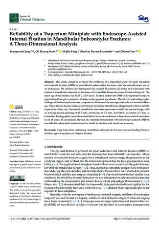
In the trapezium plate, the plate arms were designed according to the ideal line of
osteosynthesis introduced in Meyer’s study [
]. Both vertical arms were designed parallel
to the compression lines, while the upper and lower horizontal lines were designed along
the traction lines. Two types of trapezium plates were designed, which differed in the
upper arm size. The arms were similar, but were selectively used in high and low condylar
fractures. The plate was 1 mm thick, 13.2 mm long, and 4–5.2 mm wide at the top and
13.2 mm wide at the base. Four holes were located at each corner of the trapezium plate
(Figure
Figure 1.
Trapezium plate shape and dimensions. (
A
) Two subtypes of trapezium plate. (
B
) Numerical
description of each trapezium plate. (
C
) Trapezium plate applied to a rapid prototype model.
All of the patients received general anesthesia. Lidocaine with 1:100,000 epinephrine
was injected into the muco-vestibular area along the mandibular ramus. In most cases,
anchor screws were located at the upper and lower interdental alveoli for maxillomandibu-
lar fixation (MMF). A linear mucosal incision was made over the buccal vestibule and
over the anterior border of the ramus, similar to orthognathic surgery. The portion of
the mandibular angle and the lateral aspect of the ramus along the fractured subcondylar
portion were then exposed by raising the mucoperiosteal flap and masseter muscles using
a surgical curette, periosteal elevator, and ramus stripper. A stab incision was made at the
ipsilateral overlying skin, according to the level of the fracture line. The trochar system was
inserted transbuccally, providing adequate space between the bone and overlying tissue for
endoscopic instruments. Under endoscopic visualization of the fractured site, manipulation
of the proximal segment for proper reduction was attempted with a trochar shank. When
rough reduction was performed, proper occlusion was manually manipulated. To con-
firm proper reduction and occlusion, a trapezium plate was placed between the trochar
shank and the proximal segment of the bone. After temporary intermaxillary fixation

J. Clin. Med.
2022
,
11
, 207
3 of 10
via wire engagement on the corresponding upper and lower anchor screws, two holes in
the proximal segment were drilled, and screws were inserted into the upper corners of
each hole. Thorough determination of the correct anatomical reduction, especially proper
alignment of the posterior border of the ramus, was achieved endoscopically. Finally,
two holes in the distal fragment were drilled, and screws were inserted. The MMF was
removed, and occlusion was confirmed. The outcome of fracture reduction and fixation
was verified endoscopically (Karl Storz, Germany) (Figure
). If necessary, the fixation
position was adjusted by slightly loosening and retightening the screws (Figure
). Wound
closure was usually performed using 4-0 vicryl, and a silastic drain was inserted to prevent
postoperative hematoma and minor hemorrhage. A liquid diet with intermaxillary elastic
engagement was strictly maintained for 2 weeks after the operation. To ensure mouth
opening and prevent temporomandibular joint ankylosis, a soft diet was recommended
from the third week onwards. The trapezium plate was removed after a minimum of
6 months postoperatively using an intraoral and transcutaneous approach.
Figure 2.
Endoscopic view of the trapezium plate after condylar fracture fixation.
Figure 3.
3D reconstruction view. (
A
) Pre-operative 3D view. (
B
) Post-operative 3D view. Using
Mimics, an image of the condylar head collapsing inward before surgery was reproduced, and it was
confirmed that the condylar head was repositioned after surgery.

J. Clin. Med.
2022
,
11
, 207
4 of 10
Each patient was followed-up with clinical and radiographic evaluations for at least
6 months. Clinical evaluation included stable occlusion, and trismus evaluation by measure-
ments of the maximum interincisal distance, protrusion, lateral excursion, and mandibular
deviation on the mouth opening. Preoperative and postoperative radiographic evaluations
were performed using 3D facial mandible computed tomography (CT) in all patients, along
with panoramic radiography (Figure
). Adequate anatomical reduction was evaluated
according to Undt et al. [
]. Using 3D simulation in the Mimics medical program, the
ramus height was measured from the top point of the condyle to the gonion, and was
compared with the non-fractured side. Similarly, the program was used to evaluate the
angle of dislocation of the proximal fragment, compared to the contralateral side, using 3D
CT DCM files. The angle was measured from the most posterior point of the condylar head
to that of the fracture line and that of the mandibular angle (Figure
). For comparison with
the non-fractured side, the FH-plane was used, and the angle was measured by connecting
the condylion superius to the point where they met the non-operated proximal condyle by
setting a line parallel to the FH-plane and the gonion posterius (Figure
Figure 4.
X-ray and 3D facial CT view. (
A
) Pre-operative panoramic view. (
B
) Post-operative
panoramic view. (
C
) Pre-operative 3D facial computed tomography scan. (
D
) Post-operative 3D
facial computed tomography scan.

J. Clin. Med.
2022
,
11
, 207
5 of 10
Figure 5.
Ramus height and angle of dislocation measured on 3D simulation. (
A
) Pre-operative
mandible. (
B
) Post-operative mandible. CO-sup (condylion superius): The most superior point of
the condylar head. CO-post (condylion posterius): The most posterior point of the condylar head.
GO-post (gonion posterius): The most posterior point on the mandibular angle.
Figure 6.
The red plane indicates the FH-plane, orbitale to porion. The blue plane is parallel to the
FH-plane and passes through the posterior point of the subcondylar fracture line.
Statistical analysis was performed using the collected data. Descriptive statistics of
the patients’ average mouth opening and protrusive and lateral excursion of the mandible
were analyzed to evaluate the functional recovery of mandibular movement. The Wilcoxon
signed-rank test was performed to compare variables between the fractured and non-
fractured sites. The ramus height and angle of dislocation of the operated side and those of
the non-fractured side were determined as variables.
3. Results
3.1. Clinical Evaluation
The operation time ranged between 104 and 128 min, depending on the degree of
displacement or the existence of other fracture sites. The plate was easily manipulated
during the operation, and the fractured segments were adequately reduced. None of the
patients showed chronic inflammation, infection, malunion, or nonunion. The plates were
removed in all 18 patients, and no loose screws were found during removal; many of the
patients had bone accumulation over the trapezium plate.
3.2. Functional Evaluation
During the follow-up period (mean follow-up, 12.2 months; range, 6–24 months),
no patient complained of pain or occlusal discomfort. However, postoperatively, two pa-
tients complained of occlusal discomfort, which disappeared after intermaxillary elas-
tic engagement 3–5 days after admission. The mean maximum mouth opening was
24.82
±
1.99 mm immediately after the operation, 32.2
±
1.32 mm 1 month after the opera-
tion, and 41.54
±
2.21 mm 6 months after the operation.

J. Clin. Med.
2022
,
11
, 207
6 of 10
Protrusive movement and lateral excursion of the mandible varied from 4.5 to 10 mm at
the 6-month follow-up timepoint. Lateral deviation on the mouth opening to the fractured
side was observed in 11 of the 18 patients. The values of postoperative mandibular
movements (maximum mouth opening, protrusion, and lateral excursion) were almost
similar to those of the physiological mandibular movement recommended by Okeson [
The findings are summarized in Table
Table 1.
Descriptive statistics.
Variables
Study Population (
n
= 18)
Sex
n
(%)
Men
12 (66.67)
Women
6 (33.33)
Age (year)
Mean
±
SD
43.83
±
15.76
Median (min–max)
47.00 (18.00–70.00)
Post-operative MMO (mm)
Mean
±
SD
24.82
±
1.99
Median (min–max)
24.90 (21.20–27.90)
Post-operative MMO (1 month) (mm)
Mean
±
SD
32.23
±
1.32
Median (min–max)
32.50 (30.30–34.30)
Post-operative MMO (6 months) (mm)
Mean
±
SD
41.54
±
2.21
Median (min–max)
42.70 (37.20–47.80)
Mean protrusion (6 months) (mm)
Mean
±
SD
7.39
±
1.60
Median (min–max)
8.20 (4.00–10.50)
Mean lateral excursion to the right
(6 months) (mm)
Mean
±
SD
7.28
±
1.18
Median (min–max)
7.45 (4.60–9.50)
Mean lateral excursion to the left
(6 months) (mm)
Mean
±
SD
7.13
±
1.51
Median (min–max)
7.05 (4.50–10.20)
3.3. Radiographic Evaluation
Anatomical reduction was confirmed via 3D facial mandible computed tomography.
The postoperative panoramic view revealed no significant numerical gap between the
ramus heights. At 6 months follow-up, the ramus height was retained in most patients
(83%). Based on the angle of dislocation in the panoramic view, good anatomic reduction
(angle 0
◦
to 20
◦
) was achieved postoperatively in all patients. Radiography revealed no
plate fracture or plate bending in any patients.
3.4. Statistical Evaluation
The Wilcoxon signed rank test was performed between two subgroups (operated
and non-operated sides) in two categories (ramus height and angle of dislocation). At a
significance level of 0.05, negative statistical significance was found between the operated
and contralateral sides in both categories (Table
Table 2.
Comparison between the operated and non-operated sides.
Variables
Non-Operated Side (
n
= 18)
Operated Side (
n
= 18)
p
-Value *
Length (mm)
Mean
±
SD
65.68
±
6.77
65.02
±
6.48
0.205
Median (min–max)
65.33 (52.10–79.40)
66.77 (48.16–74.13)
Angle (n
◦
)
Mean
±
SD
166.31
±
7.10
164.95
±
8.26
0.099
Median (min–max)
168.10 (143.00–175.20)
165.30 (142.50–179.50)
*
p
-value by Wilcoxon signed-rank test.

J. Clin. Med.
2022
,
11
, 207
7 of 10
4. Discussion
With the development of surgical techniques and instruments, miniplates are a dedi-
cated solution for the fixation of mandibular condylar neck and base fractures. Nevertheless,
endoscopy-assisted ORIF (EAORIF) should not be indicated for all condylar fractures, as it
has limitations in cases with high condylar neck fractures or for comminuted fractures with
insufficient bone mass to tolerate screws and plating; in these cases, closed reduction is pre-
ferred. However, for cases with proper indication, ORIF of the condylar and subcondylar
fractures, rather than closed treatment, should be performed [
]. An intraoral approach
can decrease the risk of nonesthetic surgical outcomes and postoperative complications
such as Frey’s syndrome and facial nerve palsy [
]. Transoral subcondylar surgery
has overcome the limited surgical view via endoscopic assistance, and the need for a small,
manageable osteosynthesis device that can replace conventional two-plate bio-stability
is increasing [
]. Trapezium plates for subcondylar fractures have many advantages.
First, its shape designs with four arms can be placed at the compression and traction lines.
Second, they are easy to manipulate and require less time for placing, but are as stable as
the two single miniplate system biomechanics, which requires the drilling of eight holes
and screw insertion [
]. Radiographic findings confirmed proper anatomical reduc-
tion, and no permanent functional complications occurred in our patient cohort. Minor
deviations were found in five patients, but no functional or esthetic dissatisfaction was
noted. Lata et al. [
] compared delta and trapezoidal plates, and found that patients
treated with delta plates showed a significant improvement at all intervals in contrast with
the trapezoidal plate, which did not show a significant improvement in the mouth opening
from 6 weeks to 3 months. Burkhard et al. [
] found that deltoid and trapezoid plates seem
to perform equally in the treatment of condyle neck and base fractures. Hochban et al. [
emphasized that deviations from the anatomically correct position did not affect the func-
tion of the temporomandibular joint. In our patient cohort, trismus relief and restoration
of the mouth opening were observed within 6 months after operation. Six months after
surgery, the amount of MMO, protrusion, and lateral movement showed good results.
There is a time difference between the lateral and medial overrides, but it is sufficiently
overcome. In our patient cohort, the presence of additional distant fractures of the mandible
showed good results. The functional parameters of protrusive and lateral excursion of the
mandible were similar between our patients and the various treatment cohorts reported
previously [
] evaluated the complications of EAORIF of mandibular
condylar fractures. The long-term complication rate was much lower than the temporary
complication rate, except for cases where malreduction or refracture occurred. EAORIF
should be considered reliable for the treatment of condylar fractures, but intensive training
and equipment are required.
The statistical insignificance observed between the operated and non-operated sides re-
garding the ramus height and angle of dislocation indicated consistent operation outcomes,
accompanied with anatomic reduction and rigid fixation.
Plate fracture or bending and screw loosening have been observed in previous studies;
however, none of these complications occurred in our study. Occasionally, excessive bone
accumulation over the trapezium plate interfered with the process of plate and screw
removal, which implies vigorous osteogenesis around the fixation site. The two types of
trapezium plates differ in the upper horizontal arm length. Therefore, they can be applied
to most open subcondylar fractures with various fracture line locations.
The biostability and durability of the trapezium plates were evaluated by Darwich et al. [
].
Five plating techniques (single straight plate, two parallel straight plates, two divergent
straight plates, trapezoid plate, and square plate) were compared for the fixation of uni-
lateral mandibular subcondylar fractures with finite element analysis. Among these, the
trapezoid plate showed the least micro-mobility and strain on the underlying bones for
fixation [
] summarized clinical trials regarding the use of various types
of 3D condylar mini-plates (trapezoid, deltoid, rhombus, strut, nine-hole trapezoid, and
lambda). There are no convincing data that the number of reoperations depends on the

J. Clin. Med.
2022
,
11
, 207
8 of 10
type of 3D mini-plate used and clinical fractures of 3D mini-plates are extremely rare, in
contrast to the fairly frequent fractures of straight miniplates. The trapezium plate can be
applied to various subcondylar fractures. It is accurately designed according to the ideal
tension and compression line of the mandible, and can tolerate various forms of loads,
with the highest tensile strain occurring at the anterior and lateral surfaces and the highest
compressive strains on the posterior surface. Although there is a learning curve for our
protocol, the surgical outcome and time efficiency have been verified throughout the past
years. The clinical, functional, and radiologic outcomes are consistent with those from
previous studies.
However, there are possible complications of this surgical technique. In severely
dislocated and comminuted fractures, if the extraoral approach for the endoscopic-assisted
treatment of subcondylar fractures is indicated, then facial nerve damage may occur.
Subcondylar fractures with medially displaced fragments are more challenging to reduce
within the narrow sight under the endoscopic view. So, if an unskilled surgeon performs
the operation, re-fracture due to screw loosening, malocclusion, deviation, trismus, and
temporomandibular joint disease may occur.
This study has some limitations. First, the number of patients undergoing this surgery
is not enough. Second, in the 3D analysis of the position of the condyle after surgery, there
is a limit to the analysis of angles in various directions, such as the mesial and lateral side.
Moreover, the results for the follow-up of long-term clinical evaluation and functional
evaluation are insufficient.
5. Conclusions
In conclusion, the use of a trapezium plate with endoscopy-assisted ORIF can be
recommended for subcondylar fractures. However, the use of a trapezium with EAORIF
has limitations in tolerating screws and plating in cases of high condylar neck fractures
or comminuted fractures with insufficient bone mass. Trapezoid shape designs with four
arms can be placed at the compression and traction lines; thus, a sufficient fixing force can
be achieved even with a small number of screws, with equal stability to other miniplate
systems. Considering these advantages, our method can be recommended for the treatment
of patients with subcondylar fractures.
Author Contributions:
Conceptualization, C.L.; methodology, C.L.; software, Y.-Q.F. and S.D.; val-
idation, C.L.; formal analysis, S.J. and O.H.N.; investigation, S.J. and O.H.N.; resources, C.L.; data
curation, C.L.; writing—original draft preparation, S.J., O.H.N. and C.L.; writing—review and editing,
S.J., O.H.N. and C.L.; visualization, O.H.N.; supervision, C.L. All authors have read and agreed of
the published version of the manuscript.
Funding:
This research received no external funding.
Institutional Review Board Statement:
The study was conducted in accordance with the guide-
lines of the Declaration of Helsinki and was approved by the Institutional Review Board of Yonsei
University of Wonju Severance Christian Hospital, Korea (CR319018).
Informed Consent Statement:
Patient consent was waived due to the retrospective nature of this study.
Data Availability Statement:
Data are available upon reasonable request.
Conflicts of Interest:
The authors declare no conflict of interest.
References
1.
Lachner, J.; Clanton, J.T.; Waite, P.D. Open reduction and internal rigid fixation of subcondylar fractures via an intraoral approach.
Oral Surg. Oral Med. Oral Pathol.
1991
,
71
, 257–261. [
2.
Jeter, T.S.; van Sickels, J.E.; Nishioka, G.J. Intraoral Open reduction and internal rigid fixation of mandibular subcondylar fractures.
J. Oral Maxillofac. Surg.
1988
,
46
, 1113–1116. [
3.
Undt, G.; Kermer, C.; Rasse, M.; Sinko, K.; Ewers, R. Transoral miniplate osteosynthesis of condylar neck fractures.
Oral Surg.
Oral Med. Oral Pathol. Oral Radiol. Endod.
1999
,
88
, 534–543. [

J. Clin. Med.
2022
,
11
, 207
9 of 10
4.
Iizuka, T.; Lindqvist, C.; Hallikainen, D.; Mikkonen, P.; Paukku, P. Severe bone resorption and osteoarthrosis after miniplate
fixation of high condylar fractures.
Oral Surg.
1991
,
72
, 400–407. [
5.
Zachariades, N.; Papademetriou, J.; Rallis, G. Mandibular fractures treated by bone plating and intraosseous wiring: A compara-
tive study.
Revue de Stomatologie et de Chirurgie Maxillo-Faciale
1994
,
95
, 386–390.
6.
Burkhard, J.P.M.; Koba, S.; Schlittler, F.; Iizuka, T.; Schaller, B. Clinical results of two different three-dimensional titanium plates in
the treatment of condylar neck and base fractures: A retrospective study.
J. Cranio-Maxillofac. Surg.
2020
,
48
, 756–764. [
7.
Sikora, M.; Ch˛eci ´nski, M.; Sielski, M.; Chlubek, D. Use of 3D Titanium Miniplates in Surgical Treatment of Patients with Condylar
Fractures.
J. Clin. Med.
2020
,
9
, 2923. [
8.
Adhikari, M.; Bhatt, K.; Yadav, R.; Mandal, J.; Bhutia, O.; Roychoudhury, A. Fixation of subcondylar fractures of the mandible:
A randomized clinical trial comparing one trapezoidal plate with two miniplates.
Int. J. Oral Maxillofac. Surg.
2021
,
50
, 756–762.
9.
Ahuja, S.A.; Galinde, J.; Asnani, U.; Mistry, Y.A. Comparative Evaluation of Clinical Outcomes Using Delta Plates and Conven-
tional Miniplates for Internal Fixation of Mandibular Condylar Fractures in Adults.
J. Oral Maxillofac. Surg.
2018
,
76
, 1255–1266.
10.
Choi, B.H.; Yi, C.K.; Yoo, J.H. Clinical evaluation of 3 types of plate osteosynthesis for fixation of condylar neck fractures.
J. Oral
Maxillofac. Surg.
2001
,
59
, 734–737. [
11.
Zieli ´nski, R.; Kozakiewicz, M.; Konieczny, B.; Krasowski, M.; Okulski, J. Mechanical Evaluation of Titanium Plates for Osteosyn-
thesis High Neck Condylar Fracture of Mandible.
Materials
2020
,
13
, 592. [
12.
Meyer, C.; Martin, E.; Kahn, J.L.; Zink, S. Development and biomechanical testing of a new osteosynthesis plate (TCP) designed
to stabilize mandibular condyle fractures.
J. Cranio-Maxillofac. Surg.
2007
,
35
, 84–90. [
13.
Darwich, M.A.; Albogha, M.H.; Abdelmajeed, A.; Darwich, K. Assessment of the Biomechanical Performance of 5 Plating
Techniques in Fixation of Mandibular Subcondylar Fracture Using Finite Element Analysis.
J. Oral. Maxillofac.
2016
,
74
,
794e1–794e8. [
14.
Hochban, W.; Ellers, M.; Umstadt, H.E. Zur operativen reposition und fixation von unterkiefergelenkfortsatzfrakturen von enoral.
Fortschritte der Kiefer und Gesichts-Chirurgie
1996
,
41
, 80–85. [
15.
Jacobovicz, J.; Lee, C.; Trabulsy, T. Endoscopic repair of mandibular subcondylar fractures.
Plast. Reconstr. Surg.
1998
,
101
,
437–441. [
16.
Sandler, N.A. Endoscopic-assisted reduction and fixation of a mandibular subcondylar fracture: Report of a case.
J. Oral Maxillofac.
Surg.
2001
,
59
, 1479–1482. [
17.
Paeng, J.-Y.; Ahn, K.-M.; Myoung, H.; Choi, J.-Y.; Hwang, S.-J.; Kim, S.-M.; Kim, M.-J.; Lee, J.-H. Endoscopic-Assisted Open
Reduction and Internal Fixation (EAORIF) for Condylar Fracture.
J. Hard Tissue Biol.
2005
,
14
, 45–46. [
18.
Kim, D.Y.; Sung, I.Y.; Cho, Y.C.; Park, E.J.; Son, J.H. Bioabsorbable plates versus metal miniplate systems for use in endoscope-
assisted open reduction and internal fixation of mandibular subcondylar fractures.
J. Cranio-Maxillofac. Surg.
2018
,
46
, 413–417.
19.
Meyer, C.; Kahn, J.L.; Boutemi, P.; Wilk, A. Photoelastic analysis of bone deformation in the region of the mandibular condyle
during mastication.
J. Cranio-Maxillofac. Surg.
2002
,
30
, 160–169. [
20.
Okeson, J.P.
Management of Temporomandibular Disorders and Occlusion
, 8th ed.; Mosby Elsevier: St. Louis, MO, USA, 2019; pp.
324–371.
21.
Worsaae, N.; Thorn, J.J. Surgical versus nonsurgical treatment of unilateral dislocated low subcondylar fractures: A clinical study
of 52 cases.
J. Oral Maxillofac. Surg.
1994
,
52
, 353–360. [
22.
Eckelt, U.; Schneider, M.; Erasmus, F.; Gerlach, K.L.; Kuhlisch, E.; Loukota, R.; Rasse, M.; Schubert, J.; Terheyden, H. Open versus
closed treatment of fractures of the mandibular condylar process: A prospective randomized multicenter study.
J. Cranio-Maxillofac.
Surg.
2006
,
34
, 306–314. [
23.
Wilk, A.; Biotchane, I.; Rosenstiel, M. Surgical treatment of subcondylar process fractures using a rectangular plate for 3-
dimensional stabilization.
Revue de Stomatologie et de Chirurgie Maxillo Faciale
1997
,
98
, 40.
24.
Krenkel, C. Axial “anchor” screw (lag screw with biconcavewasher) or “slantes screw” plate for ostesynthesis of fractures of the
mandibular condylar process.
J. Cranio-Maxillofac. Surg.
1992
,
20
, 348–353. [
25.
Lauer, G.; Schmelzeisen, R. Endoscope-assisted fixation of mandibular condylar process fractures.
J. Oral Maxillofac. Surg.
1999
,
57
, 36–39. [
26.
Miloro, M. Endoscopic-assisted repair of subcondylar fractures.
Oral Surg. Oral Med. Oral Pathol. Oral Radiol. Endod.
2003
,
96
,
387–391. [
27.
Schön, R.; Fakler, O.; Gellrich, N.C.; Schmelzeisen, R. Five-year experience with the transoral endoscopically assisted treatment of
displaced condylar mandible fractures.
Plast. Reconstr. Surg.
2005
,
116
, 44–50. [
28.
Manoj, C.; Harshvardhan, P.; Manpreet, S.; Arpit, V.; Gagandeep, K. Evaluation of trapezoidal-shaped 3-D plates for internal
fixation of mandibular subcondylar fractures in adults.
J. Oral Biol. Craniofacial Res.
2015
,
5
, 134–139.
29.
Scott, C.; Ramakrishnan, K.; Vivek, N.; Saravanan, C.; Prashanthi, G. Does Three-Dimensional Plate Offer Better Outcome and
Reduce the Surgical Time Following Open Reduction and Internal Fixation of Adult Mandibular Unilateral Subcondylar Fractures.
A Randomized Clinical Study.
J. Oral Maxillofac. Surg.
2021
,
79
, 1330.e1–1330.e12. [

J. Clin. Med.
2022
,
11
, 207
10 of 10
30.
Lata, J.; Verma, N.; Mhajan, S. Evaluation of Efficacy of 3-Dimensional Delta Versus Trapezoidal Condylar Plates in Internal
Fixation of Mandibular Subcondylar Fractures: A Clinical Study.
Traumaxilla
2019
,
1
, 11–21. [
31.
Spiessl, B.; Schroll, K. Gesichtsschädel Bd I/I. In
Spezielle Frakturen und Luxationslehre
; Nigst, H., Ed.; Thieme: Stuttgart, Germany,
1972; pp. 43–113.
32.
Wagner, A.; Krach, W.; Schicho, K.; Undt, G.; Ploder, O.; Ewers, R. A 3-dimensional finite element analysis investigating the
biomechanical behavior of the mandible and plate osteosynthesis in cases of fractures of the condylar process.
Oral Surg. Oral
Med. Oral Pathol. Oral Radiol. Endod.
2002
,
94
, 678–686. [
33.
Kang, S.-H.; Choi, E.J.; Kim, H.W.; Kim, H.J.; Cha, I.H.; Nam, W. Complications in endoscopic-assisted open reduction and
internal fixation of mandibular condyle fractures.
Oral Surg. Oral Med. Oral Pathol.
2012
,
113
, 201–216. [