All articles - Radiology, Nuclear Medicine and Medical Imaging
-
 Мультипараметрическая ультразвуковая диагностика хронических вирусных гепатитов у детей
Prospects for the development of medicineПроблема хронических диффузных заболеваний печени, основную долю которых составляют вирусные поражения, является одной из наиболее актуальных в педиатрической гепатологии. Несмотря на использование современных методов лечения и существенное снижение острых форм заболевания всё же хронические вирусные гепатиты (ХВГ) остаются глобальными во всём мире.
-
В настоящее время ультразвук широко применяется в современных научных исследованиях и в области медицины. В связи с этим имеет важное значение изучение в биофизике способов создания ультразвука
-
 Ультразвуковая диагностика острого аппендицита у детей в серошкальном режиме
Prospects for the development of medicineНесмотря на достигнутые успехи, диагностика и лечение острого аппендицита остается актуальной проблемой современной хирургии. Экстренное УЗИ органов брюшной полости позволит объективно оценивать характер происходящих изменений в правой подвздошной области и определить выбор рационального операционного доступа.
-
 Ультразвуковая диагностика наружной гидроцефалии у детей первого полугодия жизни
Prospects for the development of medicineФормирование ультразвуковой картины послеродовой адаптации и дозревания головного мозга детей первых недель жизни находится в процессе изучения. Проведение амбулаторного ультразвукового скрининга новорожденных позволило получить объективную картину состояния головного мозга у детей в возрасте одного - двух месяцев.
-

Ранняя диагностика межпозвонкового остеохондроза позволяет своевременно начать лечение и избежать осложнений.
-
 Ультразвуковая диагностика полного удвоения желчного пузыря
Prospects for the development of medicineБолезни желчного пузыря и желчевыводящих путей у детей относятся к наиболее распространенным заболеваниям органов пищеварения. Аномалии развития и приобретенные деформации желчного пузыря могут иметь патогенетическое значение в нарушении пассажа желчи как один из причинно-значимых факторов различных патологических процессов со стороны билиарного тракта. Ультразвуковое исследование (УЗИ) является одним из основных методов диагностики аномалий развития и приобретенных деформаций желчного пузыря.
-
 Возможности цифровой рентгенографии и эхографии в диагностике доброкачественных образований молочной железы
Prospects for the development of medicineРак молочной железы (РМЖ) является одной из важнейших проблем онкологии. Многочисленными работами показано, что чем раньше выявляется опухоль, тем дольше продолжительность жизни заболевших женщин. Вместе с тем, отсутствие единой системы обследования молочной железы в настоящее время снижает возможности современных диагностических методов, не позволяет сократить сроки обследования больных и отодвигает сроки для начала лечения. Основными методами обследования молочной железы являются маммография (МГ) и ультразвуковое исследование (УЗИ).
-
 Роль цифрового рентгенологического метода в диагностике посттравматического остеомиелита у детей
Prospects for the development of medicineОстеомиелит - воспаление компактной части кости, надкостницы, костного мозга и окружающих кость мягких тканей. Развитие обусловлено попаданием в костный мозг патогенных микробов. Инвазия микробов в костную ткань происходит при открытом или огнестрельном переломе, а также при операциях на костях с последующим развитием посттравматического или послеоперационного остеомиелит а. Несмотря на определенные достижения в клинике и диагностике значений методов рентгенологических исследований в ранней диагностике заболевания остается высоким.
-
 Возможности цифровой рентгенографии в диагностике коксартроза
Prospects for the development of medicineОстеоартроз - хроническое прогрессирующее заболевание суставов, характеризующееся дегенерацией суставного хряща, изменениями в субхондральном отделе эпифизов костей и в околосуставных мягких тканях. Коксартроз - дегенерация тазобедренного сустава.
-
 Цифровой рентгенологическая и эхографическая диагностика пневмоний у детей
Prospects for the development of medicineПневмонии являются одной из наиболее часто встречающихся заболеваний у детей. По показателям заболеваемости и смертности, заболевания органов дыхания занимают первое место у детей.
-
 Возможности комплексной эхографии в диагностике обструкции верхних мочевых путей
Prospects for the development of medicineПроблема мочекаменной болезни (МКБ) является наиболее распространенной в урологической практике, которая приводит к обструкции верхних мочевых путей и требует комплексного подхода к обследованию пациентов.
-
 Роль инвазивной эхографии в диагностике кистозных образований молочной железы
Prospects for the development of medicineКисты - это одно из наиболее часто встречающихся заболеваний молочной железы. Типичным для возникновения кист является возраст менструирующих женщин (между 35 и 50 годами) с наступлением менопаузы кисты обычно регрессируют. 90 95% кист молочных желез не имеют солидного компонента (простые кисты). В 2-3% случаях кист имеют очаги пролиферации внутренней стенки (атипичные кисты).
Пункционная биопсия под контролем ультразвука с цитологическим исследованием является обязательной при выявлении атипичной кисты. -
 Мультиметрическая ультрозвуковая диагностика нефробластом у детей дошкольного возраста региона Андижанской области
Экспериментальная медицина: сегодня и в будущемСреди всех опухолевых заболеваний у детей нефробластома занимает 4 место, уступая только гемобластозам, новообразованиям ЦНС и саркомам мягких тканей. Частота её составляет от 0,4 до 1 на 100 000 детей. Чаще всего ОВ встречается у детей в возрасте 2-5 лет, редко у новорожденных и еще реже у детей старше 8 лет, как казуистика - у взрослых. Обычно ОВ возникает спорадически, а в 2% случаев она имеет семейный характер. В 10% случаев она развивается у детей с пороками развития (чаще мочеполовой системы) или генетическими синдромами. Частота заболевания среди девочек и мальчиков одинакова. В 6-10% случаев встречается билатеральное поражение почек, преимущественно у детей до 2 лет. В 2% случаев опухоль поражает подковообразную почку. Описаны случаи внеорганного расположения опухоли. Важное значение для этой патологии имеет не просто правильная, но и ранняя диагностика заболевания. Необходимо не только оценивать общее состояние больного, уточнять локализацию опухоли и степень ее распространения, с обязательным морфологическим подтверждением диагноза, но и использовать как можно больше инструментальных методов диагностики. При этом необходимо соблюдение принципа ургентности.
-
 Effect of gen-engineered biological therapy on the system of juvenil arthritis in children
in LibraryThe article demonstrates the effect of genetically engineered biological therapy on the clinical course of systemic onset juvenile arthritis in children. The analysis of the results showed that the administration of human monoclonal antibodies against the IL-6 receptor - a geneticallyengineered biological drug (tocilizumab) was confirmed by normalization of clinical symptoms and laboratory parameters, reduced disease activity, prevented oral administration of glucocorticoids and systemic initiating juvenile arthritis. provided the transition to the stage. The results obtained showed that the addition of tocilizumab to the complex treatment of systemic onset juvenile arthritis was correct, and confirmed its high efficacy in the treatment of this orphan disease in children.
-
 Комплексная ультразвуковая диагностика обструкции верхних мочевых путей
Scientific works of gifted youth and medicine of the XXI centuryПроблема мочекаменной болезни (МКБ) является наиболее распространенной в урологической практике, которая приводит к обструкции верхних мочевых путей и требует комплексного подхода к обследованию пациентов.
-
 Ультразвуковая оценка органов мошонки у детей с крипторхизмом
Scientific works of gifted youth and medicine of the XXI centuryКрипторхизм является одним из наиболее частых причин бесплодия и опухолей яичка. Медико-социальная значимость данной проблемы определяется, с одной стороны, высокой распространенностью заболевания в популяции, с другой - его выраженными негативными последствиями для мужского здоровья.
-
 Ultrasonography and magnetic resonance imaging in the diagnosis of degenerative dystrophic diseases of the hip joint
Catalog of abstractsSubject of the inquiry: 148 patients with degenerative dystrophic diseases of the hip joint, of them 103 with osteoarthritis and 45 with avascular necrosis of the femoral head.
Aim of the inquiry: improvement of the diagnosis of degenerative-dystrophic diseases of the hip using possibilities of ultrasonography and magnetic resonance imaging.
Methods of research: X-ray, ultrasonography and magnetic resonance imaging.
The results achieved and their novelty: For the first time, the role of ultrasonog-aphy and magnetic resonance imaging in osteoarthritis and avascular necrosis of the hip was established in comparison with conventional X-ray. The result have shown that sensitivity of ultrasonography in osteoarthritis was low (57.7%) in the detection of femoral head deformation. In avascular necrosis of the hip detection of femoral head deformation was revealed better - 89.7%, which can be explained by differences in the mechanism and localization of deformation in these diseases. Ultrasonography was quite sensitive to changes of the joint capsule both in osteoarthritis and avascular necrosis, the sensitivity 86.0% and 84.6% respectively. MRI was superior to X-ray in the assessment of space orientation of the joint surfaces, necrosis zone, synovitis, subchondral cysts, structural changes and joint effusion. X-ray was more preferable for the detection of ostephytosis, subchondral sclerosis and changes of the joint space in osteoarthritis.
Practical value: of the work consisted in the concretization of sonographic and MRI signs of degenerative dystrophic diseases of the hip joint and in the suggested algorithm of using radiological methods for establishing the nature of hip joint pathology.
Degree of inculcate: the results of research were introduced in the Radiology Department of First Tashkent Medical Institute and the Department of Large Joint Pathology Department of the Traumatology and Orthopedy Research Institute.
Sphere of usage: radiology, traumatology and orthopedy, rheumatology. -
 The role of magnetic resonance imaging in the comprehensive radial diagnosis of volumetric masses of the eye organ
Catalog of abstractsRelevance of the problem. The difficulties of diagnostics of orbital diseases are well known. Especially difficult is intraspecies differentiation among the multitude of tumour, pseudotumour, inflammatory, vascular, endocrine and other diseases occurring here, manifested by the symptom complex of unilateral exophthalmos [Beradze I.N., 1978; Brovkina A.F., 1993].
Malignant intraocular neoplasms are the main cause of death of patients with diseases of the organ of vision, with 45-48% of patients dying from metastases in the first 5 years after enucleation [Alekseeva I.B., 1990, Barkhash S.A.1978, Brovkina A.F..1991, 1997; Keizer R.W.. Viclvoyc G.L.,1986],
Retinoblastoma is the most frequent malignant neoplasm in children. According to different authors, the frequency of its occurrence is 1 case per 14000 - 35000 newborns. [Bobrova N.F. and Vit V.V., 1993; Brovkina A.F., 1997; Provenzale J.M., et al., 1995; Skulski M., et al., 1997; Weber A.L., Mafee M.F, 1992; Wilms G., et al., 1989]. The frequency of patients with the most malignant intraocular tumour in adults - uveal melanoma has recently reached 7-9 people per 1 million population [Brovkina A.F., 1997; Kotslyansky E.O., 1989; Yushko N.A., Peskova L.I., Kalenich L.A., 1989; Peyster R.G., Augsburger J..I., Shields J.A., 1988; Romani A.. Baldeschi L., ct al 1998; Scott I.U., 1998].
The fundamental difference in treatment tactics, depending on the stage of development, size and topography of the tumour, as well as the seriousness of the prognosis in retinoblastomas and melanomas sharply increase the requirements for the accuracy of their differential diagnosis. At the same time, the number of diagnostic errors in ocular tumours continues to be 10-30% even when complex clinical and instrumental examination is applied in specialised ophthalmological centres [Ternovoy S.K., Panfilova G.V., Rogozhin V.A., 1979; Friedman F.E., Malyuta G.D., Kodzov M.V., 1995; Song G.X., 1991].
Widely used in ophthalmological practice traditional diagnostic methods (ophthalmoscopy, gonioscopy, diaphanoscopy, fluorescence angiography, laboratory tests) are insufficient to obtain comprehensive information about the localisation, nature of growth and prevalence of volumetric pathological formations of the eye and orbit. This circumstance and not quite satisfactory results of surgical treatment are the causes of high mortality of patients [Muratova T.T., Nigmanova N.H., Kozlovskaya G.M.. 1989, Naches A.I., 1980; Cheremisin V.M., Trufanov G.E., Kholin A.V., 1991]. Untimely or erroneous recognition of pathological processes of the orbit leads to a sharp deterioration of visual functions, up to blindness, and in some cases to the death of the patient [Yuzhakov A.M., Travkin A.G., Kiseleva O.A., 1991]. All this determines the importance of timely and accurate diagnosis of diseases of the orbit, on the one hand, and the difficulty of such diagnosis - on the other [Gabunia R.I., Kolesnikova E.K., Tumanov L.B., 1982].
The fact that the orbit is closed from direct inspection and palpation by bone walls and the eyeball, indicates the advantage of radial diagnostics in comparison with other methods of examination. In the arsenal of clinicians there is a great variety of methods of clinical-radial diagnostics of orbital pathology, however, at present the information in the literature about their resolving capabilities and significance in comparative aspect is incomplete and not fully studied. The priority of using one or another instrumental investigation, their sequence and expedient combination have not been determined yet. This makes it difficult to choose the optimal standardised approach for diagnosis and adequate treatment [Cheremisin V.M., Trufanov G.E., 1993, Weber A.L., Sabates N.R., 1996; Wenig V.M., Mafee M.F., 1998].
Thus, the study of these and other questions, contributing to the improvement of diagnostics and treatment of patients with neoplasms of the eye and ocular cavity, should be recognised as urgent urgent.
Purpose of the study. Comparative evaluation of magnetic resonance tomography capabilities and development of algorithms for complex radial diagnostics of volumetric formations of the visual organ. To solve this goal we set the following tasks.
1. To study the normal picture of the magnetic resonance image of the visual organ in comparison with other methods of visualisation.
2. To find out the possibilities of magnetic resonance tomography, ultrasound and computed tomography in detection and evaluation of intraocular neoplasms.
3. To determine the role and place of magnetic resonance tomography in differential diagnostics of volumetric pathological formations of the eye cavity in comparison with other radial methods of research.
4. To determine the indications and to develop an algorithm for the complex application of radiography, ultrasound, computer and magnetic resonance tomography for diagnostics of volumetric formations of the eye organ.
Scientific novelty.
The present work is the first to give a detailed and detailed description of the complex clinical and radiation examination, with generalisation and standardisation of magnetic resonance, computer and ultrasound semiotics of volumetric pathological formations of the eye and eye cavity. The conducted clinical and instrumental investigations allowed to determine the diagnostic value and resolving capabilities of each of the applied methods. The ultrasound, CT and MRI signs of volumetric formations of the eye organ were studied, clarified and supplemented taking into account the use of low-field magnetic field and general-purpose ultrasound apparatus. The developed standardised diagnostic algorithm of examination of patients with this pathology is new, thanks to which the pre-oppositional diagnosis of tumour and other diseases of the visual organ is improved and the total radiation load on the patient is reduced.
Conclusions
1. MPT will provide an opportunity to study the weight of the soft tissue and anatomical components of the ocular cavity, up to the optic nerve sheath and perineural liquor space, the orbital apex and chiasmal-sellar region, as well as to assess the condition of adjacent structures of the brain and facial skull. The method is limited in the evaluation of changes in the bony walls of the orbital cavity.
2. MRI is inferior in detecting characteristic signs of retinoblastoma (presence of calcification). The sensitivity of MRI was 66.6%, while for ultrasound and CT these values were 96.1 and 100%, respectively. But when the tumour spreads rstrobulbarly outside the eyeball (at 3-4 stages) the informativeness of MRI increases significantly. In uveal melanoma the sensitivity and specificity of MRI reaches 100%.
3. Both MRI and CT have a high detection rate (98.1% and 95.8% respectively) of benign orbital tumours of both primary and secondary origin. However, MRI is the preferred method of investigation. MRI is especially informative when a cranioorbital tumour and pseudotumour are suspected. The sensitivity of the method is 90.9% and 91.6%, respectively
4. In some cases ultrasound can be used to differentiate between encapsulated and diffuse neoplasms, which facilitates the diagnosis. However, when the pathological process is localised near the orbital apex, the diagnostic value of ultrasound decreases. In such cases it is advisable to use MRI.
5. In detection of primary and secondary malignant tumours of the orbital cavity both MRI and CT are quite informative (sensitivity 97,2% and 95,4% respectively), but the most comprehensive information about the state of bone walls will be provided by CT. When the process spreads intracranially, the value of MRI increases significantly, especially with the use of contrast enhancement.
6. The developed algorithm of complex clinical and radiation examination of patients with the use of ultrasound, CT and MRI is the most effective in the diagnosis of volumetric pathological formations of the eye and eye cavity, allowing to reduce to an adequate minimum the total radiation load on the patient and diagnostic period, excluding duplication of research techniques and choosing the most informative in each case, which in turn allows to develop appropriate treatment tactics and reduce the level of disability of the patient. -
 Ультразвуковое исследование пороков развития головного мозга плодаАномалии головного мозга привлекают пристальное внимание исследователей во всем мире не только в связи с высокой частотой встречаемости, но и потому, что они являются причиной перинатальных потерь. В настоящее время ультразвуковое исследование входит в комплекс обязательного обследования беременных, которое, в первую очередь, направлено на своевременное пренатальное выявление врожденных и наследственных заболеваний.
Сегодняшние научные исследования в глазах молодежи -
 Rentgenografik tasvirlarga dastlabki ishlov berish dasturiy majmuasiMazkur maqola rentgenografik tasvirlarga ishlov berish yondashuv va algoritmlari asosida ishlab chiqilgan dasturiy majmuani tuzilmasi, talablari va imkoniyatlari bayoniga bag‘ishlangan bo'lib, unda dasturiy majmuani tashkil etuvchi modullar va ulami tasniflari hamda dasturiy majmuadan foydalanish uchun kompyuter konfiguratsiyasi kcltirilgan.
Новый Узбекистан: наука, образование и инновации -
 Gistologik tasvir chegarasiga tegib turgan sohalarni olib tashlash usuliMazkur ishda gistologik tasvirlami bo‘sag‘aviy segmentlash usuli tadqiq qilinib, tasvir chegaralarida bo'lgan va tasvirda to‘liq nomoyon bo‘lmagan sohalami olib tashlash usuli taklif etilgan. Usui eksperimental tadqiqotlar orqali tekshirib ko’rilgan. Taklif qilingan usul asosida yaratilgan dasturiy vosita yordamida olib borilgan tajribaviy tadqiqotlar yoritilgan.
Новый Узбекистан: наука, образование и инновации -
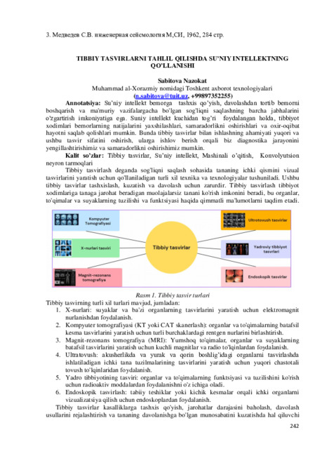 Tibbiy tasvirlarni tahlil qilishda su’niy intellektning qo’llanishiSu’niy Intellekt bemorga tashxis qo'yish, davolashdan tortib bemorni boshqarish va ma'muriy vazifalargacha bo'lgan sog'liqni saqlashning barcha jabhalarini o'zgartirish imkoniyatiga ega. Suniy Intellekt kuchidan tog’ri foydalangan holda, tibbiyot xodimlari bemorlarning natijalarini yaxshilashlari, samaradorlikni oshirishlari va oxir-oqibat hayotni saqlab qolishlari mumkin. Bunda tibbiy tasvirlar bilan ishlashning ahamiyati yuqori va ushbu tasvir sifatini oshirish, ularga ishlov berish orqali biz diagnostika jarayonini yengillashtirishimiz va samaradorlikni oshirishimiz mumkin.
Новый Узбекистан: наука, образование и инновации -
 Sonography and magnetic resonance imaging of the hand joints in rheumatoid arthritis
Catalog of abstractsSubject of the inquiry: 116 patients with rheumatoid arthritis of the hand joints and 25 healthy subjects.
Aim of the inquiry: improvement of the diagnosis of rheumatoid arthritis of the hand joints using sonography and magnetic resonance imaging.
Methods of inquiry: X-ray, sonography, magnetic resonance imaging and magnetic resonance imaging with contrast enhancement.
The results achieved and their novelty: For the first time data were presented on the role of sonography, magnetic resonance imaging and magnetic resonance imaging with contrast enhancement in the diagnosis of rheumatoid arthritis of the hand joints. The findings have shown that sonography in hand joint rheumatoid arthritis allowed detection of changes in soft tissues, synovial capsule, joint surfaces and ligaments. Diagnostic value was given of sonography in revealing characteristic sonographic signs of rheumatoid arthritis. Magnetic resonance imaging was highly informative radiological method to detect synovitis, changes of synovial capsule and subchondral cysts. Magnetic resonance imaging with contrast enhancement reliably detected the degree of activity and severity of the rheumatoid process.
Practical value: consisted in revealing and describing characteristic sonographic and MR1 signs of hand joint rheumatoid arthritis and in the developed radiological algorithm of the disease.
Degree of embed: the results of the investigation were introduced in the practice of rheumatology and radiology departments of the First Tashkent Medical Institute, in teaching process of the Radiology Chair of the First Tashkent Medical Institute.
Sphere of usage: radiology, rheumatology, traumatology and orthopedics. -
 Influence of radio frequency electromagnetic radiation on the cardiovascular system (clinical-experimental study)
Catalog of abstractsThe urgency and relevance of the theme of dissertation. Among the factors of the external electromagnetic radiation is invisible and is not perceived by the senses, not eye-catching factor standing on human health. Dangerous is the impact of radio frequency electromagnetic radiation (RF-EMR) on the body of people working under the impact of radiation, especially on the nervous and cardiovascular system (CVS), the organs of blood, endocrine, metabolic processes, and organs of digestion. With long-term occupational exposure revealed functional changes in the organs of 60% of those diseases SSA 60%, gastrointestinal disease in 40%.
It should be noted that the protection of public health from the effects of electromagnetic fields (EMF) is one of the serious problems. WHO is seriously concerned about the problem of protection of public health under conditions of EMF. Since 1994, the organization conducts a special program «WHO International EMF Project» (International Project WHO on electromagnetic fields). According to the WHO EMF under certain conditions can cause a change in behavior, the development of Parkinson's disease and Alzheimer's disease, memory loss, cancer and many other diseases.
At the same time, the evaluation of complex research to study the effect of different RF-EMR exposures on the immune and cardiovascular system (CVS), holding morphofunctional and biochemical tests on experimental animals and in people working in the factories pollution jobs this kind of radiation, development of clinical guidelines for the management of these patients in order to prevent the RF-EMR influence on the body arc topical issues of medical science and practice.
The available literature is not revealed details characterizing RF-EMR influence on the functional state of the vascular endothelium, indicators of immune system, coagulation factors, the state of cell metabolism et aL, contributes to the progression of CVD in individuals working in conditions of exposure to RF-EMR.
According to many researchers, chronic exposure to electromagnetic radiation (EMR) in vulnerable people develops multiple organ pathology. Studies of biological action of RF-EMR arc of great interest to scientists and the public. Publications in the media accurately reflect current trends in these studies.
This dissertation work is to organize high-quality implementation of the tasks set out in the Decree of the President of the Republic of Uzbekistan № U11-3923 dated 19.09.2007 «On main directions of further deepening reforms and implementing the State program of development of public health».
The aim of research work is to develop the tactics of the patients, the criteria for early diagnosis and prevention approaches violations of the cardiovascular system caused by electromagnetic radiation of radio frequencies in individuals occupationally exposed to direct and permanent effects of this radiation.
Scientific novelty of the research work is as follows:
developed a method for predicting the formation and progression of cardiovascular disease on people with occupational risk exposure to radio frequency electromagnetic radiation;
for the first time on experimental model evaluated the pathogenic mechanisms and depth cardio vascular system disorders, immune system and certain metabolic effects during radio frequency electromagnetic radiation exposure depending on different exposures and the radiation power;
for the first time proven the nature and severity of degenerative disorders of the cardio vascular system with the persons working under the direct and permanent radio frequency electromagnetic radiation exposure and the interrelation of these disorders with some clinical and instrumental and biochemical parameters;
improved integrative index based on a malondialdehyde over superoxide dismutase ratio, proved its high sensitivity and specificity and the possibility of use as a predictor of the development and progression of violations by the cardio vascular system under the influence radio frequency electromagnetic radiation.
CONCLUSION
1. In experimental animals under RF-EMR influence the morphological and functional CVS disorders develop from minor microcirculatory disorders, lung edema and myocardial hypertrophy, fiber pulping fibre muscle to severe violations of their own blood the heart, blood vessels perdiapedeza expressed structural abnormalities of the heart tissue, plasmorrhages appearance, stasis and aggregation of formed of blood, which increases with the power and duration of irradiation.
2. RF-EMR in experimental animals causes depression of cellular immunity and causes marked changes in carbohydrate and energy metabolism, the extent of which is limited by the power and duration of exposure, as well as the nature of the adaptive-adaptive processes and may determine destructive and degenerative processes in the body.
3. From the cardiovascular system in individuals with direct and constant RF-EMR exposure to develop expressed destructive and degenerative disorders in terms of ABL to TAG tissues of heart and blood vessels, the degree of which increases with the length of service under RF-EMR and arc not detected by routine clinical and instrumental CVS research methods.
4. RF-EMR impact on CVS in people with professional risk appears small manifestation of clinical and functional disorders in a variety of arrhythmias, and metabolic changes that do not reflect the true destructive and degenerative disorders by the CVS. As diagnostic tests should take into account the increase in the duration of the P wave, an increase of Makruz index, negative RV1, reduction EOS QRS, depression ST V5-6, QRS V5, the degree of change which correlates with the level of ABL to the TA, reflecting the severity of tissue destruction of the heart and blood vessels.
5. Persons working under the direct and constant RF-EMR exposure, work experience have a close relationship with the degree of activation of lipid peroxidation and antioxidant protection factor reduction, raising and lowering the index phospholipase deformation of red blood cells, increasing hyper-aggregation of platelets, which allows predicting the development and progression of multiple organ pathology, including CVS.
6. In individuals with direct and constant RF-EMR exposure is a significant inhibition of cellular immunity factors, progressive with increasing length of their work.
7. As the risk of violations of the CVS should use indicators such as the increase in part-time work under RF-EMR exposure more than 5 years, the growth of MDA and the degree of platelet aggregation, reduction in the rate of platelet aggregation, EDI and SOD, which with a high degree of confidence (P <0,01 - <0.001) correlated with the level of ABL to TAG infarction, reflecting the degree of destruction in it.
8. In persons with direct and constant RF-EMR exposure index TA heart tissue can be used as a method for early diagnosis of the CVS. Exceeding this figure is 5 or more times should be regarded as a diagnostic level, and to recommend to patients holding a routine treatment for the relief and prevention of progression -
 Neuroimaging and hemodynamic criterias for acute cerebral circulatory disorders in conditions of emergency medical care…
Catalog of abstractsThe urgency and relevance of the dissertation topic. According to the World Health Organization cerebral stroke is a major cause of mortality in developed countries, and in the structure of total mortality takes 2-3 position. The total number of patients with stroke or transient ischemic attacks in the anamnesis in the world more than 50 million. Ischemic stroke (IS) - Important medical and social problem and a major cause of hospitalization, disability and mortality in developed countries. In despite of the close attention of scientists and specialists around the world the problem of early diagnosis of ischemic stroke and adequate cerebral reperfusion remains topical.
The aim of research work is development of radial methods of prediction of ischemic stroke flow depending on neuroimaging and hemodynamic patterns in the acute phase of the disease.
The scientific novelty' of the research work is the following:
modified criteria of embologenic plaques;
studied "ultrasound morphology" of atherosclerotic plaques of carotid arteries in patients with ischemic stroke, modified criteria of embologenity of plaques;
demonstrated relationship between hemodynamics by the middle cerebral artery and the size of focus of brain lesions;
studied changes in echocardiographic parameters in patients with ischemic stroke;
suggested ultrasound criteria of acute and chronic occlusion of the internal carotid artery, and identified significant relationship between the nature of the occlusion and stroke outcome;
shown, that flow of ischemic stroke depends on the hemodynamic flow over the middle cerebral artery and cerebrovascular reserve indicators in the acute period;
developed criteria of radiation diagnosis, allowing to carry out more detailed selection of patients for thrombolysis.
It is revealed, that cardiac pathology and, in particular, the potential sources of embolism not correlated with flows of hemodynamics along the middle cerebral artery and the size of cerebral ischemia focus.
The algorithm of diagnostic procedures in acute ischemic stroke was proposed.