


Article
Accuracy of the Provisional Prosthesis Scanning
Techniqueversus a Conventional Impression Technique on
Completely Edentulous Arches
Chunui Lee
, Shavkat Dusmukhamedov
†
, Yi-Qin Fang, Seung-Mi Jeong and Byung-Ho Choi *
,‡
Citation:
Lee, C.;
Dusmukhamedov, S.; Fang, Y.-Q.;
Jeong, S.-M.; Choi, B.-H. Accuracy of
the Provisional Prosthesis Scanning
Techniqueversus a Conventional
Impression Technique on Completely
Edentulous Arches.
Appl. Sci.
2021
,
11
, 7182. https://doi.org/10.3390/
Academic Editor: Antonio Scarano
Received: 13 July 2021
Accepted: 2 August 2021
Published: 4 August 2021
Publisher’s Note:
MDPI stays neutral
with regard to jurisdictional claims in
published maps and institutional affil-
iations.
Copyright:
© 2021 by the authors.
Licensee MDPI, Basel, Switzerland.
This article is an open access article
distributed under the terms and
conditions of the Creative Commons
Attribution (CC BY) license (https://
creativecommons.org/licenses/by/
4.0/).
Department of Dentistry, Yonsei University Wonju College of Medicine, Wonju 26426, Korea;
chunuilee@naver.com (C.L.); mr.shavkat595@bk.ru (S.D.); qin0302@naver.com (Y.-Q.F.);
smj3@yonsei.ac.kr (S.-M.J.)
*
Correspondence: choibh@yonsei.ac.kr
† Chunui Lee and Shavkat Dusmukhamedov have equally contributed to this work and should be considered
co-first authors.
‡ Current address: Department of Oral and Maxillofacial Surgery, Yonsei University Wonju College of Medicine,
162 Ilsandong, Wonju 26426, Korea.
Abstract:
Purpose: In this study, we aimed to compare the marginal fit of fixed dental restorations
fabricated with the provisional prosthesis scanning technique versus a conventional impression
technique and to determine the effect of both variables on the accuracy outcome. Materials and
Methods: Twelve identical polyurethane edentulous maxillary models were equally divided into two
groups: control (conventional impression group) and test (provisional prosthesis scanning group).
After obtaining the impression using the above-mentioned methods and further preparing the final
prosthesis, the passivity of the metal framework prosthesis was checked using a single screw test, i.e.,
only one screw was fixed on the terminal right abutment, and all others were empty. The marginal
fit of the final prosthetic frameworks screwed onto the implants on the terminal left abutment was
measured at the terminal right sight by periapical radiographs obtained immediately after metal
framework placements in both groups. The medians derived from the two groups were compared
using the Mann–Whitney test. In all tests, a
p
-value < 0.05 indicated statistical significance. Results:
In the provisional prosthesis scanning group, the median marginal fit discrepancy was 170
µ
m
(range 120–190). In the conventional impression group, the median marginal fit discrepancy was
1080
µ
m (range 1040–1100). There was a significant difference in the implant-framework marginal
gap fit discrepancy between these two groups. Conclusion: Prostheses fabricated with the provisional
prosthesis scanning technique are significantly more accurate than those fabricated with conventional
impression techniques.
Keywords:
accuracy of implant prosthesis; provisional prosthesis scanning; conventional implant
impressions; edentulous arch
1. Introduction
In implant dentistry, passive fitting of an implant-supported fixed prosthesis is essen-
tial to ensure correct and successful oral rehabilitation, especially in cases of immediate
placement and implant loading [
]. There are several clinical and laboratory variables
that affect the accuracy of an implant cast [
], but the most significant factor is the im-
pression procedure. Heckmann et al. [
] reported that 50% of errors in terms of accuracy
are because of the impression technique performed by the clinicians, while the remaining
50% are related to inaccurate laboratory procedures. The development of CAD/CAM
systemshas resulted in new and more accurate methods that have replacedconventional
techniques, particularly in implant prosthetic dentistry. In the digital workflow for full-
arch fixed screw-retained restoration, final restorations can be fabricated by correlation
techniques [
]. Several studies have recommended the advantage of digital impression
Appl. Sci.
2021
,
11

Appl. Sci.
2021
,
11
, 7182
2 of 10
methods as compared with conventional methods [
], however, there are relatively
few studies reporting on the precision of final prostheses fabricated by digital workflow in
edentulous patients.
The marginal fit of a final prosthesis is one of the most important factors for quality
assessment of successful prosthetic treatment [
]. Numerous studies have evalu-
ated the value of passive fit using multiple methods to demonstrate the importance of
this point [
]. A misfit at the implant-abutment junction results in complications,
including screw loosening/fracture, ceramic veneer fracture/wear/chipping, and crestal
bone loss [
]. Hence, attempts should be made to ensure that an accurate master cast
is produced to generate an accurately fitting implant-supported fixed final prosthesis.
The role of an implant and final prosthesis interconnection on the accuracy of implant
casts generated with provisional prosthesis scanning technique for edentulous jaws has
still not been fully investigated. Thus, additional studies are necessary to evaluate the
accuracy of this method as compared with those of conventional techniques.
In this study, we aimed to compare the accuracy of full-arch prosthesis frameworks
fabricated with the provisional prosthesis scanning versus a conventional impression
technique, in completely edentulous cases. The null hypothesis was that the provisional
prosthesis scanning technique would provide more accurate frameworks than the conven-
tional impression technique.
2. Materials and Methods
2.1. Conventional Impression Group
Here, we used a polyurethane edentulous maxillary model with a soft tissue replica.
Nine implants (UFII, DIO Inc., Atlanta, GA, USA) were placed in the canine, first premolar,
second premolar, first molar, second molar, and right upper incisor, thereby creating a
master model representing an edentulous maxilla with nine implants (Figure
). The
pick-up impression was obtained after connecting impression copings (DIO Inc.) to each
of the nine implants (Figures
and
). After obtaining the impression, implant analogs
(DIO Inc.) were connected to the copings and a dental model was made by pouring stone
(GC FUJIROCK, Tokyo, Japan) (Figure
). Subsequently, digital impressions were obtained
using an intraoral scanner (TRIOS, 3Shape, Copenhagen, Denmark) (Figure
). A metal
framework was designed using the digital impressions (Dental System, 3ShapeA/S, Copen-
hagen, Denmark) (Figure
). The designed framework was fabricated with a CAD/CAM
milling machine (Arum 5x-200, Doowon, Daejeon, Republic of Korea). This process was
repeated six times to produce six titanium frameworks. The fabricated frameworks were
placed on the model. Periapical radiographs (BEMEMS, Seoul, South Korea) were used to
evaluate framework fit.
Appl. Sci.
2021
,
11
, x FOR PEER REVIEW
3 of 12
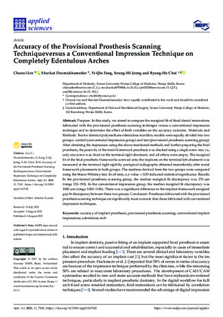
Figure 1.
Placed implants with multiple abutments.
Figure 2.
Connected impression copings.
Figure 1.
Placed implants with multiple abutments.

Appl. Sci.
2021
,
11
, 7182
3 of 10
Appl. Sci.
2021
,
11
, x FOR PEER REVIEW
3 of 12
Figure 1.
Placed implants with multiple abutments.
Figure 2.
Connected impression copings.
Figure 2.
Connected impression copings.
Appl. Sci.
2021
,
11
, x FOR PEER REVIEW
4 of 12
Figure 3.
Obtaining the pick-up impression.
Figure 4.
Creation of a stone model using implant analogs attached to copings.
Figure 3.
Obtaining the pick-up impression.
Appl. Sci.
2021
,
11
, x FOR PEER REVIEW
4 of 12
Figure 3.
Obtaining the pick-up impression.
Figure 4.
Creation of a stone model using implant analogs attached to copings.
Figure 4.
Creation of a stone model using implant analogs attached to copings.

Appl. Sci.
2021
,
11
, 7182
4 of 10
Appl. Sci.
2021
,
11
, x FOR PEER REVIEW
5 of 12
Figure 5.
Scan image of the stone cast.
Figure 6.
Designed metal framework.
2.2. Provisional Prosthesis Scanning Group
An identical polyurethane edentulous maxillary model with a soft tissue replica and
nine implants was used for each model. The cylinders were connected to the implant
(Figure 7). The rubber dam was placed beneath these cylinders (DIO Inc.) (Figure 8), and
a provisional prosthesis, with cylinder access holes that were designed using IO scan data
Figure 5.
Scan image of the stone cast.
Appl. Sci.
2021
,
11
, x FOR PEER REVIEW
5 of 12
Figure 5.
Scan image of the stone cast.
Figure 6.
Designed metal framework.
2.2. Provisional Prosthesis Scanning Group
An identical polyurethane edentulous maxillary model with a soft tissue replica and
nine implants was used for each model. The cylinders were connected to the implant
(Figure 7). The rubber dam was placed beneath these cylinders (DIO Inc.) (Figure 8), and
a provisional prosthesis, with cylinder access holes that were designed using IO scan data
Figure 6.
Designed metal framework.
2.2. Provisional Prosthesis Scanning Group
An identical polyurethane edentulous maxillary model with a soft tissue replica and
nine implants was used for each model. The cylinders were connected to the implant
(Figure
). The rubber dam was placed beneath these cylinders (DIO Inc.) (Figure
), and a
provisional prosthesis, with cylinder access holes that were designed using IO scan data
and CBCT scan data, was bonded with cylinders by injecting a composite luting cement
(VERICOM CO., Anyang, South Korea) (Figure
). After filling the provisional prosthesis
access holes, the denture was removed with preliminary bonded cylinders to complete this
process more precisely. Some laboratory tools (DIO Inc.) (Figure
) were used to protect
the base of the cylinders during the polishing process and to keep screw access holes open
to easily fill the empty spaces between the cylinder and denture by injecting acryl resin
(composite luting cement). The placed provisional prosthesis was scanned (TRIOS, 3Shape
A/S, Copenhagen, Denmark), exported as standard tessellation language files, and saved.
Once the virtual model of the jaws was created with the dental implant in position, a
virtual digital framework was designed using CAD software (Dental System, 3ShapeA/S,
Copenhagen, Denmark) (Figure
). The designed final metal framework was fabricated
(Arum 5x-200, Doowon, Daejeon, Republic of Korea) and positioned. This process was
repeated six times to produce six titanium frameworks.

Appl. Sci.
2021
,
11
, 7182
5 of 10
Appl. Sci.
2021
,
11
, x FOR PEER REVIEW
6 of 12
and CBCT scan data, was bonded with cylinders by injecting a composite luting cement
(VERICOM CO., Anyang, South Korea) (Figure 9). After filling the provisional prosthesis
access holes, the denture was removed with preliminary bonded cylinders to complete
this process more precisely. Some laboratory tools (DIO Inc.)(Figure 10) were used to
protect the base of the cylinders during the polishing process and to keep screw access
holes open to easily fill the empty spaces between the cylinder and denture by injecting
acryl resin (composite luting cement). The placed provisional prosthesis was scanned
(TRIOS, 3Shape A/S, Copenhagen, Denmark), exported as standard tessellation language
files, and saved. Once the virtual model of the jaws was created with the dental implant
in position, a virtual digital framework was designed using CAD software (Dental
System, 3ShapeA/S, Copenhagen, Denmark) (Figure 5). The designed final metal
framework was fabricated (Arum 5x-200, Doowon, Daejeon, Republic of Korea) and
positioned. This process was repeated six times to produce six titanium frameworks.
Figure 7.
Cylinders connected to the multiple abutments of the implant.
Figure 7.
Cylinders connected to the multiple abutments of the implant.
Appl. Sci.
2021
,
11
, x FOR PEER REVIEW
7 of 12
Figure 8.
Rubber dam placed beneath the cylinders.
Figure 9.
Provisional prosthesis with cylinder access holes.
Figure 8.
Rubber dam placed beneath the cylinders.
Appl. Sci.
2021
,
11
, x FOR PEER REVIEW
7 of 12
Figure 8.
Rubber dam placed beneath the cylinders.
Figure 9.
Provisional prosthesis with cylinder access holes.
Figure 9.
Provisional prosthesis with cylinder access holes.

Appl. Sci.
2021
,
11
, 7182
6 of 10
Appl. Sci.
2021
,
11
, x FOR PEER REVIEW
8 of 12
Figure 10.
Laboratory tools that facilitate the bonding process to keep the cylinder access holes open.
2.3. Examination
The fit of the full-arch frameworks was tested on the master model using a single
screw test (SST), i.e., only one screw was fixed on the last right abutment, and all others
were empty. The marginal fit of the final prosthetic frameworks screwed onto the
implants was checked on the last left abutment by periapical radiographs obtained after
metal framework placement in both groups. All periapical radiographies were
standardized, i.e., performing by the same surgeon, with the same radiographic machine
and angulation (90°, perpendicular to implant axis).The gap between the implant’s
abutment and base of prosthetic framework was measured in “µm”.
The medians derived from the models (two groups) were compared using the Mann–
Whitney test. In all tests, a
p
-value < 0.05 indicated statistical significance.
3. Results
A total of 12 identical polyurethane edentulous maxillary models with a soft tissue
replica were used, and 12 metal frameworks were manufactured using six models for each
group.
In the provisional prosthesis scanning group, the median marginal fit discrepancy
was 170 µm (range, 120–190).In the conventional impression group, the mean marginal fit
discrepancy was 1080 µm (range, 1040–1100). There was a significant difference in the
implant-framework marginal fit discrepancy between the two groups (Table 1).
Table 1.
Marginal fit discrepancy between the conventional impression group (CIG) and provisional
prosthesis scanning group (PPSG).
CIG
Mean±SM
PPSG
Mean±SM
Mann–Whitney
Test
Marginal fit discrepancy
(µm)
170 ± 50
1080 ± 40
p
< 0.05
4. Discussion
Figure 10.
Laboratory tools that facilitate the bonding process to keep the cylinder access holes open.
2.3. Examination
The fit of the full-arch frameworks was tested on the master model using a single
screw test (SST), i.e., only one screw was fixed on the last right abutment, and all others
were empty. The marginal fit of the final prosthetic frameworks screwed onto the implants
was checked on the last left abutment by periapical radiographs obtained after metal
framework placement in both groups. All periapical radiographies were standardized,
i.e., performing by the same surgeon, with the same radiographic machine and angulation
(90
◦
, perpendicular to implant axis). The gap between the implant’s abutment and base of
prosthetic framework was measured in “
µ
m”.
The medians derived from the models (two groups) were compared using the Mann–
Whitney test. In all tests, a
p
-value < 0.05 indicated statistical significance.
3. Results
A total of 12 identical polyurethane edentulous maxillary models with a soft tissue
replica were used, and 12 metal frameworks were manufactured using six models for
each group.
In the provisional prosthesis scanning group, the median marginal fit discrepancy
was 170
µ
m (range, 120–190). In the conventional impression group, the mean marginal
fit discrepancy was 1080
µ
m (range, 1040–1100). There was a significant difference in the
implant-framework marginal fit discrepancy between the two groups (Table
Table 1.
Marginal fit discrepancy between the conventional impression group (CIG) and provisional
prosthesis scanning group (PPSG).
CIG
Mean
±
SM
PPSG
Mean
±
SM
Mann–Whitney
Test
Marginal fit discrepancy (
µ
m)
170
±
50
1080
±
40
p
< 0.05
4. Discussion
In this study, we aimed to compare the marginal fit of fixed dental restorations fabri-
cated with the provisional prosthesis scanning technique versus a conventional impression
technique and to determine the effect of both variables on the accuracy outcome. Achieving
passive fit of implant prosthesis is a key factor in the long-term success of treatment [
Our study showed that the fit of metal frameworks fabricated with the provisional pros-
thesis scanning technique was significantly more accurate than those fabricated with the
conventional impression open-tray technique. The results of a few previous studies have
demonstrated superior accuracy, in terms of trueness and precision of fully digital impres-
sions for full-arch restorations as compared with those of conventional techniques [

Appl. Sci.
2021
,
11
, 7182
7 of 10
Our results are in agreement with those by Jokstad et al. [
] who reported that the mean
vertical fit discrepancy of a prosthesis with a fully digital workflow was 169
µ
m.
Clinical studies have reported that an acceptable fit discrepancy is within the range
from 100 to 200
µ
m [
]. According to Jokstad et al. [
], a fit discrepancy < 200
µ
m
was considered to be clinically acceptable. The results of the present study lie within
these ranges for the provisional prosthesis scanning group, while those of the conven-
tional impression group lie outside the reported ranges of the acceptable misfit. The
above-mentioned findings highlight the potential advantages of the so-called complete
digital workflow.
Impression accuracy and the fit of the definitive prosthesis depend on the phases of the
process. Various factors can contribute to a misfit of the final prosthesis, such as the accuracy
of implant impression, master cast fabrication, and prosthesis fabrication procedures [
conventional techniques, each step (connection of impression copings, obtaining impres-
sion, connection of implant analogs, mechanical tolerance in each step, creating a stone cast,
scanning, merging process, designing, and milling) has its own errors, and accumulating
all of these has a significant effect on the accuracy of the final fit [
]. In
contrast, the provisional prosthesis scanning technique requires fewer steps (i.e., creating
provisional prosthesis, scanning, designing, and milling); consequently, the number of
error sources is fewer as compared with the conventional techniques [
]. This can
explain the significant marginal fit discrepancy between these two methods in our study.
Understanding the impact of these factors helps to ensure the fabrication of an accurate
master cast, which reduces the risk of framework misfit.
Using fully digital workflow minimizes complications associated with framework
misfit. In the present study, a relatively high variation of precision in the provisional
prosthesis scanning group can be caused by cylinder displacement. One of the common
reasons is cylinder damage which can occur during the prosthesis polishing process. To
eliminate its effect on the final prosthesis accuracy, the cylinder base should be protected
using polishing caps during the polishing process. Another key factor that hinders the full
sitting of cylinders is an ill-fitting prosthesis screw access hole with cylinders connected
to the implant during the bonding process. In this case, the final accuracy depends on
the skills of the surgeon and their precision in applying adequate mechanical power and
fixing/bonding in the correct angle, which plays a key role in achieving a satisfactory final
result. The next important aspect, which depends on the skills/methods of the surgeon, is
the scanning process, in which the surgeon scanning technique is considered to be more
crucial than the influence of various intraoral scanners on the accuracy of the results [
To assess the effect of misfit in clinical studies, most authors have attempted to measure
the gap between the framework and abutments. The importance of a vertical fit has been
emphasized in several studies [
]. However, there is still no standard protocol to
assess the fit of dental restorations [
].There are several methods for assessing the fit
of screw-retained implant prostheses that have been used by others [
]; however,
SST continues to be a simple and popular method to use both clinically and in dental
laboratories [
].Moreover, this method is considered to be highly informative for the
estimation of workflow accuracy despite its ordinariness. Thus, in the present study,
qualitative assessment of the vertical microgap was performed with SST.
The findings of the current study can be beneficial in the clinical setting for both
surgeons and patients. First, less time is required for the whole process with a reduction in
the preparation efforts by the surgeon (total, 9 h; designing, 3 h; milling, 5.5 h; and finishing
in patient mouth, 0.5 h) and there is a decrease in the number of patient visits.
The provisional prosthesis scanning technique is a relatively new method involving a
fully digital workflow. To the best of our knowledge, there have been no published studies
on the role of the provisional prosthesis scanning method in improving the accuracy of
full-arch implant prosthesis on the edentulous ridge. This limits the comparison of current
findings with the results of other studies. Nevertheless, the results of the present study for
the provisional prosthesis scanning group lie within the clinically acceptable range, and

Appl. Sci.
2021
,
11
, 7182
8 of 10
the findings were derived from a model experiment. Further clinical studies are required
to confirm these outcomes in the clinical sphere.
5. Conclusions
In general, our findings suggest that prostheses fabricated with the provisional pros-
thesis scanning technique are significantly more accurate than those fabricated with conven-
tional impression techniques. Nevertheless, a model experiment does not always produce
predictable and possible uncontrolled cause and effect out comes in natural conditions.
Hence, further in vivo investigations are required to determine whether the results of this
study are consistent with clinical findings.
Author Contributions:
S.D., writing—original draft preparation, and investigation; C.L., conceptual-
ization, funding acquisition, and formal analysis; Y.-Q.F., software; S.-M.J., resources and supervision;
B.-H.C., writing—project administration, review and editing, and supervision. All authors have read
and agreed to the published version of the manuscript.
Funding:
This research did not receive any specific grant from funding agencies in the public,
commercial, or not-for-profit sectors.
Institutional Review Board Statement:
Not applicable.
Informed Consent Statement:
Not applicable.
Conflicts of Interest:
The authors declare no conflict of interest.
References
1.
Jemt, T.; Hjalmarsson, L. In vitro measurements of precision of fit of implant-supported frameworks. A comparison between
“virtual” and “physical” assessments of fit using two different techniques of measurements.
Clin. Implant. Dent. Relat. Res.
2011
,
14
, e175–e182. [
2.
Papaspyridakos, P.; Lal, K. Computer-assisted design/computer-assisted manufacturing zirconia implant fixed complete pros-
theses: Clinical results and technical complications up to 4 years of function.
Clin. Oral Implant. Res.
2012
,
24
, 659–665.
3.
Papaspyridakos, P.; Chen, C.-J.; Chuang, S.-K.; Weber, H.-P.; Gallucci, G.O. A systematic review of biologic and technical
complications with fixed implant rehabilitations for edentulous patients.
Int. J. Oral Maxillofac. Implant.
2012
,
27
, 134–139.
4.
Papaspyridakos, P.; Gallucci, G.O.; Chen, C.J.; Hanssen, S.; Naert, I.; Vandenberghe, B. Digital versus conventional implant
impressions for edentulous patients: Accuracy outcomes.
Clin. Oral Implant. Res.
2016
,
27
, 465–472. [
5.
Di Fiore, A.; Monaco, C.; Brunello, G.; Granata, S.; Stellini, E.; Yilmaz, B. Automatic digital design of the occlusal anatomy of
monolithic zirconia crowns compared to dental technicians’ DigitalWaxing: A controlled clinical trial.
J. Prosthodont.
2021
,
30
,
104–110. [
6.
Karlsson, S. The fit of Procera titanium crowns: An in vitro and clinical study.
Acta Odontol. Scand.
1993
,
51
, 129–134. [
7.
Od
é
n, A.; Andersson, M.; Krystek-Ondracek, I.; Magnusson, D. Five-year clinical evaluation of Procera AllCeram crowns.
J.
Prosthet. Dent.
1998
,
80
, 450–456. [
8.
Heckmann, S.M.; Karl, M.; Wichmann, M.G.; Winter, W.; Graef, F.; Taylor, T.D. Cement fixation and screw retention: Parameters
of passive fit. An in vitro study of three-unit implant-supported fixed partial dentures.
Clin. Oral Implant. Res.
2004
,
15
, 466–473.
9.
Zhang, R.; Ding, Q.; Sun, Y.; Zhang, L.; Xie, Q. Assessment of CAD-CAM zirconia crowns designed with 2 different methods: A
self-controlled clinical trial.
J. Prosthet. Dent.
2018
,
120
, 686–692. [
10.
Di Fiore, A.; Meneghello, R.; Graiff, L.; Savio, G.; Vigolo, P.; Monaco, C.; Stellini, E. Full arch digital scanning systems performances
for implant-supported fixed dental prostheses: A comparative study of 8 intraoral scanners.
J. Prosthodont. Res.
2019
,
63
, 396–403.
11.
Bilmenoglu, C.; Cilingir, A.; Geckili, O.; Bilhan, H.; Bilgin, T. In vitro comparison of trueness of 10 intraoral scanners for
implant-supported complete-arch fixed dental prostheses.
J. Prosthet. Dent.
2020
,
124
, 755–760. [
12.
Mangano, F.G.; Admakin, O.; Bonacina, M.; Lerner, H.; Rutkunas, V.; Mangano, C. Trueness of 12 intraoral scanners in the
full-arch implant impression: A comparative in vitro study.
BMC Oral Health
2020
,
20
, 1–21. [
13.
Besimo, C.; Jeger, C.; Guggenheim, R. Marginal adaptation of titanium frameworks produced by CAD/CAM techniques.
Int. J.
Prosthodont.
1998
,
10
, 154–160.
14.
May, K.B.; Russell, M.M.; Razzoog, M.E.; Lang, B.R. Precision of fit: The ProceraAllCeram crown.
J. Prosthet. Dent.
1998
,
80
,
394–404. [
15.
De Francesco, M.; Stellini, E.; Granata, S.; Mazzoleni, S.; Ludovichetti, F.S.; Monaco, C.; Di Fiore, A. Assessment of fit on ten
screw-retained frameworks realized through digital full-arch implant impression.
Appl. Sci.
2021
,
11
, 5617. [

Appl. Sci.
2021
,
11
, 7182
9 of 10
16.
Abduo, J.; Bennani, V.; Waddell, N.; Lyons, K.; Swain, M. Assessing the fit of implant fixed prostheses: A critical review.
Int. J.
Oral Maxillofac. Implant.
2010
,
25
, 506–515.
17.
Kan, J.Y.; Rungcharassaeng, K.; Bohsali, K.; Goodacre, C.J.; Lang, B.R. Clinical methods for evaluating implant framework fit.
J. Prosthet. Dent.
1999
,
81
, 7–13. [
18.
Di Fiore, A.; Meneghello, R.; Savio, G.; Sivolella, S.; Katsoulis, J.; Stellini, E. In vitro implant impression accuracy using a new
photopolymerizing sdr splinting material.
Clin. Implant. Dent. Relat. Res.
2015
,
17
, e721–e729. [
19.
Jemt, T.; Book, K. Prosthesis misfit and marginal bone loss in edentulous implant patients.
Int. J. Oral Maxillofac. Implant.
1996
,
11
, 142–150.
20.
Eckert, S.E.; Meraw, S.J.; Cal, E.; Ow, R.K. Analysis of incidence and associated factors with fractured implants: A retrospective
study.
Int. J. Oral Maxillofac. Implant.
2000
,
15
, 155–163.
21.
Abduo, J.; Judge, R.B. Implications of implant framework misfit: A systematic review of biomechanical sequelae.
Int. J. Oral
Maxillofac. Implant.
2014
,
29
, 608–621. [
22.
Alshawaf, B.; Kudara, Y.; Weber, H.P. Management of technical complications during full-mouth implant rehabilitation with
hybrid prostheses over a 7-year period.
Compend. Contin. Educ. Dent.
2018
,
39
, 1–4.
23.
Box, V.H.; Sukotjo, C.; Knoernschild, K.L.; Campbell, S.D.; Afshari, F.S. Patient-Reported and Clinical Outcomes of Implant-
Supported Fixed Complete Dental Prostheses: A Comparison of Metal-Acrylic, Milled Zirconia, and Retrievable Crown Prostheses.
J. Oral Implant.
2018
,
44
, 51–61. [
24.
Gonzalez, J.; Triplett, R.G. Complications and clinical considerations of the implant-retained zirconia complete-arch prosthesis
with various opposing dentitions.
Int. J. Oral Maxillofac. Implant.
2017
,
32
, 864–869. [
25.
Albader, B.; Alhelal, A.; Proussaefs, P.; Garbacea, A.; Kattadiyil, M.; Lozada, J. Digitally Milled Metal Framework for Fixed
Complete Denture with Metal Occlusal Surfaces: A Design Concept.
Int. J. Periodontics Restor. Dent.
2017
,
37
, 180–188. [
26.
Jemt, T. Failures and complications in 391 consecutively inserted fixed prostheses supported by Branemark Implants in edentulous
jaws: A study of treatment from the time of prosthesis placement to the first annual checkup.
Int. J. Oral Maxillofac. Implant.
1991
,
6
, 89–102.
27.
Malo, P.; Nobre, M.; Guedes, C.; Almeida, R. Outcomes of immediate function implant prosthetic restorations with mechanical
complications: A retrospective clinical study with 5 years of follow-up.
Eur. J. Prosthodont. Restor. Dent.
2017
,
25
, 26–34.
28.
Yilmaz, B.; Gilbert, A.B.; Seidt, J.D.; McGlumphy, E.A.; Clelland, N.L. Displacement of implant abutments following initial and
repeated torqueing.
Int. J. Oral Maxillofac. Implant.
2015
,
30
, 1011–1018. [
29.
Balshi, T.J. An analysis and management of fractured implants: A clinical report.
Int. J. Oral Maxillofac. Implant.
1996
,
11
, 666.
30.
Amin, S.; Weber, H.P.; Finkelman, M.; El Rafie, K.; Kudara, Y.; Papaspyridakos, P. Digital vs. conventional full-arch implant
impressions: A comparative study.
Clin. Oral Implant. Res.
2017
,
28
, 1360–1367. [
31.
Ahlholm, P.; Sipilä, K.; Vallittu, P.; Jakonen, M.; Kotiranta, U. Digital Versus Conventional Impressions in Fixed Prosthodontics: A
Review.
J. Prosthodont.
2018
,
27
, 35–41. [
32.
Abdel-Azim, T.; Elathamna, E.; Lin, W.; Zandinejad, A.; Morton, D. The Influence of Digital Fabrication Options on the Accuracy
of Dental Implant–Based Single Units and Complete-Arch Frameworks.
Int. J. Oral Maxillofac. Implant.
2014
,
29
, 1281–1288.
33.
Albdour, E.A.; Shaheen, E.; Vranckx, M.; Mangano, F.G.; Politis, C.; Jacobs, R. A novel in vivo method to evaluate trueness of
digital impressions.
BMC Oral Health
2018
,
18
, 117. [
34.
Alikhasi, M.; Siadat, H.; Nasirpour, A.; Hasanzade, M. Three-Dimensional Accuracy of Digital Impression versus Conventional
Method: Effect of Implant Angulation and Connection Type.
Int. J. Dent.
2018
,
2018
, 1–9. [
35.
Zimmermann, M.; Koller, C.; Rumetsch, M.; Ender, A.; Mehl, A. Precision of guided scanning procedures for full-arch digital
impressions in vivo.
J. Orofac. Orthop.
2017
,
78
, 466–471. [
36.
Jokstad, A.; Shokati, B. New 3D technologies applied to assess the long-term clinical effects of misfit of the full jaw fixed prosthesis
on dental Implant.
Clin. Oral Implant. Res.
2014
,
26
, 1129–1134. [
37.
Tiossi, R.; Rodrigues, R.C.S.; Mattos, M.D.G.C.D.; Ribeiro, R.F. Comparative analysis of the fit of 3-unit implant-supported
frameworks cast in nickel-chromium and cobalt-chromium alloys and commercially pure titanium after casting, laser welding,
and simulated porcelain firings.
Int. J. Prosthodont.
2008
,
21
, 234–244.
38.
Wettstein, F.; Sailer, I.; Roos, M.; Hämmerle, C.H.F. Clinical study of the internal gaps of zirconia and metal frameworks for fixed
partial dentures.
Eur. J. Oral Sci.
2008
,
116
, 272–279. [
39.
Papaspyridakos, P.; Chen, C.-J.; Gallucci, G.O.; Doukoudakis, A.; Weber, H.-P.; Chronopoulos, V. Accuracy of Implant Impressions
for Partially and Completely Edentulous Patients: A Systematic Review.
Int. J. Oral Maxillofac. Implant.
2014
,
29
, 836–845.
40.
Joda, T.; Bragger, U. Time-efficiency analysis of the treatment with monolithic implant crowns in a digital workflow: A randomized
controlled trial.
Clin. Oral Implant. Res.
2016
,
27
, 1401–1406. [
41.
Joda, T.; Brägger, U. Patient-centered outcomes comparing digital and conventional implant impression procedures: A random-
ized crossover trial.
Clin. Oral Implant. Res.
2016
,
27
, e185–e189. [
42.
Favero, R.; Volpato, A.; De Francesco, M.; Di Fiore, A.; Guazzo, R.; Favero, L. Accuracy of 3D digital modeling of dental arches.
Dent. Press J. Orthod.
2019
,
24
, 038e1–037e7. [

Appl. Sci.
2021
,
11
, 7182
10 of 10
43.
De Torres, E.M.; Barbosa, G.A.S.; Bernardes, S.R.; De Mattos, M.D.G.C.; Ribeiro, R.F. Correlation between vertical misfits and
stresses transmitted to implants from metal frameworks.
J. Biomech.
2011
,
44
, 1735–1739. [
44.
Al-Otaibi, H.N.; Akeel, R.F. The Effects of Two Torque Values on the Screw Preload of Implant-Supported Prostheses with Passive
Fit or Misfit.
Int. J. Oral Maxillofac. Implant.
2014
,
29
, 1058–1063. [
45.
MassignanBerejuk, H.; Hideo Shimizu, R.; Aparecida de Mattias Sartori, I.; Valgas, L.; Tiossi, R. Vertical microgap and passivity
of fit of three-unit implant-supported frameworks fabricated using different techniques.
Int. J. Oral Maxillofac. Implant.
2014
,
29
,
152–159.
46.
Colpani, J.T.; Borba, M.; Della Bona, A. Evaluation of marginal and internal fit of ceramic crown copings.
Dent. Mater.
2013
,
29
,
174–180. [
47.
Oka, Y.; Sasaki, J.-I.; Wakabayashi, K.; Nakano, Y.; Okamura, S.-Y.; Nakamura, T.; Imazato, S.; Yatani, H. Fabrication of a
radiopaque fit-testing material to evaluate the three-dimensional accuracy of dental prostheses.
Dent. Mater.
2016
,
32
, 921–928.
48.
Schaefer, O.; Kuepper, H.; Thompson, G.A.; Cachovan, G.; Hefti, A.F.; Guentsch, A. Effect of CNC-milling on the marginal and
internal fit of dental ceramics: A pilot study.
Dent. Mater.
2013
,
29
, 851–858. [
49.
Praça, L.; Pekam, F.C.; Rego, R.O.; Radermacher, K.; Wolfart, S.; Marotti, J. Accuracy of single crowns fabricated from ultrasound
digital impressions.
Dent. Mater.
2018
,
34
, e280–e288. [
50.
Zeller, S.; Guichet, D.; Kontogiorgos, E.; Nagy, W.W. Accuracy of three digital workflows for implant abutment and crown
fabrication using a digital measuring technique.
J. Prosthet. Dent.
2019
,
121
, 276–284. [
51.
Mai, H.N.; Lee, K.E.; Ha, J.-H.; Lee, D.-H. Effects of image and education on the precision of the measurement method for
evaluating prosthesis misfit.
J. Prosthet. Dent.
2018
,
119
, 600–605. [
52.
Park, J.-M.; Hämmerle, C.H.; Benic, G.I. Digital technique for in vivo assessment of internal and marginal fit of fixed dental
prostheses.
J. Prosthet. Dent.
2017
,
118
, 452–454. [