


Биология ва тиббиёт муаммолари 2018, №2 (100) 163
UDC: 611.311.-573.7.017.6
FEATURES OF STRUCTURAL ALTERATION IN PERIODONTAL COMPLEX COMPONENTS AT
THE EARLY STAGE OF THE EXPERIMENTAL BACTERIAL-IMMUNE PERIODONITIS
DEVELOPMENT
A.YE. DEMKOVYCH
I. Horbachevsky Ternopil State Medical University, Ukraine, Ternopil
ТАЖРИБАДА БАКТЕРИАЛ-ИММУН ПАРОДОНТИТ РИВОЖЛАНИШИНИНГ ЭРТА
БОСҚИЧИДА ПАРОДОНТАЛ КОМПЛЕКС СТРУКТУРАЛИ АЛМАШИНУВ
КОМПОНЕНТЛАРИНИНГ ХУСУСИЯТЛАРИ
А.Е. ДЕМКОВИЧ
И.Я. Горбачевский номидаги Тернополь давлат медицина университети, Украина, Тернополь шаҳри
ОСОБЕННОСТИ СТРУКТУРНОЙ ПЕРЕСТРОЙКИ КОМПОНЕНТОВ ПАРОДОНТАЛЬНОГО
КОМПЛЕКСА НА РАННЕМ ЭТАПЕ РАЗВИТИЯ ЭКСПЕРИМЕНТАЛЬНОГО
БАКТЕРИАЛЬНО-ИММУННОГО ПАРОДОНТИТА
А.Е. ДЕМКОВИЧ
Тернопольский государственный медицинский университет им. И.Я. Горбачевского,
Украина. г. Тернополь
Пародонтдаги яллиғланиш жараёнлари стоматологик касалликлар ичида кенг тарқалган бўлиб,
қайталаниб кечиш билан характерланади ва беморлар ҳаёт сифатини ниҳоятда пасайтиради.
Пародонтал комплекс тўқимасида яллиғланиш жараёни ривожланишида мавжуд бўлган структурали
ва патоморфологик ўзгаришларнинг нисбийлиги ушбу касалликда деструктив белгиларнинг
ривожланиш босқичларини тўлиқ ифодаламайди, шунинг учун бу касаллик чуқур тадқиқотга муҳтож.
Тадқиқот мақсади – тажрибада бактериал-иммун пародонтит ривожланишининг 7-чи суткасида
пародонтал комплекс структурали компонентларининг хусусиятларини ўрганиш. Тадқиқот
натижасида соғлом ҳайвонларда хусусий пластинканинг бириктирувчи тўқимаси коллаген толалардан
иборат эканлиги, баланд бириктирувчи тўқимали сўрғичлар ҳосил қилиши ва эпителийга зич ёпишиши
аниқланди. Гистологик таҳлил натижасига кўра, тажрибадаги бактериал-иммун пародонтит
ривожланишининг 7-чи суткасида бутун пародонт бўйлаб типик яллиғланиш жараёни тарқалганлиги
маълум бўлди. Аммо бу жараённинг яққоллиги пародонтнинг структурали компонентларида ҳар хил
эканлиги аниқланди. Ҳўжайра элементлари ичида кўпинча фибробластлар учради, кам ҳолатда
гистиоцитлар, лимфоцитлар, якка плазмоцитлар ва моноцитлар учради. Тадқиқотнинг бу босқичида
фибробластлар миқдорининг ошиши кузатилди, булар коллаген толалар резорбция функциясини
бажариб, фаол остеобластлар миқдорини камайтирди. Бу барча патоморфологик ўзгаришлар
яллиғланиш жараёни ривожланиши белгиларидан дарак беради.
Калит сўзлар:
Пародонтит, яллиғланиш, периодонт, пролиферация, коллаген толалар.
The inflammatory process in the periodontium is the most widespread among dental diseases, that is
characterized by a recurrent course and significantly reduces the quality of life patients. Existing data about
structural and pathomorphological changes in the tissues of the periodontal complex for the of inflammation
development does not explain largely the patterns of the destructive phenomena development in this pathology.
and therefore require additional research. The purpose of this study was to determine the peculiarities of struc-
tural components of periodontal complex and their changes on the 7
th
day of the experimental bacterial-
immune periodontitis development. In the study of intact animals, it was found that connective tissue of its own
plate is represented by collagen fibers, forms high connective tissue papillae and closely adheres to the epithe-
lium. The histological study showed that on the 7th day of the experimental bacterial-immune periodontitis de-
velopment of typical and spread inflammatory reaction onto all periodontium was observed. However, the se-
verity of this reaction was different in the structural components of the periodontium. Fibroblasts were the most
often found among cellular elements, less frequently – histiocytes, lymphocytes, single plasmacytes and mono-
cytes. It was an increase of the fibroblasts number, that perform the resorptive functionof collagen fibers and
reduce number of active osteoblasts. All these pathomorphological changes testified about of the inflammatory
process development.
Key words:
Periodontitis, inflammation, periodontium, proliferation, collagen fibers.
Introduction.
The periodontal diseases by the
character of clinical course concern predominantly to
the chronic and complete inflammatory-destructive
changes in tissues that hold teeth in the alveolar bone,
and lead to progressive growth of connective tissue.
The data of epidemiological studies indicate that the
currency of periodontal diseases in the world ranges
from 5-20% and with age increases to 75% [1, 2]. In

Features of structural alteration in periodontal complex components at the early stage of the
…
164 2018, №2 (100) Проблемы биологии и медицины
recent years, generalized inflammatory diseases of
periodontium attract increased interest of researchers
and clinicians, because after 35 years age they lead to
extraction of teeth, increase the risk of associated sys-
temic pathology development. It is known that exact-
ly inflammatory processes that develop in the perio-
dontal complex are most often the main cause of teeth
loss [3, 4].
Features of the structural organization of tis-
sues that are part of the periodontium, promote to
damage with mechanical, chemical, bacterial and
immune factors, and the integrality of the structure
depends on maintaining appropriate level of metabol-
ic, microcirculatory processes, neuroendocrine regu-
lation [5, 6].
In the diagnostics of periodontal diseases to
clinical and morphological research is put the task of
determination degree, activity and spread of destruc-
tive process. On its base is defined an objective as-
sessment of the severity of the injury and an adequate
treatment tactic is worked out [7, 8]. In this regard,
expediently an experimental study of structural
changes in the soft and bone tissues of periodontium,
that forms under influence of pathogenetic processes,
which underlie of inflammatory and dystrophic dis-
orders in the periodontal complex [9, 10].
At present, scientific researches have received
enough material concerning structural changes as a
result inflammation in periodontal tissues, but the
consequentness and lawfullness of its formation are
far from being studied.
The purpose of this research is to determine
peculiarities of changes in the structural components
of the periodontal complex in the early period (on the
7
th
day) of the experimental bacterial-immune perio-
dontitis development and to give them a prognostic
assessment.
Materials and methods.
The experimental
bacterial-immune periodontitis was induced in the
experimental animals by introducing complex mix-
tures of microorganisms diluted in egg protein into
periodontal tissue [11]. Simultaneously with the in-
jections of the microbial pathogen, a complete
Freund’s adjuvant was injected in the rat's paw to
enhance the immune response. The experimental
studies were performed with the use of clinically
healthy rats weighing 150-200 g in the conditions of
vivarium. The animals were in a standard diet bal-
anced by the main elements of nutrition. Systemati-
cally healthy rats of the same age were used as con-
trols. The reseach was performed in accordance with
sanitary-hygienic norms and GLP requirements. The
experiments were conducted in conformity with the
general rules and provisions of the "European Con-
vention for the Protection of Vertebrate Animals
Used for Experimental and Other Scientific Purpos-
es" (Strasbourg, 1986), and the "General Ethical
Principles of Animal Experimentation" (Kyiv, 2001).
For evaluation of the degree structural changes
in the tissues of the maxillofacial area was used a
morphological study. The experimental animals were
sacrificed on the 7
th
day through decapitation under
thiopental anesthesia. The fragments of the mandible
tissues, in particular, the periodontal complex were
removed, washed in saline from the blood and fixed
in 10% neutral formalin solution. The material was
poured in paraffin blocks. The transverse sections on
a microtome were made in the thickness of 5-6 mi-
crons. The resulting preparations were stained with
hematoxylin and eosin [12].
Results and discussion.
Considering that his-
tological norm of periodontal components is known
and described in the literature [13, 15], however ex-
isting individual variability of tissues there is necessi-
ty to study them in the group of intact animals.
The mucous membrane of the rat’s gums is
covered with a multilayer plane epithelium with ex-
pressed signs of keratinization and parakeratosis be-
sides corneal layer was uneven thickness and has
view homogeneous eosinophilic strip. The usual phe-
nomenon was small areas of superficial desquamation
of the epithelium.
The granular and spinal layers located below
had not layer-by-layer differentiation, as opposed to
basal, which clearly defines the boundary of the epi-
thelial lining and its own plate of the mucous mem-
brane. The dystrophic changes of the epithelial cells
in this group were presented formations with weak to
moderately expressed. In such cases in the cytoplasm
of the keratinocytes was visualized vacuoles of dif-
ferent sizes and different amounts.
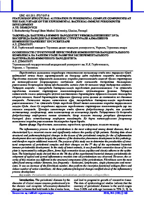
The connective tissue of its own plate was rep-
resented by collagen fibers, which formed high con-
nective tissue papillae and tightly adjacent to the epi-
thelium. The papillary layer was presented with a
loose connective tissue. The collagen fibers in deeper
part had more compact, dense, location and transited
into the periosteum of the cortical plate of the alveo-
lar processes. Among the cellular elements, fibro-
blasts were found most frequently, lesser frequently –
histiocytes, lymphocytes, single plasmacytes and
monocytes (Figure 1).
The own plate was intensively vascularized,
especially in the superficial layers. Numerous blood
capillaries of the surface layer had narrowed lumen,
almost did not contain form elements and therefore
poorly visualized. The hemocapillar wall contain s
plane endothelial cells that located on the basement
membrane. The periodontium was represented by
sidelong directed bundles of oxyphilic collagen fi-
bers, between that layers of loose connective tissue
with thin wall vessels were visualized. The cellular
composition was diverse, in particular, it included
epithelial and osteogenic basophils, undifferentiated
mesenchymal cells, except of fibroblasts, which were
the predominant type of cells (Figure 2).

A.Ye. Demkovych
Биология ва тиббиёт муаммолари 2018, №2 (100) 165
Figure 1
– Structural organization of the rat’s gum of the control group.
Staining with hematoxylin and eosin. × 200
Figure 2
– Structural organization of the rat’s periodontium of the control group. Staining with hematoxylin
and eosin. × 200
The cement of the tooth roots is constructed
with collagen fibers, which contained layers of amor-
phous matter. Cellular elements of the cement ap-
peared closer to the top of the root – cementoblasts
and cementocytes. Vessels in this structure were not
found.
The alveolar process, which surrounds the
tooth root, represents a thin bone plate, to which the
periodontal ligament attached. The bone plate, in
one’s turn, consist of plate bone tissue, it formed os-
teomas and was infiltrated with periodontal fibers,
vessels and nerves.
Own alveolar bone of the alveolar wall passed
into a supporting alveolar bone, covered with a corti-
cal plate and formed by compactly composed bone
plates. The osteocytes had an elongated shape and
were placed individually in clearly-contoured gaps in
the matrix.
The matrix itself was stained poorly but even-
ly. Orsene fibers had a multi-directional placement,
due to which the nearest layers of the fabric had dif-
ferent optical properties. The presented vessels in cut
were moderately blood-contented. The superficial
layer of the periosteum was represented by collagen

Features of structural alteration in periodontal complex components at the early stage of the
…
166 2018, №2 (100) Проблемы биологии и медицины
fibers, located parallely to the surface of the bone,
with a small number of fibroblasts.
The deep – osteogenic layer was visualized un-
clearly and contained thin spindle-like osteogenic
cells. The osteoclastes and osteoblasts appeared rare-
ly. Sites between plate of spongy bones were filled in
with red and yellow bone marrow rarely (Figure 3).
The analysis of tissue changes on the 7
th
day of
the experimental bacterial-immune periodontitis was
characterized by development of extensive inflamma-
tory process during whole periodontal period. How-
ever, the severity of this reaction was different in the
structural components of periodontum.
So, pathological changes in the gum mucus
were found for that period. The epithelial lining, as a
rule, became uneven thickness at that stage, which
arose mainly due to desquamation of epithelial cells.
The epithelium reacted with proliferation to the in-
flammatory process in a certain areas.
At the same time with noted above changes,
the epithelial plate not infrequently became visually
interrupted and preserved papillae of the surface layer
its own plate reached to the surface of the epithelium.
The keratinous r was also uneven thickness and inter-
rupted, it was raised above the osteal layer.
Layer-by-layer differentiation of the epithelium
was retained, but unclear especially in the surface
layers. Dystrophic changes of the cell occured on
the
spot. Their cytoplasm became light, vacuolated, and
the picnotic nucleus was displaced to periphery. Such
changes were interpreted as so-called balloon dystro-
phy.
The cells with a hyperbasophilic festoon nu-
cleus, surrounded by an empty ring-shaped space oc-
curred in the suprabasal and basal layers. The cells
with similar changes contrasted sharply with intact
keratinocytes. The presence of these cells suggested
strengthening of induced apoptosis.
Many lymphocytes were found among the
keratinocytes. The clear line of the basal layer was
blurred at the expense of inflammatory infiltration in
the papillae of its own plate. In the infiltrate were
dominated neutrophils. The pappilae of their own
plate were different height and thickness, somewhere
polished with polymorphocytic infiltration (Figure 4).
The bundles of periodontiumal collagen fibers
somewhat were loosened and lost their clarity, their
contours were uncleared and became homogeneous.
The oxyphilic stainig somewhat changed onto baso-
philic. The amorphous component of its own plate,
which became more enlightened and heterogeneous
as compared with the previous observation period. Its
areas expanded parallely.
Thus, on the base of that results, it may be con-
sider that all signs of disorganization of connective
tissue components occured. Both surface and deep
layers were revealed lymphahistiocytic infiltrates
with an admixture of neutrophils.
The capillaries and small blood vessels of the
arterial type were uneven blood-filled with a predom-
inance of hypertrophy and stasis. The veins were also
mainly congested (Figure 5).
The bundles of periodontal collagen fibrils
were somewhat loosened and lost their clarity. The
layers of loose connective tissue that situated between
them were enlarged both at the expense of the inter-
cellular fluid, and at the expense of cellular elements
that were found in that structure in the norm.
Somewhere in the infiltrate were found neutro-
phils. It should be noted that increase separately of
number fibroblasts, performing the function of re-
sorption of collagen fibres, promoted to remodeling
of periodontium already at the initial stages of exper-
imental study. The microcirculatory stream was une-
venly blood-filled, with moderately expressed signs
of hemodynamic disorders: stasis, erythrocytic
sludge.
There were minimal changes in the cement of
the tooth root for this experimental period that char-
acterized the initial signs of disorganization of the
connective tissue in the form of fibre structure viola-
tion and swelling of the collagen fibers.
Some disturbances in the alveolar bone struc-
ture after 7 days of the experiment were not found.
The alveolar bone tissue in the experimental animals
remained characteristic structure. The bodies of the
osteocytes localized in the gaps, the walls of which
were contoured somewhat weaker than in the control
animals. The cells became more oval shape, the nu-
clei were hyperchromic, that caused condensation of
chromatin. The osteoid between the gaps was homo-
geneous and somewhat enlightened.
The osein fibrils in the plates were clearly ori-
ented. Only moderate blood filling was observed in
the blood vessels. There were gigantic oxyphilic mul-
tinuclear cells – osteoclast localized near of the ves-
sels in the superficial areas of the bone. The matrix
around them looked slightly enlightened. The bone
plates were thinned and found focal hollow – lacunar
resorption. The cell nucleuses were also hypochro-
matic in the periosteal osteogenic layer. The numbers
of active osteoblasts were decreased (Figure 6).

A.Ye. Demkovych
Биология ва тиббиёт муаммолари 2018, №2 (100) 167
Figure 3
– Structural organization of the bone tissue rat’s alveolar process of the control group.
Staining with hematoxylin and eosin. × 200
Figure 4
– Histological structure of the rat’s gum after 7 days of the experiment.
Dense round cell infiltration of the mucous membrane own plate. Balloon dystrophy of keratinocytes.
Staining with hematoxylin and eosin. × 100.

Features of structural alteration in periodontal complex components at the early stage of the
…
168 2018, №2 (100) Проблемы биологии и медицины
Figure 5
– Histological structure of the rat’s gum own plate after 7 days of experiment.
Swelling and homogenization of the collagen fibers, dense leukocytic infiltration, predominant venous
hyperemia and leukostasis in the vessels. Staining with hematoxylin and eosin. × 200
Figure 6
– Histological structure of the rat’s alveolar bone process after 7 days of the experiment.
Cell proliferation of the mesenchyma. Initial signs of lacunar resorption of bone plates.
Staining with hematoxylin and eosin. × 200
Conclusion:
1.
Progressing inflammatory
changes in the structural components of the animal
periodontal complex arise for early period (on the 7
th
day of the study) of the experimental bacterial-
immune periodontitis development that display by
disorganization and destruction of connective tissue
and walls of tooth alveolus’s, structural reconstruc-
tion of epithelial gum lining and its own plate, cellu-
lar infiltration and microcirculatory disorders. 2. The
character of pathomorphological changes may be a
sign of deep destructive-dystrophic processes that
form clinical picture of periodontitis with protracted
course.

A.Ye. Demkovych
Биология ва тиббиёт муаммолари 2018, №2 (100) 169
References:
1.
Булкина Н. В., Моргунова В. М. Современные
аспекты этиологии и патогенеза воспалительных
заболеваний пародонта. Особенности клиниче-
ских проявлений рефрактерного пародонтита
//Фундаментальные исследования. – 2012. – №. 2-
2.
2.
Демкович А. Є., Бондаренко Ю. І. Патогене-
тичні основи моделювання пародонтиту у тварин
//Здобутки клінічної і експериментальної медици-
ни. – 2015. – Т. 22. – №. 1.
3.
Arimatsu K, Yamada H, Miyazawa H, Minaqawa
T, Nakajima M, Ryder MI, Gotoh K, Motooka D,
Nakamura S, Iida T, Yamazaki K: Oral pathobiont
induces systemic inflammation and metabolic chang-
es associated with alteration of gut microbiota. Sci
Rep 6(4), 4828 (2014)
4.
Borisenko A. (2013): Periodontal disease. Medi-
cine, Kyiv, 455 p
5.
Dimitrova A, Kolenko Y: Evaluating the effec-
tiveness of various immunomodulators in complex
treatment generalized periodontitis in young adults
(18-25 years). J Modern Dentistry 2, 38-39 (2013)
6.
Demkovych A, Bondarenko Yu, Hasiuk PA: Oxi-
dative modification of proteins in the process of ex-
perimental periodontitis development. Interventional
Medicine and Applied Science 9(4), 218-221 (2017)
7.
Demkovych AYe, Bondarenko YuI: Pathogenetic
basis periodontitis modeling in rats. Achiev of Clin
and Exper Med 1(22), 54-57 (2015)
8.
Di Benedetto A, Gigante I, Colucci, S, Grano M:
Periodontal disease: linking the primary inflammation
to bone loss. Clin Dev Immunol 2013, 503754
(2013).
9.
Hyde S, Dupuis V, Mariri BP, Dartevelle S: Pre-
vention of tooth loss and dental pain for reducing the
global burden of oral diseases. Int Dent J 2, 19-25
(2017)
10.
Ioannidou E: The Sex and Gender Intersection in
Chronic Periodontitis. Front Public Health 5, 189
(2017)
11.
Chapple IL, Van der Weijden F, Doerfer C, Herre-
ra D, Shapira L, Polak D: Primary prevention of peri-
odontitis: managing gingivitis. J Clin Periodontol 16,
71-76 (2015)
12.
Livingstone D, Murthy V, Reddy VK, Pillai A:
Prosthodontic rehabilitation of a patient with aggres-
sive periodontitis. BMJ Case Rep 5, 2015 (2015)
13.
Slots J: Periodontitis: facts, fallacies and the fu-
ture. Periodontol 2000 75(1), 7-23 (2017)
14.
Hodovana O: Investigation of the level of mineral
density of skeletal osseous tissue in patients with per-
iodontal tissue diseases. Lik Sprava 7-8, 123-128
(2015)
15.
Chan JK: The wonderful colors of the hematoxy-
lin-eosin stain in diagnostic surgical pathology. Int J
Surg Pathol 22(1), 12-32 (2014)
16.
Menicanin D, Hynes K, Han J, Grothos S, Bartold
PM: Cementum and Periodontal Ligament Regenera-
tion. Adv Exp Med Biol 881, 207-236 (2015)
17.
Sokos D, Everts V, de Vries TJ: Role of periodon-
tal ligament fibroblasts in osteoclastogenesis: a re-
view. J Periodontal Res 50(2), 152-159 (2015)
ОСОБЕННОСТИ СТРУКТУРНОЙ
ПЕРЕСТРОЙКИ КОМПОНЕНТОВ
ПАРОДОНТАЛЬНОГО КОМПЛЕКСА НА
РАННЕМ ЭТАПЕ РАЗВИТИЯ
ЭКСПЕРИМЕНТАЛЬНОГО
БАКТЕРИАЛЬНО-ИММУННОГО
ПАРОДОНТИТА
А.Е. ДЕМКОВИЧ
Тернопольский государственный медицинский
университет им. И.Я. Горбачевского,
Украина, г. Тернополь
Воспалительные процессы в пародонте от-
носятся к числу наиболее распространенных сто-
матологических заболеваний, характеризуется
рецидивирующим течением и существенно сни-
жает качество жизни пациентов. Существующие
данные относительно структурных и патоморфо-
логических изменений в тканях пародонтального
комплекса в процессе развития воспаления не в
полной мере объясняют закономерности развития
деструктивных явлений при данной патологии, а
потому нуждаются в дополнительных исследова-
ний. Целью данного исследования было изучить
особенности структурных компонентов пародон-
тального комплекса и их изменений на 7-е сутки
развития
экспериментального
бактериально-
иммунного пародонтита. При исследовании ин-
тактных животных установлено, что соедини-
тельная ткань собственной пластинки представ-
лена коллагеновыми волокнами, образует высо-
кие соединительнотканные сосочки и плотно
прилегает к эпителию. Гистологическое исследо-
вание показало, что на 7-е сутки развития экспе-
риментального бактериально-иммунного паро-
донтита наблюдалось развитие типичной и рас-
пространенной на весь пародонт воспалительной
реакции. Однако выраженность этой реакции бы-
ла различной в структурных компонентах паро-
донта. Среди клеточных элементов наиболее ча-
сто встречаются фибробласты, реже – гистиоци-
ты, лимфоциты, единичные плазмоциты и моно-
циты. На этом этапе исследования происходило
увеличение количества фибробластов, которые
выполняя функцию резорбции коллагеновых во-
локон и уменьшение количества активных осте-
областов. Все эти патоморфологические измене-
ния указывают на признаки развития воспали-
тельного процесса.
Ключевые слова:
Пародонтит, воспаление,
периодонт, пролиферация, коллагеновые волокна.