
All articles
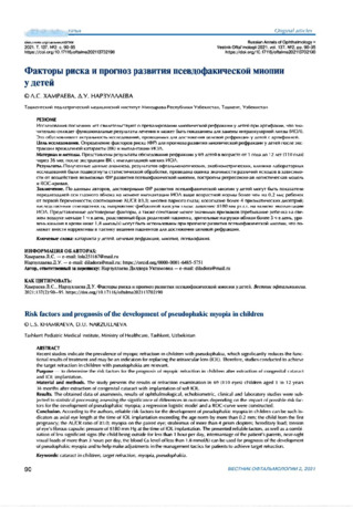
Risk factors and prognosis of the development of pseudophakic myopia in children
Recent studies indicate the prevalence of myopic refraction in children with pseudophakia, which significantly reduces the functional results of treatment and may be an indication for replacing the intraocular lens (IOL). Therefore, studies conducted to achieve the target refraction in children with pseudophakia are relevant.
Purpose — to determine the risk factors for the prognosis of myopic refraction in children after extraction of congenital cataract and IOL implantation.
Material and methods. The study presents the results of refraction examination in 69 (110 eyes) children aged 1 to 12 years 36 months after extraction of congenital cataract with implantation of soft IOL.
Results. The obtained data of anamnesis, results of ophthalmological, echobiometric, clinical and laboratory studies were subjected to statistical processing assessing the significance of differences in outcomes depending on the impact of possible risk factors for the development of pseudophakic myopia; a regression logistic model and a ROC-curve were constructed.
Conclusion. According to the authors, reliable risk factors for the development of pseudophakic myopia in children can be such indicators as axial eye length at the time of IOL implantation exceeding the age norm by more than 0.2 mm; the child from the first pregnancy; the AL/CR ratio of 23.0; myopia on the paired eye; strabismus of more than 4 prism diopters; hereditary load; tension of eye's fibrous capsule: pressure of SI 80 mm Hg at the time of IOL implantation. The presented reliable factors, as well as a combination of less significant signs (the child being outside for less than 1 hour per day, intermarriage of the patients parents, near-sight visual loads ot more than 3 hours per day, the blood Ca level of less than 1.8 mmol/L) can be used for prognosis of the development of pseudophakic myopia and to help make adjustments in the management tactics for patients to achieve target refraction.

Retrospective analysis of the structure of closed-eye injuries in children
AIM: This study aimed to analyze retrospectively the status of closed-eye injuries in children based on clinic data of the Tashkent Pediatric Medical Institute (TashPMI) from 2018 to 2022 .
MATERIAL AND METHODS: The study analyzed TashPMI clinic’s reporting records for 2018–2022 .
RESULTS: During the reporting period, 5,938 patients with various diseases of the eye and its appendages were treated . Of these, 1,438 (24 .2%) patients were diagnosed with eye traumas and their complications . Among complications of closed eye injury, retinal detachment and traumatic cataracts accounted for 8 .3% and 7 .5%, respectively. Closed eye injury were more common among boys (57 .2%) aged 5–14 years (64 .4%) . From 2018 to 2022, the number of patients with post-contusion retinal detachment declined, despite a persistent upward trend in the number of hospitalized patients with blunt traumas . This decline was likely due to the timely diagnosis and treatment of these injuries in the acute period . CONCLUSION: For 2018–2022 in TashPMI clinic, patients with closed-eye injuries accounted for 7% of the total number of hospitalized patients and 28 .9% of the total number of injuries . The number of patients with closed-eye injuries tended to increase over 5 years, both in relation to the total number of hospitalized patients and the total number of patients with injuries and their complications for each year separately . The results of the retrospective analysis of closed-eye injuries in children based on TashPMI clinic data demonstrated the urgency of treating ophthalmic injuries in children, which requires prevention, prompt first aid, and specialized high-tech assistance.
Retinal dystrophy in high myopia
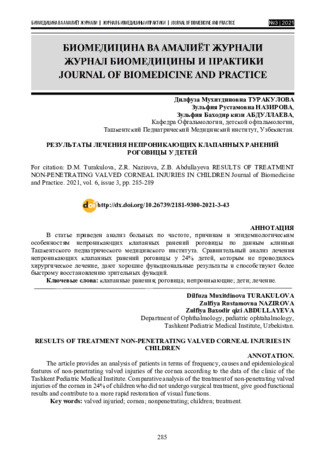
Results of treatment non-penetrating valved corneal injuries in children
The article provides an analysis of patients in terms of frequency, causes and epidemiological features of non penetrating valved injuries of the cornea according to the data of the clinic of the Tashkent Pediatric Medical Institute. Comparative analysis of the treatment of non-penetrating valved injuries of the cornea in 24% of children who did not undergo surgical treatment, give good functional results and contribute to a more rapid restoration of visual functions.
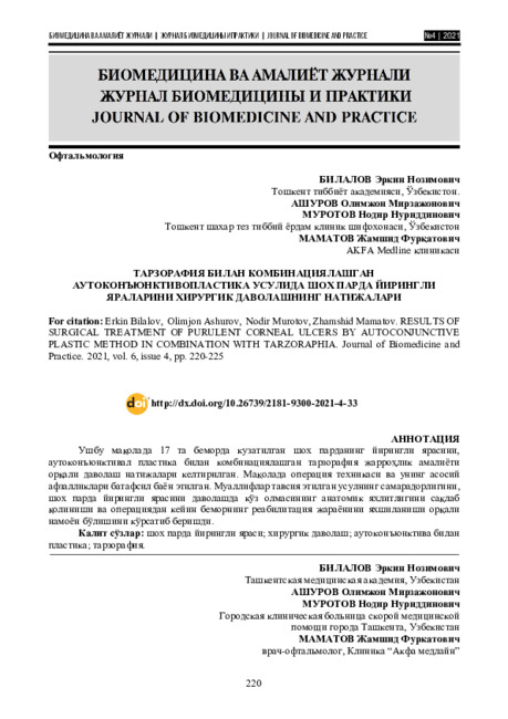
Results of surgical treatment of purulent corneal ulcers by autoconjunctive plastic method in combination with tarzoraphia
This article presents the results of surgical treatment of purulent corneal ulcers in 17 patients by the method of autoconjunctival plasty in combination with tarsorraphy. The article describes in detail the technique of the operation and its main advantages. The authors have shown the effectiveness of the proposed method, which is expressed in the fact that in ease of purulent corneal ulcers, the proposed method allows you to preserve the anatomical integrity of the eyeball and improve the patient's rehabilitation process after surgery.
Results of surgical correction of high anisometropia in patients with strabismus
RESULTS OF LENSECTOMY WITH IMPLANTATION OF TWO BACK-TO-BACK IOLS IN HIGH-GRADE MYOPIA
Material and methods. The study included 68 patients (114 eyes) diagnosed with high myopia, which were divided into two groups: the main group included 40 patients (58 eyes) who underwent LE with implantation of two IOLs, the control group consisted of 28 patients (56 eyes), with which the LE was performed by the traditional method.
Results and discussion.During implantation of 2 IOLs, an increase in the volume of the lens bag to 1.15 ± 0.05 mm and a lesser displacement of the vitreous body occur, in contrast to a lensectomy with one IOL (LT 0.52 ± 0.02).
Conclusion. Analysis of the research results allows us to recommend the removal of a clear crystalline lens with the implantation of two lOLs using the back-to-back method with high myopia, with the aim of creating stability of the anatomical and topographic relative position of intraocular structures and the possibility of reducing the risk of retinal detachment, in contrast to the traditional method of surgical treatment.
Reasons for late surgical treatment of patients with congenital cataracts
On the basis of the eye department of the TashPMI clinic, a retrospective study of the case
histories of 42 (84 eyes) patients with congenital cataracts was carried out. The age of the patients
ranged from 1 month to 13 years. All patients underwent a comprehensive clinical, ophthalmological, laboratory and instrumental examination and consultations of related specialists. The reasons for the late surgical treatment of children with congenital cataracts are: late diagnosis associated with clinical
forms of cataracts that are difficult to detect during external examination, the presence of concomitant somatic pathology, which is a contraindication to surgical treatment.
Reactive course of the postoperative period in a child with a combined eyeball injury
The article presents a clinical case of observation of the reactive course of the
postoperative period in a child with a combined injury of the eyeball, who was on inpatient treatment
in the department of ophthalmology of the clinic of the Tashkent Pediatric Medical Institute. The
results of the study showed: adequate specialized surgical treatment of combined eye trauma in
combination with intensive therapy allowed to stop the exudative process in the eye chambers, in the
vitreous body, which allowed to stabilize the reactive course of the postoperative period, preserve the
eye as a functional organ and prevent the development of post-traumatic uveitis.
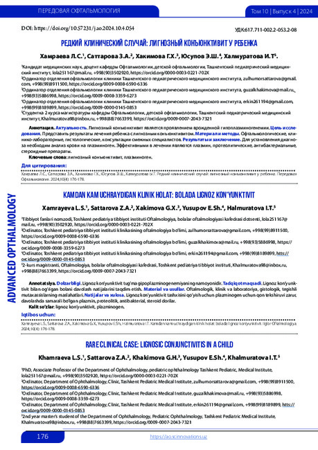
Rare clinical case: lignosic conjunctivitis in a child
Relevance. Lignous conjunctivitis is a manifestation of congenital hypoplasminogenemia. Purpose of the study. To present the results of treatment of a child with lignous conjunctivitis. Material and methods. Ophthalmological, clinical and laboratory, histo-logical, consultations of related specialists. Results and conclusion. To establish the diagnosis of ligneous conjunctivitis, a blood test for plasminogen is necessary, plasmin, proteolytic, antibacterial, and steroid drugs are effective in the treatment.
QUESTIONS OF PATHOGENESIS AND PREVENTION OF MYOPIA
Primary glaucoma
The most important medical and social problem in modern ophthalmology is the early diagnosis and treatment of glaucoma. Despite the known advances in the diagnosis and treatment of this disease, it remains one of the main causes of irreversible vision loss in people around the world. In the 70s of the last century, the total number of patients with glaucoma in the world was about 20 million (Nesterov A.P., 2007). At the moment, their number has increased to 105 million, and, according to WHO forecasts, by 2030. it can double (Kuroedov A.V., Gorodnichiy V.V., 2007). In Russia in 2003, about 800 thousand patients with glaucoma were registered (Kuroedov A.V., Gorodnichiy V.V., 2007). Glaucoma took the 1st place in the structure of blindness and primary visual disability (Libman E.S., Chumaeva E.A., 2004; Son I.M., Kalininskaya A.A., Sergeeva E.S., Chernenko E. .G., 2009).
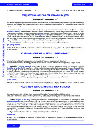
Predictors of complications in artifakia in children
Purpose of the study. To study risk factors for the development of complications in pseudophakic eyes. Material and methods. We carried out a retrospective and prospective analysis of the case histories of 65 children (65 eyes) diagnosed with pseudophakia, who were in the hospital in the eye department of the clinic of the Tashkent Pediatric Medical Institute over the past 5 years. The results of the study showed that the most significant risk factors for the development of intra- and postoperative complications are the following factors: FAI, neutrophil- leukocyte index (0.94±0.5), true intraocular pressure 20.5±1.2 mm Hg. Art., thickness of the choroid 1.02 ± 0.33 mm, protein in the CV above 3.5 ± 0.09 g/l, and in the blood below 62 ± 1.3 g/l
Peculiarities of the course of the intra- and postoperative period in conditionally frequently ill children after iol implantation
To study the results of IOL implantation in children with frequent respiratory diseases - conditionally frequently ill children (FCCI) taking into account the parameters of the composition of the chamber moisture of the eye. Material and methods: A retrospective and prospective analysis of the case histories of 50 children (50 eyes) aged from 1 to 5 years, who were treated in the eye department of the TashPMI clinic, was carried out. All patients underwent ophthalmological, clinical and laboratory studies: biochemical studies of blood and chamber moisture of the eye (EC). Results: The children were divided into 2 groups: the 1st group - 28 UCBD, the 2nd group (control) - 22 patients with no pathology from the somatic status. In patients of the 1st group, intraoperative complications occurred 1.8 times more often than in patients of the 2nd group, postoperative complications - 2.5 times more often. Of the late postoperative complications in patients of the 1st group, there was fibrosis of the posterior lens capsule (61%), poste-rior synechia (18%), and IOL dislocation (14%), which were indications for repeated surgical interventions. Conclu-sions: UCBD has a higher percentage of early postoperative inflammatory and late proliferative reactions. In patients of the 1st group, a significant increase in the protein content in the chamber moisture and a significant decrease in the protein level in the blood before cataract extraction were also revealed.
Our experience of medical treatment of age-related macular degeneration
Optimization of the method of treatment of phlegmon of a lachrymal bag in young child
A differentiated approach in the treatment of phlegmon of the lacrimal sac, taking into account
the etiopathogenesis of the disease and the presence of concomitant pathology, helps to reduce the recurrence of the disease, the need for additional treatment, as well as a decrease in the inpatient number of patients.
Optical and biometric indicators of the eye in children with juvenile glaucoma combined with myopia
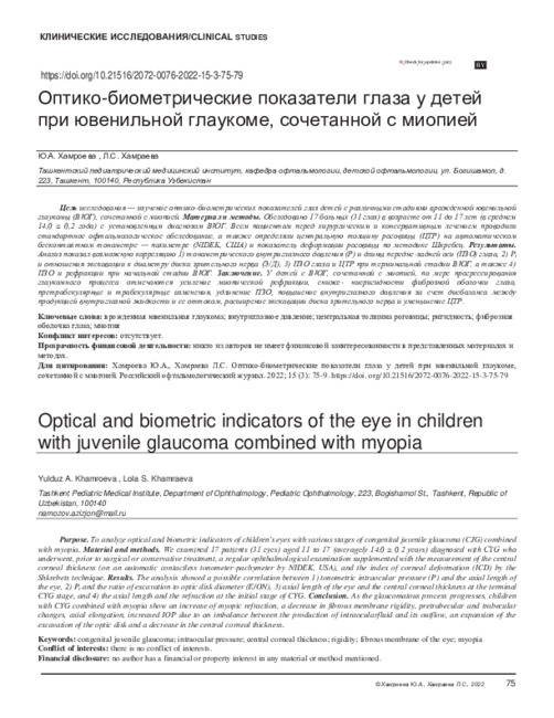
Optical and biometric indicators of the eye in children with juvenile glaucoma combined with myopia
Purpose. To analyze optical and biometric indicators of children's eyes with various stages of congenital juvenile glaucoma (CJG) combined with myopia. Material and methods. We examined 17 patients (31 eyes) aged 11 to 17 (averagely 14.0 ± 0.2 years) diagnosed with CYG who underwent, prior to surgical or conservative treatment, a regular ophthalmological examination supplemented with the measurement of the central corneal thickness (on an automatic contactless tonometer-pachymeter by NIDEK, USA), and the index of corneal deformation (ICD) by the Shkrebets technique. Results. The analysis showed a possible correlation between 1) tonometric intraocular pressure (P) and the axial length of the eye, 2) P t and the ratio of excavation to optic disk diameter (E/ON), 3) axial length of the eye and the central corneal thickness at the terminal CYG stage, and 4) the axial length and the refraction at the initial stage of CYG. Conclusion. As the glaucomatous process progresses, children with CYG combined with myopia show an increase of myopic refraction, a decrease in fibrous membrane rigidity, pretrabecular and trabecular changes, axial elongation, increased IOP due to an imbalance between the production of intraocularfluid and its outflow, an expansion of the excavation of the optic disk and a decrease in the central corneal thickness.
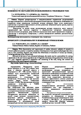
Opportunity ultrasonography in hemorrhage vitreous humor
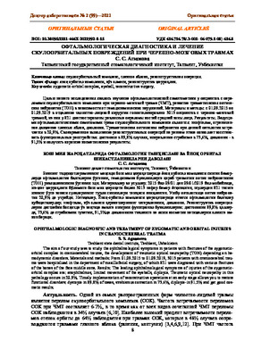
OPHTHALMOLOGIC DIAGNOSTIC AND TREATMENT OF ZYGOMATIC AND ORBITAL INJURIES IN CRANIOCEREBRAL TRAUMA
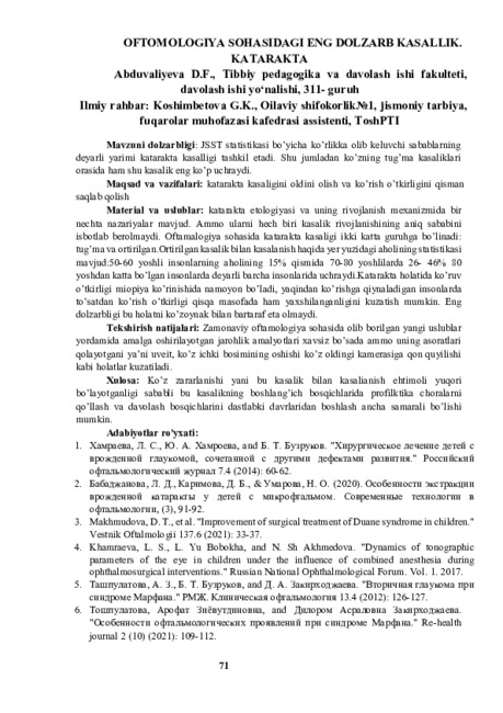
Oftomologiya sohasidagi eng dolzarb kasallik. Katarakta
Ocular manifestations of CAVID-19 complications in a teenager
A clinical case of EYE MANIFESTATIONS of complications is presented in a teenager who has had COVID-19. The studies were carried out in the intensive care unit and intensive care unit (ICU) of the clinic of the Tashkent Pediatric Medical institute. The patient underwent standard laboratory and instrumental studies, as well as an ophthalmological examination, including visometry, biomicroscopy, ophthalmoscopy. This clinical case showed that COVID-19 infection was complicated polymorphic ophthalmic symptoms indicating involvement in the process lacrimal gland, mucous membranes of the eyeball and eyelids, cornea of the lacrimal sac, retrobulbar tissue, while from the side of the eyes these complications are expressed in dry keratoconjunctivitis (Sjögren's syndrome).
Nosological structure of ophthalmic diseases
For parents, a child of any age seems vulnerable, so adults take care of him, they want protection from all difficulties. Unfortunately, a person is not strong and some diseases are very dangerous for the life of children. Some pathologies pass quickly and affect the future life, otherwise others will significantly affect the future life of the child. In order to reduce the impact of pathologies on the child's body, it is possible to diagnose the disease in the early stages, it is necessary to identify and immediately begin treatment. Among these diseases are ophthalmic we can not do without the introduction of diseases. If a child has vision problems from an early age, it can lead to a delay in the development of the child in the future. Ophthalmic diseases the main part: eye injuries, glaucoma, cataracts, glaucoma, retinopathy, myopia, cataract diseases, etc. More than 20% of diseases in ophthalmological practice, depending on the injury, damage the orbit and the eyeball. From an eye injury, then in 13% of cases, subatrophy of the eye develops, in 25% anophthalmos may occur. As for the characteristics of injuries, 10% of children suffer from damage to the organ of vision. This leads to various pathologies of the eye, in 30-60% of cases it can lead to one- or two-sided blindness. The most important traumatic factors in children are: knives, bullets, stones and clubs, hockey sticks, spears, nails, wire, etc. Glaucoma is one of these diseases. The disease also requires special attention. Reason: Prevalence of glaucoma in children Occurs in 1:10,000-1:12,000 cases. Its share in eye pathology is 0.1%. enough. More than 75% of glaucoma cases are bilateral. In parallel, there was glaucoma in 5 to 15% among children, blind and non-blind schoolchildren. Blindness in children, the proportion of this pathology ranges from 2 to 15%. in the Russian Federation Congenital glaucoma accounts for 10.1% of childhood blindness.
New Ways to Measure Intraocular Pressure in Children with Microphthalmia and Blepharophimosis
Childhood glaucoma is a chronic vision threatening condition that may significantly impact an individual’s psychosocial well-being. The prognosis of glaucoma largely depends on early and accurate diagnosis and successful treatment, including control of increased. The purpose is to determine the effectiveness of modified tonometers (Maklakova) NGm2-"OF TP" in performing tonometry and tonography in infants and children with microphthalmos and blepharophimosis. We have proposed modified models of Maklakoff tonometers weighing 5, 10, and 15 g, made of silver, the weights on both sides have contact plates, which are made of medical plastic and have a diameter of the applanation area d=8 mm. Methods: Tonometry and tonography using the simplified method of Nesterov A.P. were carried out in 30 (60 eyes) children aged from birth (0) to 16 years. Results: Our recommended design of weights showed results that do not differ from those obtained using standard Maklakov tonometers. At the same time, modified weights provide the opportunity to determine tonometric IOP and eye hydrodynamics in newborns and patients with microphthalmia and/or blepharophimosis, which greatly facilitates the work of pediatric ophthalmologists and reduces examination time.
Modern condition of the morbidity of the organ of vision in children
affect not only the structure of the organ of vision, but also disrupt its activity, which is expressed by the development of various pathologies.

