


Technical Note
Digital Denture Fabrication: A Technical Note
Shavkat Dusmukhamedov, Chu-Nui Lee
, Seung-Mi Jeong and Byung-Ho Choi *
Citation:
Dusmukhamedov, S.; Lee,
C.-N.; Jeong, S.-M.; Choi, B.-H. Digital
Denture Fabrication: A Technical
Note.
Appl. Sci.
2021
,
11
, 8093.
https://doi.org/10.3390/app11178093
Academic Editors: Yong-Deok Kim
and Luca Testarelli
Received: 25 July 2021
Accepted: 25 August 2021
Published: 31 August 2021
Publisher’s Note:
MDPI stays neutral
with regard to jurisdictional claims in
published maps and institutional affil-
iations.
Copyright:
© 2021 by the authors.
Licensee MDPI, Basel, Switzerland.
This article is an open access article
distributed under the terms and
conditions of the Creative Commons
Attribution (CC BY) license (https://
creativecommons.org/licenses/by/
4.0/).
Department of Dentistry, Wonju College of Medicine, Yonsei University, 162 Ilsandong, Wonju 26426, Korea;
mr.shavkat595@bk.ru (S.D.); chunuilee@naver.com (C.-N.L.); smj3@yonsei.ac.kr (S.-M.J.)
*
Correspondence: choibh@yonsei.ac.kr
Abstract:
Fabricating a complete denture in a conventional manner may be complicated and difficult.
The purpose of this article was to describe the benefits of a fully digital workflow and fabrication
procedure of complete dentures based on digital impressions of edentulous jaws. The digital data
for the workflow were acquired using an intraoral scanner and were then used to design the den-
ture base and teeth. The resulting data were exported to a 3D printer or a milling machine for
denture fabrication.
Keywords:
digital denture; edentulous jaw; intraoral scanner; CAD/CAM
1. Introduction
In the past decades, dentists have used the conventional method for complete denture
fabrication, which is complicated, difficult, and time consuming [
]. The conventional
method uses functional impression, cast pouring, wax rim fabrication, mounting the
models in an articulator, and curing the denture. The conventional process is associated
with human processing errors, inaccuracies, and multiple clinical appointments. Recently,
computer-aided design and computer-aided manufacturing (CAD/CAM) technology
was applied to the fabrication of complete dentures. The introduction and evolution
of computer-aided technology can overcome the complications related to the conven-
tional method, thereby facilitating the fabrication process [
]. The process of fabricating
complete dentures with digital technology involves digitization of clinical information
registered from the patient’s mouth, digital designing of complete dentures on computer
software, and an automatized process of manufacturing. The high accuracy of digitalized
dental fabrication has been proved by several studies compared to conventional meth-
ods [
]. This chapter describes the CAD/CAM fabrication of complete dentures based
on digital impressions of edentulous jaws obtained using an intraoral scanner.
2. Advantages of Digital Denture
•
More Accurate
The CAD/CAM fabrication process is a more accurate and repro-
ducible denture fabrication technique in comparison to the conventional method;
•
More Efficient
The CAD/CAM fabrication process needs a reduced total number of
appointments (two or three visits) in comparison to the conventional protocol (more
than five visits) and a significantly reduced clinical treatment time for the digital
fabrication process (approximately 3.5 h less compared to the conventional protocol);
•
Improved Retention
Digital dentures are better in retention, fit, and stability com-
pared to conventional dentures [
]. The improved retention of digital dentures
could be explained by their superior fit and absence of polymerization shrinkage;
•
Teeth Arrangement
In the digital denture workflow, the teeth are arranged on the
digital denture design software, and the digital preview of the teeth setup of complete
dentures is reviewed and easily modified;
•
Remake Dentures
The most beneficial feature of CAD/CAM dentures in comparison
to conventional dentures is the stored digital files that allow the fabrication of new
Appl. Sci.
2021
,
11

Appl. Sci.
2021
,
11
, 8093
2 of 14
prostheses when dentures are lost or damaged. Therefore, the electronic achieving of
all clinical data from the patient along with the design of the manufactured prostheses
enables making spare or new dentures, in case of breakage or loss, without clinical
appointments [
•
Better Fracture Resistance
The material used for digital denture fabrication was
significantly improved in its properties [
]. Increased toughness, ultimate strength,
and higher elastic modulus provide clinical benefits regarding the denture base design
with minimal thickness, without the common occurrence of denture fractures.
The process of digital denture fabrication may have some disadvantages related to
additive cost. Nevertheless, the above-mentioned benefits and especially increased level of
patient satisfaction can help to overcome the other disadvantages.
3. Digital Denture Fabrication Procedure
First, a trial denture is fabricated. Thereafter, the trial denture is used as a custom tray
for the final impression. Finally, a final denture is fabricated by scanning the impression.
The digital denture fabrication procedure is as follows:
3.1. Step 1. Trial Denture Fabrication
The procedure for fabricating a trial denture is as follows:
(1)
Trial denture design After the images of the edentulous ridge and opposite teeth with
the interarch relationship are obtained by recording the digital impression and all
the jaw relationships are recorded using an intraoral scanner, trial denture designing
begins using the 3Shape Dental software (3Shape, Copenhagen, Denmark). Anatomic
structures (incisal papilla, retro-molar pad, and tuberosities) are identified on the
edentulous image to correctly place the teeth (Figure
A). Thereafter, the limit of the
future denture base is drawn (Figure
B). The occlusal plane is determined by consid-
ering the jaw relationships, the curve of Spee, and the curve of Wilson. Subsequently,
denture teeth are arranged using the denture designing software (Figure
C). The
software contains a library of teeth of different brands and shapes and a function
with an automatic setup of tooth position, axis, and height that are matched with the
arches and the occlusal plane (Figure
(2)
Fabricating trial denture After the design is finalized, the digital STL files are sent
to the three-dimensional (3D) printing machine. The trial denture is 3D printed as a
monolithic denture, teeth, and base in one unit (Figure
(3)
Verifying jaw relation with trial denture At the trial placement, the horizontal and
vertical jaw relationships are evaluated with the trial denture.
Verifying vertical relationship:
The vertical jaw relationship is evaluated by consid-
ering the following aspects:
-
Midline regarding the maxillary midline being aligned with the center of the nose;
-
Resting interocclusal distance;
-
Phonetics and esthetic appearance;
-
Vertical dimension at the rest position;
-
Facial measurements;
-
Lip length in relation to the teeth;
-
Lip support;
-
Smile line;
-
Occlusal plane parallel to the interpupillary line.
VerifyingCentric Relation (CR):
After verifying the vertical relationship, the patient
is guided into CR. Any error in CR will be apparent when the teeth slide over each other;
(4)
Occlusal adjustment After confirming the CR, occlusal adjustment is carried out to
achieve a bilateral balanced occlusion.

Appl. Sci.
2021
,
11
, 8093
3 of 14
Figure 1.
Cont
.

Appl. Sci.
2021
,
11
, 8093
4 of 14
Figure 1.
Cont
.

Appl. Sci.
2021
,
11
, 8093
5 of 14
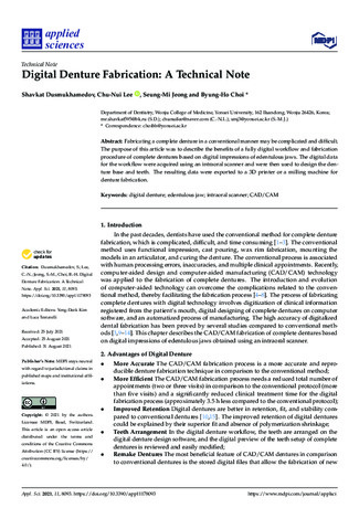
Figure 1.
Trial denture fabrication. (
A
): Anatomic structures are identified on the edentulous ridge image. (
B
): Limit of the
future denture base is drawn. (
C
): Denture teeth are arranged. (
D
): Denture base is designed. (
E
): Printed trial denture.
(
F
): Trial denture is printed as a monolithic denture, teeth, and base in one unit.

Appl. Sci.
2021
,
11
, 8093
6 of 14
3.2. Step 2. Final Impression
(1)
Border molding The trial denture is checked in the mouth to ascertain that the bor-
ders are approximately 2 mm short of the vestibular reflections. Border molding is
performed by adding a modeling compound to the trial denture borders and moving
the tongue, lips, and cheeks for adjustment (Figure
A,B). The trial denture is adapted
closely to the tissues of the vestibule before making the final impression;
(2)
Recording final impression The final impression is made with the border-molded trial
denture (Figure
C). After obtaining the impression, the denture is removed from the
mouth and is scanned using an intraoral scanner (Figure
D). The denture scanning
area should include all areas of the denture, such as the denture base, border, teeth,
and palate (Figure
3.3. Step 3. Final Denture Fabrication
Digital images of the trial denture obtained by scanning the final impression are
used to design the definitive denture [
] (Figure
A). If any modifications in the jaw
relationship or teeth arrangement are required, these are made in the digital images. After
the modifications are made, the definitive denture is fabricated during either milling or 3D
printing (Figure
B).In the case of using a 3D printer, it can print the artificial teeth and the
pink denture base separately or both in a single unit [
] (Figure
A–C). When the artificial
teeth and denture base are printed in one unit, tissue-colored composite resin is applied
onto the denture flange (Figure
A,B).In the case of using a milling machine, the denture
base is milled from a pink block of prepolymerized cross-linked polymethyl methacrylate
(PMMA) disks. The teeth are also milled from the PMMA disk. The milled teeth are
securely bonded onto the milled denture base (Figure
A,B). The monolithic denture is
more accurate than the bonded denture because of the lack of bonding processing errors.
Figure 2.
Cont
.

Appl. Sci.
2021
,
11
, 8093
7 of 14
Figure 2.
Cont
.

Appl. Sci.
2021
,
11
, 8093
8 of 14
Figure 2.
Final impression using trial denture. (
A
): Trial denture borders are approximately 2 mm short of the vestibular
reflections. (
B
): Border molding is performed by adding modeling compound to the trial denture borders. (
C
): Final
impression is obtained with the border-molded trial denture. (
D
): The trial denture is scanned using an intraoral scanner.
(
E
): Scan image of the trial denture.

Appl. Sci.
2021
,
11
, 8093
9 of 14
Figure 3.
Final denture fabrication. (
A
): Final denture is designed using the digital images of the trial denture obtained by
scanning the final impression. (
B
): Printed final denture.

Appl. Sci.
2021
,
11
, 8093
10 of 14
Figure 4.
Cont
.

Appl. Sci.
2021
,
11
, 8093
11 of 14
Figure 4.
Bonded printed denture. (
A
): Printed denture base. (
B
): Printed artificial teeth. (
C
): Printed artificial teeth are
bonded onto the printed denture base.
Figure 5.
Cont
.

Appl. Sci.
2021
,
11
, 8093
12 of 14
Figure 5.
Monolithic printed denture. (
A
): Artificial teeth and denture base are printed in one unit. (
B
): Tissue-colored
composite resin is applied onto the denture flange.
Figure 6.
Cont
.

Appl. Sci.
2021
,
11
, 8093
13 of 14
Figure 6.
(
A
): Milled denture base. (
B
): Milled artificial teeth.
Author Contributions:
S.D.—writing—original draft preparation; C.-N.L.—conceptualization;
B.-H.C.—writing—project administration, review and editing, and supervision; S.-M.J.—resources
and supervision. All authors have read and agreed to the published version of the manuscript.
Funding:
This research did not receive any specific grant from funding agencies in the public,
commercial, or not-for-profit sectors.
Institutional Review Board Statement:
Not applicable.
Informed Consent Statement:
Not applicable.
Conflicts of Interest:
The authors declare no conflict of interest.
References
1.
Neumeier, T.T.; Neumeier, H. Digital immediate dentures treatment: A clinical report of two patients.
J. Prosthet. Dent.
2016
,
116
,
314–319. [
2.
Kanazawa, M.; Inokoshi, M.; Minakuchi, S.; Ohbayashi, N. Trial of a CAD/CAM system for fabricating complete dentures.
Dent.
Mater. J.
2011
,
30
, 93–96. [
3.
Goodacre, C.J.; Garbacea, A.; Naylor, W.P.; Daher, T. CAD/CAM fabricated complete dentures: Concepts and clinical methods of
obtaining required morphological data.
J. Prosthet. Dent.
2012
,
107
, 34–46. [
4.
Miyazaki, T.; Hotta, Y.; Kunii, J.; Kuriyama, S.; Tamaki, Y. A review of dental CAD/CAM: Current status and future perspectives
from 20 years of experience.
Dent. Mater. J.
2009
,
28
, 44–56. [
5.
Alghazzawi, T.F. Advancements in CAD/CAM technology: Options for practical implementation.
J. Prosthodont. Res.
2016
,
60
,
72–84. [
6.
Christensen, G.J. Impressions are changing: Deciding on conventional, digital or digital plus in-offlce milling.
J. Am. Dent. Assoc.
2009
,
140
, 1301–1304. [
7.
Ting-Shu, S.; Jian, S. Intraoral digital impression technique: A review.
J. Prosthodont.
2015
,
24
, 313–321. [
8.
Christensen, G.J. The challenge to conventional impressions.
J. Am. Dent. Assoc.
2008
,
139
, 347–349. [
9.
Pereira, A.L.C.; de Medeiros, A.K.B.; de Sousa Santos, K.; de Almeida,
É
.O.; Barbosa, G.A.S.; Carreiro, A.D.F.P. Accuracy of
CAD-CAM systems for removable partial denture framework fabrication: A systematic review.
J. Prosthet. Dent.
2021
,
125
,
241–248. [

Appl. Sci.
2021
,
11
, 8093
14 of 14
10.
Goodacre, B.J.; Goodacre, C.J.; Baba, N.Z.; Kattadiyil, M.T. Comparison of denture base adaptation between CAD-CAM and
conventional fabrication techniques.
J. Prosthet. Dent.
2016
,
116
, 249–256. [
11.
Kattadiyil, M.T.; Goodacre, C.J.; Baba, N.Z. CAD/CAM complete dentures: A review of two commercial fabrication systems.
J. Calif. Dent. Assoc.
2013
,
41
, 407–416.
12.
Artopoulos, A.; Juszczyk, A.S.; Rodriquez, J.M.; Clark, R.K.; Radford, D.R. Three-dimensional processing deformation of three
denture base materials.
J. Prosthet. Dent.
2013
,
110
, 481–487. [
13.
Soltanzadeh, P.; Suprono, M.S.; Kattadiyil, M.T.; Goodacre, C.; Gregorius, W. An in vitro investigation of accuracy and fit of
conventional and CAD/CAM removable partial denture frameworks.
J. Prosthet. Dent.
2019
,
28
, 547–555. [
14.
Lee, C.; Dusmukhamedov, S.; Fang, Y.Q.; Jeong, S.M.; Choi, B.H. Accuracy of the provisional prosthesis scanning technique
versus a conventional impression technique on completely edentulous arches.
Appl. Sci.
2021
,
11
, 7182. [
15.
AlHelal, A.; AlRumaih, H.S.; Kattadiyil, M.T.; Baba, N.Z.; Goodacre, C.J. Comparison of retention between maxillary milled and
conventional denture bases: A clinical study.
J. Prosthet. Dent.
2017
,
117
, 233–238. [
16.
Lo, R.L.; Salamini, A. Removable complete digital dentures: A workflow that integrates open technologies.
J. Prosthet. Dent.
2018
,
119
, 727–732.
17.
Lee, D.H.; Lee, J.S. Comparison of flexural strength according to thickness between CAD/CAM denture base resins and
conventional denture base resins.
J. Dent. Rehabil. Appl. Sci.
2020
,
36
, 183–195. [
18.
Sun, Y.; Tian, W.; Zhang, T.; Chen, P.; Li, M. Strength and toughness enhancement in 3D printing via bioinspired tool path.
Mater.
Des.
2020
,
185
, 108239. [
19.
Gilboa, I.; Cardash, H.S. An alternative approach to the immediate overdenture.
J. Prosthodont.
2009
,
18
, 71–75. [
20.
Shah, F.K.; Gebreel, A.; Elshokouki, A.H.; Habib, A.A.; Porwal, A. Comparison of immediate complete denture, tooth and
implant-supported overdenture on vertical dimension and muscle activity.
J. Adv. Prosthodont.
2012
,
4
, 61–71. [
21.
Caputi, S.; Murmura, G.; Ricci, L.; Varvara, G.; Sinjari, B. Immediate denture fabrication: A clinical report.
Ann. Di Stomatol.
2014
,
4
, 273–277.
22.
Soni, A. Use of loose fitting copper bands over extremely mobile teeth while making impressions for immediate dentures.
J. Prosthet. Dent.
1999
,
81
, 638–639. [
23.
Fang, J.H.; An, X.; Jeong, S.M.; Choi, B.H. Digital immediate denture: A clinical report.
J. Prosthet. Dent.
2018
,
119
, 698–701.