


JOURNAL OF HEPATO-GASTROENTEROLOGY RESEARCH | ЖУРНАЛ ГЕПАТО-ГАСТРОЭНТЕРОЛОГИЧЕСКИХ ИССЛЕДОВАНИЙ
№3,2 (том II) 2021
75
Belyх N. A,
MD, PhD, Dr Med Sci, Associate Professor,
Head of the Department of Faculty and Polyclinic Pediatrics with the Course of Pediatric of Postgraduate
Education
Ryazan State Medical University, Ryazan, Russian Federation
Buloхova E,
Professor of the Department of Child Diseases and Hospital Pediatrics
Ryazan State Medical University, Ryazan, Russian Federation
ASSESSMENT OF THE RELATIONSHIP BETWEEN LIPID AND CARBOHYDRATE METABOLISM
INDICATORS AND VITAMIN D STATUS IN CHILDREN WITH DIFFERENT BODY MASS INDEX
ANNOTATION
Overweight children represent a particularly vulnerable group for vitamin D deficiency. was to study the
relationship between lipid and carbohydrate metabolism indicators and VD status in children, depending on the div
mass index (BMI). A cross-sectional (one-step) study carried out on a sample of 154 children with different weight of 8-
10 years old (girls - 74, boys - 80). There were identified three groups of research participants: group 1 - 44 obese, 2
group - 58 overweight, 3 group - 52 children with normal div weight. For all children, the serum 25(OH)D,
parathyroid hormone (PTH), calcium (Ca), phosphorus (P), alanine aminotransferase (ALT), aspartate aminotransferase
(AST), cholesterol (CS), triglycerides (TG), beta-lipoproteins (ß-LP), glucose, insulin determined, and Homeostasis
Model Assessment of Insulin Resistance (HOMA-IR) calculated.
VD deficiency in obese children was found almost 2.3
times more often than in overweight (p = 0.002) and 2.8 times more often than in children with normal div weight (p
= 0.001). Indicators of lipid and carbohydrate metabolism were within physiological limits. However, in obese children
they significantly exceeded the indicator of healthy children (p <0.05). Children with VD deficiency (25(OH)D<20
ng/ml) had statistically significantly higher medians of serum PTH, TC, TG, ALT, AST, glucose, insulin, HOMA-IR and
lower serum P and Ca compared with children with optimal VD status (p <0.05). The medians of serum ALT, AST, TC,
ß-LP, TG, glucose, insulin and HOMA-IR in obese children with VD deficiency was statistically significantly higher
compared in healthy children with VD deficiency and optimal VD status.
VD deficiency is an important predictor of
obesity complications and it exacerbates the risk of cardiometabolic disorders in children who are obese in the early
school years.
Key words:
children, obesity, vitamin D, vitamin D status, cardiometabolic disorders.
Белых Н.А,
доктор медицинских наук, доктор медицинских наук, доцент,
Заведующий кафедрой факультетской и поликлинической педиатрии с курсом педиатрии
последипломного образования
Рязанский государственный медицинский университет, Рязань, Российская Федерация
Булохова Е,
доцент кафедры детских болезней и госпитальной педиатрии
Рязанский государственный медицинский университет, Рязань, Российская Федерация
ОЦЕНКА ВЗАИМОСВЯЗИ ПОКАЗАТЕЛЕЙ ЛИПИДНОГО И УГЛЕВОДНОГО ПРОФИЛЯ С
УРОВНЕМ ОБЕСПЕЧЕННОСТИ ОРГАНИЗМА ВИТАМИНОМ D У ДЕТЕЙ В ЗАВИСИМОСТИ ОТ
ИНДЕКСА МАССЫ ТЕЛА
АННОТАЦИЯ
Дети с избыточной массой тела (МТ) представляют особо уязвимую группу по гиповитаминозу D.
Поперечное (одномоментное) исследование проведено на выборке 154 детей с разными весоростовыми
показателями в возрасте 8-10 лет (девочек - 74, мальчиков - 80). Выделено 3 группы участников исследования: 1

JOURNAL OF HEPATO-GASTROENTEROLOGY RESEARCH | ЖУРНАЛ ГЕПАТО-ГАСТРОЭНТЕРОЛОГИЧЕСКИХ ИССЛЕДОВАНИЙ
№3,2 (том II) 2021
76
группа - 44 ребенка с ожирением, 2 группа – 58 детей с избыточной массой тела, 3 группа – 52 человека с
нормальной массой тела. Всем детям определяли в сыворотке крови уровень 25(ОН)D, паратгормона (ПТГ),
кальция (Са), фосфора (Р), общего холестерина (ХС), триглицеридов (ТГ), бета-липопротеидов (ß-ЛП), глюкозы,
инсулина, активность АЛТ, АСТ, а также рассчитывали индекс инсулинорезистентности (HOMA-IR). Дефицит
витамина D у детей с ожирением встречался почти в 2,3 раза чаще, чем у детей с избыточной массой тела
(р=0,002) и в 2,8 раза чаще, чем у детей с нормальной массой тела (р=0,001). Показатели липидного и
углеводного обменов находились в физиологических пределах. Однако у детей с ожирением они значимо
превышали показатель здоровых детей (р<0,05). Дети с дефицитом VD имели статистически значимо более
высокие медианы ПТГ, ХС, ТГ, глюкозы, инсулина, активности АЛТ, АСТ, НОМА-IR и более низкую
концентрацию Р и Са по сравнению с детьми, имеющими оптимальный VD статус (р<0,05). Медианы АЛТ,
АСТ, ХС, ß-ЛП, ТГ, глюкозы и HOMA-IR у детей с ожирением и дефицитом VD были статистически значимо
выше, чем у здоровых детей с дефицитом VD и с оптимальной концентрацией 25(ОН)D в сыворотке крови.
Дефицит витамина D является важным предиктором формирования осложнений ожирения и усугубляет риск
развития кардиометаболических расстройств у детей, страдающих ожирением в младшем школьном возрасте.
Ключевые слова:
дети, ожирение, витамин D, дефицит витамина D, кардиометаболические
расстройства.
The growing prevalence of obesity in the child
population is one of the problems of modern health care.
According to World Health Organization (WHO, 2018)
forecasts, the number of obese children by the end of
2025 may exceed 70 million only in the age group from 0
to 5 years old [1]. Childhood obesity has serious lifelong
consequences. In the short term, such children are
accompanied by psychological disorders (depression,
anxiety and low self-esteem, a number of emotional and
behavioral disorders), they are more likely to suffer from
asthma, diseases of the musculoskeletal system [2]. In the
future, they have an increased risk of metabolic disorders
and
cardiovascular
pathology,
such
as
arterial
hypertension, dyslipidemia, atherosclerosis [3]. In the
long term, childhood obesity increases of the risk of
developing cardiovascular diseases, diabetes mellitus,
some types of cancer and diseases of the musculoskeletal
system, which can lead to disability and premature death
[4].
In parallel with obesity, the problem of low
vitamin D (VD) status in child and adolescent population
is becoming more and more urgent. At present,
hypovitaminosis D among the child population recorded
in many countries of the world, including the Russian
Federation [5-7].
For a long time, the regulation of calcium and
phosphorus homeostasis considered the main effect of
VD. However, in recent years, VD viewing as a hormone
that has receptors in most div tissues and performs
many “non-classical” effects. Non-skeleton effects of VD
include of regulation of cell proliferation and cell
differentiation, inhibition of renin and angiogenesis
synthesis, contributing of insulin production, activation of
macrophage formation, etc. [8]. Overweight children
represent a particularly vulnerable group for vitamin D
deficiency, which, in recent years, has been associated
with health risks similar to obesity [9]. Therefore,
according to Mirhosseini N. et al. (2018), VD deficiency
may play an important role in the development of
cardiovascular diseases [10]. There is also an opinion
about of the positive effect of VD subsidy on metabolism
in adults with chronic cardiovascular disease. Schroten N.
et al. (2013) observed a decrease in plasma renin activity
in 101 patients with stable heart failure after 6 weeks of
taking 2000 IU VD [11]. The VINDICATE study group
(Vitamin D treating patients with chronic heart failure)
noted a significant improvement in cardiac function in
229 patients with chronic heart failure after taking VD
4000 IU daily for 1 year [12]. In contrast, several
metaanalyzes and systematic reviews have not found a
positive effect of VD on the course of cardiovascular
disease. Ford J. et al. (2014), for example, expressed
insufficient data to support the use of VD as a supplement
to reduce the incidence of cardiovascular disease [13]. In
their systematic review, Wang L. et al. (2010) noted a
statistically insignificant decrease in the incidence of
cardiovascular diseases when taking moderate doses of
VD. Mao P. et al. (2013) also found that neither VD
supplementation nor calcium supplementation affected
the incidence of myocardial infarction or stroke [14].
However, most modern studies substantiate the negative
effect of low serum 25(OH) D levels on the state of the
cardiovascular system, and associate this primarily with
the role of micronutrients in the regulation of the renin-
angiotensin-aldosterone system (RAAS). Thus, the renin
gene has a VD sensitive element that has a regulatory
effect on the transcription and production of renin, which,
in turn, acting on angiotensin, triggers a number of
processes that promote the formation of angiotensin II,
which acts as a vasoconstrictor [15].
There are few data on the role of VD deficiency
as a risk factor for the onset and progression of
cardiovascular disorders in primary school children. In
this regard, the study of this problem is interesting,
especially among obese children, who form a risk group
for the development of chronic pathology.
Aim
: to study of the relationship between lipid
and carbohydrate metabolism indicators and VD status in
children, depending on the div mass index (BMI).
Materials and methods.
A cross-sectional (one-
step) study carried out on a sample of 154 children with
different weight and height indicators. Among the
surveyed children there were 74 girls (48.0%) and 80
boys (52.0%) of primary school age (the average age –
9.4±0.7 years). All children permanently live in Ryazan.
Study inclusion criteria: absence of acute or
exacerbation of chronic diseases at the time of inclusion
in the study; lack of intake of vitamin and mineral
complexes for at least 6 months. Before inclusion in the
study, the absence of chronic diseases of the kidneys,
liver, gastrointestinal tract, as well as the signed informed
consent of the child's parent to his participation in the
study.
The studies carried out on the bases of the City
Child Polyclinic No. 1, Regional Child Hospital and
Central Research Laboratory of the RyazSMU. The local

JOURNAL OF HEPATO-GASTROENTEROLOGY RESEARCH | ЖУРНАЛ ГЕПАТО-ГАСТРОЭНТЕРОЛОГИЧЕСКИХ ИССЛЕДОВАНИЙ
№3,2 (том II) 2021
77
ethics committee of the RyazGMU approved the study
protocol. The parents had appropriate information about
their participation in the study and their informed consent
obtained.
Trained health workers in accordance with a
standardized protocol developed by WHO [16] performed
anthropometric measurements during a preventive
medical examination. The physical growth assessed using
the WHO AnthroPlus (2009) [17]. There were calculated
the following parameters: Weight-for-Age Z-score
(WAZ), div mass-to-age index (BMI-for-Age Z-score,
BAZ). The interpretation of the obtained Z-score values
carried out according to the following criteria:
malnutrition - with <–2 SDS, under nutrition from -
2<SDS<-1, norm - -1<SDS<+1, overweight - +1<SDS<+
2, obesity - with SDS> +2 [18].
According to the anthropometry data, there were
formed 3 groups: 1
st
group - obese children (n=44, 22
girls and 22 boys), 2
nd
group - overweight children (n=58,
18 girls, 40 boys), 3
rd
group – healthy children (n=52, 34
girls, 18 boys).
Serum 25(OH)D level, parathyroid hormone
(PTH), glucose, insulin, triglycerides (TG), transaminase
activity (alanine aminotransferase, ALT and aspartate
aminotransferase, AST), β-lipoprotein (β-LP) level,
cholesterol (CS), calcium (Ca), phosphorus (P) were
tested in all children. A procedural nurse in a
manipulation room located in the Regional Child
Hospital carried out blood sampling on an empty
stomach, from the ulnar vein. Serum 25(OH)D was
evaluated by the enzyme-linked immunoabsorbent assay
(DIAsource 25OH Vitamin D Total ELISA Kit,
Diasource, Spain) and values <20 ng/ml were considered
deficient, 20-30 ng/ml insufficient, and >30 ng/ml -
sufficient [19]. The PTH content by the method of
immunoradiometric
analysis
(IRMA
PTH
kits,
IMMUNOTECH, Czech Republic) and insulin by the
immunochemiluminescent method on a Roche Cobas
e8000 602 analyzer (Roche Cobas, Switzerland) was
determined. The serum Ca, P, β-LP, TG, CS, glucose,
ALT, AST on a Mindray BS-400 biochemical analyzer
(Mindray, China) was measured. The insulin resistance
index (HOMA-IR) was calculated (normally below 3.2
U) [20].
The STATISTICA 12 software package used for
statistical analysis. Continuous variables presented as
medians with an interquartile range (25-75 percentiles).
The analysis of the normal distribution of the values of
the studied features performed using the Shapiro-Wilk
test. When comparing continuous variables across
groups, the Kruskal-Wallis test used (for paired
comparisons, the Mann-Whitney test). The degree of
relationships assessed by calculating the pairwise
Spearman correlation coefficients (r). The χ
2
test used to
determine the relationship between the two categorical
variables. p< 0.05 was considered significant
.
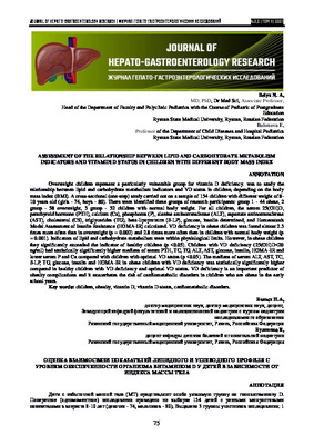
Results.
VD deficiency occurred in 76 (49.4%)
of the examined children, deficiency - in 30 (19.5%), and
normal provision was found only in 48 (31.1%) children.
Obese children have VD deficiency in 2.3 times more
often than overweight (p=0.002) and in 2.8 times more
often than healthy (p=0.001) (Fig.1). The normal
provision of VD in overweight children detected almost 2
times less often than in healthy children. Among the
surveyed group 1, normal concentration of 25 (OH) D in
the blood serum not detected in any child. There were no
statistically significant gender differences among the
assessed groups (p> 0.05).
Figure 1. Vitamin D status in participants depending on BMI (%)
The median values of mineral, lipid and
carbohydrate metabolism compared depending on BMI in
the compared groups (Table 1). Serum PTH level was
within the reference values; statistically significant
differences between the groups were no found (p>0.05).
The median of serum Ca in was normal - 2.46 [2.36;
2.54] mmol/L. Me Ca in obesity child was statistically
significantly lower than in 2
nd
and 3
rd
groups (p<0.05).
Was revealed that with an increase of BMI, the serum Ca
level significantly decreased (r=0.51, p<0.05), and in 7
(32.0%) participants with the highest BMI was found of
hypocalcaemia (p=0.014). Serum P in all children was
within the physiological norm. However, in the 1
st
and 2
nd
group the serum P was statistically significantly lower

JOURNAL OF HEPATO-GASTROENTEROLOGY RESEARCH | ЖУРНАЛ ГЕПАТО-ГАСТРОЭНТЕРОЛОГИЧЕСКИХ ИССЛЕДОВАНИЙ
№3,2 (том II) 2021
78
than in 3
rd
group (p<0.05). A negative correlation
between BMI and P level was found (r=-0.51, p<0.05).
Serum ALT, AST, CHS, TG, β-LP, glucose and insulin
was within physiological limits. However, the median
ALT activity in children in 1
st
group was 1.8 times higher
than in 2
nd
group (p<0.001) and was more than 2.5 times
lower than in healthy children (p<0.001). Moreover, in
obese boys, this indicator was higher compared to girls in
this group (p = 0.003). The median serum AST activity in
obese children also exceeded the value in the overweight
children (p<0.001) and normal BMI (p<0.001). Serum
CS, TG and β-LP had a direct moderate correlation with
BMI (p<0.05), and the medians of these indicators in
obese children significantly exceeded the values in
overweight
and
healthy
children
(p<0.005).
Table 1.
Indicators of mineral, lipid and carbohydrate metabolism depending on the div mass index of children
Indicator
Reference
values
Group 1
BMI-for-Age Z-
score
>+2 SDS
(n=22)
Group 2
BMI-for-Age
+1< Z-score<+2
SDS
(n=29)
Group 3
BMI-for-Age
-1<Z-score<+1
SDS
(n=26)
Р
k-w
1-2
Р
k-w
1-3
Р
k-w
2-3
25(ОН)D,
ng/ml
>30 ng/ml
12,5 [5,7; 19,1]
23,6 [11,3; 34,5] 32,6 [15,9; 44,4]
0,014
0,001
0,080
PTH,
pg/ml
10,0–65,0
28,3 [23,2; 38,3] 25,1 [20,9; 32,6] 27,2 [19,9; 33,5]
0,210
0,562
0,227
Са,
mmol/l
2,3–2,8
2,3 [2,2; 2,4]
2,5 [2,4; 2,5]
2,5 [2,5; 2,7]
0,031
0,000
0,021
Р, mmol/l
1,1–2,0
1,2 [1,1; 1,2]
1,2 [1,2; 1,3]
1,3 [1,2; 1,5]
0,011
0,001
0,362
ALT, U/l
< 40,0
35,0 [32,0; 38,0] 20,0 [18,0; 24,0] 13,0 [11,0; 16,0]
0,000
0,000
0,000
AST, U/l
< 40,0
34,0 [32,0; 36,0] 22,0 [20,0; 26,0] 21,0 [17,0; 25,0]
0,000
0,000
0,851
CS,
mmol/l
2,8-5,5
4,8 [4,4; 5,2]
4,4 [4,0; 4,5]
3,9 [3,8; 4,4]
0,003
0,001
0,018
ß-LP, U/l
35,0-55,0
45,0 [40,0; 50,0] 40,0 [37,0; 42,0] 35,0 [32,0; 36,0]
0,021
0,000
0,000
TG,
mmol/l
0,3-1,5
1,4 [1,3; 1,5]
0,7 [0,5; 0,9]
0,5 [0,5; 0,7]
0,000
0,000
0,020
Glucose,
mmol/l
3,4-6,1
4,3 [4,1; 4,5]
4,1 [3,8; 4,4]
3,6 [3,4; 3,7]
0,152
0,000
0,000
Insulin,
μU / ml
3,0-20,0
15,5 [14,9; 16,0]
10,8 [9,0; 13,3]
7,8 [5,0; 9,9]
0,000
0,000
0,010
HOMA-IR
<3,2
2,9 [2,8; 3,2]
2,0 [1,7; 2,5]
1,3 [0,8; 1,5]
0,000
0,000
0,000
Note: BMI - div mass index;
HOMA-IR increased with increasing BMI. At
the same time, in 5 (23.0%) obese children HOMA-IR
exceeded the permissible normal values (p=0.057),
despite the normal isolated levels of glucose and insulin.
Children with VD deficiency had a higher BMI.
PTH, CS, TG, glucose, insulin, the activity of ALT and
AST, as well as HOMA-IR in them exceeded those in
children with normal VD status (p<0.05), but the serum P
and Ca was lower (p<0.05) (Table 2).
Children with insufficient VD status have
statistically significantly higher BMI, TG, ALT, AST,
HOMA-IR and a reduced level of Ca compared with
children optimally provided with vitamin D (p <0.05).
There were no statistically significant differences in the
level of β-LP between the groups (p> 0.05). PTH and TG
in children with VD deficiency was 1.3 times (p<0.05)
higher than in children with insufficient VD status. At
decrease serum 25(OH)D increase PTH, Ca, P, β-LP, TG,
glucose, insulin, activity ALT, AST and HOMA-IR (Table
3). Thus, these changes indicate that vitamin D deficiency
in children 8-10 years old is a risk factor for
cardiometabolic disorders at an older age.
The medians of ALT, AST, CS, ß-LP, TG,
glucose, insulin, and HOMA-IR in obese children with
VD deficiency were statistically significantly higher than
in healthy children with VD deficiency and a sufficient
VD status.
The discussion.
Vitamin D deficiency is quite
common in childhood and is more common among obese
children. The data obtained coincide with the results of
studies of previous years [21, 22]. It is believed that the
relationship of anthropometric and biochemical markers
of cardiovascular risks with the high prevalence of
vitamin D deficiency is indirect, because this is a
consequence of a sedentary lifestyle, decreased activity,
stay indoors and poor nutrition, which lead to the
progressive accumulation of fat mass. So, in the works of
Skinner A. et al. (2015) and Durá-Travé T. et al. (2017) it
was obesity rather than insufficient VD provision that
positively correlated with dyslipidemia [23, 24].
Nevertheless, various authors describe the existence of
strong correlations between low VD-status and various
components of the lipid metabolism [25, 26].

JOURNAL OF HEPATO-GASTROENTEROLOGY RESEARCH | ЖУРНАЛ ГЕПАТО-ГАСТРОЭНТЕРОЛОГИЧЕСКИХ ИССЛЕДОВАНИЙ
№3,2 (том II) 2021
79
Table 2.
Anthropometric and biochemical parameters depending on vitamin D status
Indicator
Serum 25(OH)D
Р
k-w
1-2
Р
k-w
1-3
Р
k-w
2-3
< 20 ng/ml
(n=76)
20–29 ng/ml
(n=30)
> 30 ng/ml
(n=48)
BMI z-score
2,0 [1,01; 2,9]
1,4 [1,0; 1,9]
0,8 [-0,3; 1,0]
0,150
0,000
0,001
PTH, pg/ml
32,1
[25,5; 39,3]
24,5
[20,1; 36,2]
23,2
[18,3; 29,0]
0,041
0,009
0,649
Са, mmol/l
2,4 [2,3; 2,5]
2,5 [2,4; 2,6]
2,7 [2,5; 2,7]
0,001
0,000
0,129
Р, mmol/l
1,2 [1,1; 1,2]
1,3 [1,2; 1,3]
1,7 [1,7; 1,8]
0,006
0,000
0,000
ALT, U/l
28 [19; 36]
24 [18; 30]
14 [11,5; 18]
0,407
0,000
0,001
AST, U/l
29 [22; 34]
25 [22; 31]
20 [18; 24]
0,272
0,000
0,007
TC, mmol/l
4,4 [3,7; 4,8]
4,3 [3,9; 4,6]
4,1 [3,8; 4,4]
0,963
0,035
0,035
ß-LP, U/l
40 [36; 46]
39 [36; 42]
36 [33; 40]
0,296
0,059
0,217
TG, mmol/l
1,2 [0,5 1,4]
0,9 [0,5; 1,1]
0,6 [0,5; 0,8]
0,041
0,017
0,015
Glucose,
mmol/l
4,1 [3,7; 4,4]
4,2 [4; 4,4]
3,6 [3,45; 3,8]
0,696
0,001
0,001
Insulin, μU/ml
14,5 [8,1; 15,6]
13,1 [9,5;
15,0]
9,0 [7,3; 10,4]
0,150
0,000
0,015
HOMA-IR
2,6 [1,5; 2,9]
2,6 [1,8; 2,7]
1,4 [1,2; 1,7]
0,827
0,000
0,002
Note: BMI - div mass index;
Table 3.
Spearman's correlation coefficients between 25(OH)D level and z-score BMI/age and biochemical parameters
Indicator
z-score BMI / age
25(ОН)D, ng/ml
r
р
r
р
z-score BMI / age
1,000
≥0,05
-0,480
<0,05
25(ОН)D, ng/ml
-0,480
<0,05
1,000
≥0,05
PTH, pg/ml
0,122
≥0,05
-0,441
<0,05
Са, mmol/l
-0,512
<0,05
0,799
<0,05
Р, mmol/l
-0,512
<0,05
0,873
<0,05
ALT, U/l
0,816
<0,05
-0,471
<0,05
AST, U/l
0,626
<0,05
-0,427
<0,05
TC, mmol/l
0,448
<0,05
-0,216
≥0,05
ß-LP, U/l
0,616
<0,05
-0,234
<0,05
TG, mmol/l
0,717
<0,05
-0,332
<0,05
Glucose, mmol/l
0,817
<0,05
-0,365
<0,05
Insulin, μU/ml
0,740
<0,05
-0,341
<0,05
HOMA-IR
0,850
<0,05
-0,400
<0,05
Ertugrul D. et al. (2011) suggested that in adults
it is dyslipidemia that negatively affects the level of
25(OH)D, and not vice versa, since the use of statins
improves the lipid profile and the concentration of
25(OH) D simultaneously [27]. Studies by Song Y. et al.
(2013) and Durá-Travé T. et al. (2020) show that low
25(OH)D level are associated with a high prevalence of
intolerance glucose and the development of type 2
diabetes [28, 29]. Since VD receptors also founded in the
tissue of the pancreas, and Ca plays an important role in
the secretion of insulin by ß-cells, it is very likely that
VD deficiency increases the risk of carbohydrate
metabolic disorders.
Conclusions.
Obesity is more associated with
the risks of impaired lipid and carbohydrate metabolism
than VD deficiency. However, insufficient VD status is an
important predictor of comorbid pathology and
aggravates the risk of cardiometabolic disorders in obese
children already at primary school age. Medical
professionals,
including
pediatricians,
pediatric
endocrinologists, cardiologists, should be aware of the
possible consequences of VD deficiency in obese
children, as well as timely adjust the VD status when the
level of the div's supply with this micronutrient
decreases.
References/ Список литературы
1.
World Health Organization (WHO). Nutrition: Global Targets 2025. Geneva: WHO; 2018.
http://www.who.int/nutrition/global-target-2025. Accessed 2 Mar 2021
2.
Kansra A., Lakkunarajah S., Jay M. Childhood and Adolescent Obesity: A Review. Front. Pediatr. 2021;
Vol. 8 (581461): 1-16. doi: 10.3389/fped.2020.581461
3.
Chung S., Onuzuruike A, Magge S. Cardiometabolic risk in obese children. Ann N Y Acad Sci. 2018; Vol.
1411 (1): 166–183. doi: 10.1111/nyas.13602
4.
Cesare M., Sorić M., Bovet P., et al. The epidemiological burden of obesity in childhood: a worldwide

JOURNAL OF HEPATO-GASTROENTEROLOGY RESEARCH | ЖУРНАЛ ГЕПАТО-ГАСТРОЭНТЕРОЛОГИЧЕСКИХ ИССЛЕДОВАНИЙ
№3,2 (том II) 2021
80
epidemic requiring urgent action. BMC Medicine. 2019; Vol. 17 (212): 1-21. https://doi.org/10.1186/s12916-019-1449-
8
5.
Zakharova I.N., Klimov L.Ya., Maltsev S.V., et al. Security of vitamin d and correction of its
insufficiency in children of early age in the Russian Federation (fragment of the national program). Prakticheskaya
medicina [Practical medicine]. 2017; Vol. 5 (106): 22-8. (In Russian)
6.
Belykh N.A., Blokhova E.E. Obesity and micronutrient disbalance in children. Science of the young
[Eruditio Juvenium]. 2019; Vol. 7 (3): 429-38. (In Russian) doi: 10.23888/HMJ201973429-438
7.
Zakharova I.N., Tvorogova T.M., Gromova O.A., et al. Vitamin D Insufficiency in Adolescents: Results
of Year-Round Screening in Moscow. Pediatricheskaya farmakologiya [Pediatric pharmacology]. 2015; Vol. 12 (5):
528-531. (In Russian).
https://doi.org/10.15690/pf.v12i5.1453
8.
Dreval' A.V., Kryukova I.V., Barsukov I.A., et al. Extra-osseous effects of vitamin D (a review). RMJ.
2017; Vol. 1: 53–6. (In Russian)
9.
Filatova T.E., Nizov A.A., Davydov V.V. Experience of treatment of male hypertension with obesity,
fasting hyperglycemia and deficiency of vitamin D. Rossijskij mediko-biologicheskij vestnik im. akademika I.P.
Pavlova [I.P.Pavlov Russian Medical Biological Herald]. 2017; Vol. 25 (1): 69-75. (In Russian) doi:
10.
Mirhosseini N., Rainsbury J., Kimball S. Vitamin D Supplementation, Serum 25(OH)D Concentrations
and Cardiovascular Disease Risk Factors: A Systematic Review and Meta-Analysis. Front. Cardiovasc. Med. 2018;
Vol. 5 (87): 1-35. doi: 10.3389/fcvm.2018.00087
11.
Schroten N., Ruifrok W., Kleijn L., et al. Short-term vitamin D3 supplementation lowers plasma renin
activity in patients with stable chronic heart failure: An open-label, blinded end point randomized prospective trial
(VitD-CHF trial). Am Heart J. 2013; Vol. 166: 357-64.
12.
Witte K., Byrom R., Gierula J., et al. Effects of vitamin D on cardiac function in patients with chronic
HF: The VINDICATE study. J Am Coll Cardiol. 2016; Vol. 67 (22): 2593-603.
13.
Ford J., MacLennan G., Avenell A., et al. Cardiovascular disease and vitamin D supplementation: trial
analysis, systematic review, and meta-analysis. Am J Clin Nutr. 2014; Vol. 100 (3): 746–55. doi:
10.3945/ajcn.113.082602
14.
Mao P., Zhang C., Tang L., et al. Effect of calcium or vitamin D supplementation on vascular outcomes:
a metaanalysis of randomized controlled trials. Int J Cardiol. 2013; Vol. 169 (2): 106–11. doi:
10.1016/j.ijcard.2013.08.055
15.
Kolesnikov A.N., Dubovaya A.V., Udovitchenko Yu.V. Participation of Vitamin D in Pathogenesis of
Cardiovascular Diseases. Ros Vestn Perinatol i Pediatr. 2018; Vol. 63 (5): 43–50. (In Russian). doi: 10.21508/1027–
4065–2018–63–5–43–50
16.
WHO Regional Office for Europe: Copenhagen, Denmark. WHO European Childhood Obesity
Surveillance
Initiative.
Protocol.
2016.
[Accessed
2021
Mar
1].
Available
from:
http://www.euro.who.int/__data/assets/pdf_file/0018/333900/COSI-protocolen.pdf?ua=1
17.
WHO. AnthroPlus for Personal Computers Manual: Software for Assessing Growth of the World’s
Children and Adolescents; WHO: Geneva, Switzerland, 2009. [Accessed 2020 Nov 1]. Available online:
http://www.who.int/entity/growthref/tools/who_anthroplus_manual.pdf
18.
Peterkova V.A., Nagaeva E.V., Shiryaeva T.Yo. Assessment of the physical development of children and
adolescents. Normative-methodical and reference materials. Monthly supplement to the journal "Information Bulletin of
Health of the Samara Region". 2018; Vol. 194 (1): 1-75. (in Russian)
19.
Union of Pediatricians of Russia. National program «Vitamin D deficiency in children and adolescents of
the Russian Federation: modern approaches to correction». Moscow: Pediatr", 2018: 96 р. (in Russian)
20.
Zil’berman L.I., Kuraeva T.L., Peterkova V.A., the expert board of the Russian Association of
Endocrinologists. Federal clinical recommendations on diagnostics and treatment of type 2 diabetes mellitus in the
children and adolescents. Problemy endokrinologii [Problems of Endocrinology]. 2014; Vol. 5: 57-68. (in Russian). doi:
10.14341/probl201460557-68
21.
Migliaccio1 S., Nisio A., Mele C., et al. Obesity and hypovitaminosis D: causality or casualty?
International Journal of Obesity Supplements. 2019; Vol. 9 (1): 20–31.
https://doi.org/10.1038/s41367-019-0010-8
22.
Beketova N.A., Pavlovskaya E.V., Kodentsova V.M., Vrzhesinskaya O.A., Kosheleva O.V., Sokolnikov
A.A., Strokova T.V. Biomarkers of vitamin status in obese school children. Voprosy pitaniia [Problems of Nutrition].
2019; 88 (4): 66–74. doi: 10.24411/0042-8833-2019-10043 (in Russian)
23.
Skinner A., Perrin E., Moss L., et al. Cardiometabolic Risks and Severity of Obesity in Children and
Young Adults. N. Engl. J. Med. 2015; Vol. 373 (14): 1307–1317.
24.
T., Gallinas-Victoriano F., Chueca-Guindulain M., et al. Prevalence of hypovitaminosis D
and associated factors in obese Spanish children. Nutr. Diabetes. 2017; Vol. 7 (3): 248. doi:
25.
Okbay Güneş A., Alikaşifoğlu M., Erginoz E., et al. The relationship between cardiometabolic risks and
vitamin D levels with the degree of obesity. Turk Pediatri Ars. 2019; Vol.54 (4): 256–263.
26.
Mellati A., Sharifi F., Faghihzadeh S., et al. Vitamin D status and its associations with components of
metabolic syndrome in healthy children. J. Pediatr. Endocrinol. Metab. 2015; Vol. 28, (5-6): 641–48. doi:
10.1515/jpem-2013-0495
27.
Ertugrul D., Yavuz B., Cil H., et al. STATIN-D Study: Comparison of the Influences of Rosuvastatin and
Fluvastatin Treatment on the Levels of 25 Hydroxyvitamin D. Cardiovasc. Ther. 2011; Vol. 29, (2): 146–52. doi:
10.1111/j.1755-5922.2010.00141.x
28.
Song Y., Wang L., Pittas A., et al. Blood 25-hydroxy vitamin D levels and incident type 2 diabetes: A
metaanalysis of prospective studies. Diabetes Care. 2013; Vol. 36, (5): 1422–28. doi: 10.2337/dc12-0962
29.
Durá-Travé T., Gallinas-Victoriano F., Peñafiel Freire D., et al. Hypovitaminosis D and Cardiometabolic
Risk Factors in Adolescents with Severe Obesity. Children. 2020; Vol. 7, (2): 1-11. doi:10.3390/children7020010/