

PLATELET-RICH PLASMA IMPROVES ESTHETIC POSTOPERATIVE OUTCOMES
Acta Chir Plast 2021; 63(3): 118– 126
118
doi: 10.48095/ccachp2021118
Platelet-rich plasma improves esthetic
postoperative outcomes of maxillofacial
surgical procedures
Y. Menchisheva
1
, U. Mirzakulova
1
, D. Usupova
2
, G. Yermukhanova
3
, Z. Rysbayeva
4
1
Department Surgical Dentistry, State Hospital №5, S. D. Asfendiyarov Kazakh National Medical University, Almaty, Kazakhstan
2
Department of the Diseases and Traumatology of the Maxillofacial Region, Tashkent State Dental Institute, Tashkent, Uzbekistan
3
Department of Pediatric Dentistry, S. D. Asfendiyarov Kazakh National Medical University, Almaty, Kazakhstan
4
Department of Clinical Disciplines, Al Farabi Kazakh National University, Almaty, Kazakhstan
Summary
Background:
Postoperative facial scars after plastic and reconstructive surgery are visible results that can seriously affect the quality of life
of recovering patients. Currently, platelet-rich plasma (PRP) is widely used in medicine to improve tissue regeneration.
Purpose:
To analyze
the esthetic outcomes of using PRP in the late postoperative period of maxillofacial surgical interventions.
Material and methods:
A total of
100 patients aged 18–60 years who were undergoing plastic and reconstructive surgery in the maxillofacial region were included in this study.
The patients were randomly divided in two groups. Fifty patients in the treatment group received PRP injections at the time of surgery. Patients
in the control group did not receive any injections. PRP was injected intradermally after suturing the wound. Evaluation of treatment outcomes
was carried out by planimetry, the Image J programme during 1 month after surgery and by the Patient and Observer Scar Assessment Scale
30 and 90 days after the surgical procedure. The Dermatological Quality of Life Index was used to assess the negative impact of treatment
outcomes on various aspects of the patient’s life.
Results:
The change of scar width was twice less pronounced in the treatment group. The
patients in the treatment group were more satisfied with the results of the treatment and had a higher quality of life. The treatment group
exhibited less scaring at all time points than the control group 3 months after surgery.
Conclusions:
The use of PRP had a pronounced beneficial
therapeutic effect in influencing the esthetic outcomes of surgical interventions.
Key words
platelet-rich plasma – scars – quality of life – plastic surgery – outcome measurement
surgery (treatment of disc tissue pathol-
ogy, spinal cord injury) [12,13], ophthal-
mology (treatment of symptomatic dry
eye, corneal ulcers and ocular burns)
[14,15], dentistry and oral surgery (heal-
ing after tooth extraction, treatment
of periodontitis, use in implantology)
[16,17]. PRP is used in dermatology for
purposes including the treatment of ul-
cers, scars, and alopecia [18].
Thus, the clinical use of PRP is treat-
ment of soft tissue injuries, burns and
hard to heal wounds. Also, PRP might be
applied to initiate repair of bone lesion
in case of reduced osteoblasts prolifera-
tion or delayed chondrogenesis [19].
bronectin, increase vascular permeabil-
ity and stimulate angiogenesis [3].
PRP is widely applied in different clin-
ical applications to promote healing of
damaged tissues. Most of the studies
described in the literature are devoted
to the use of PRP in traumatology and
sports medicine (repairing of acute mus-
cle, tendon, ligament, nerve and carti-
lage injury and relieve pain in tendoni-
tis, arthritis, ligament sprains and tear)
[4–7], gynecology (use of PRP in vulvar
lesions, genital prolapse and genital fis-
tulas, in gynesthetics treatment) [8,9],
surgery (healing of acute and chronic
wounds, burns, defects) [10,11], neuro-
Introduction
Platelet-rich plasma (PRP) is the fraction
of plasma containing higher concentra-
tions of platelets compared to whole
blood [1]. The platelets are rich in growth
factors which boosts healing and repair
process [2]. Growth factors contained
in PRP – platelet-derived growth factor
(PDGF), transforming growth factor β
(TGF-β), vascular endothelial growth fac-
tor (VEGF or PDAF), insulin-like growth
factor (IGF) and epidermal growth factor
(EGF) – activate cells migration, stimu-
late fibroblasts, osteoblasts, endothelial
cells and keratinocytes proliferation, en-
hance the production of collagen and fi-
Menchisheva Y., Mirzakulova U., Usupova D., et al. Platelet-rich plasma improves esthetic postoperative outcomes of maxillofacial surgical procedures. Acta Chir Plast.
2021, 63(3): 118–126.
proLékaře.cz | 22.2.2022 | login: menchisheva.y@kaznmu.kz

PLATELET-RICH PLASMA IMPROVES ESTHETIC POSTOPERATIVE OUTCOMES
Acta Chir Plast 2021; 63(3): 118– 126
119
purposes. This randomized controlled
trial was based on the revised CONSORT
statement [35]. The study is pre-regis-
tered in clinical trials registry.
The inclusion criteria were for patients
undergoing plastic and reconstructive
operation in the maxillofacial area with
high risk for scaring. Exclusion criteria
were used for patients with platelet dys-
function syndrome, haemodynamic in-
stability, local infection at the site of the
procedure, systemic use of corticoster-
oids within 2 weeks, recent fever, and
cancer. To identify patients considered
to be at high risk for scaring, we used re-
gression analysis of the results of clin-
ical and laboratory research methods.
By the stepwise logistic regression, we
identified factors that significantly in-
crease the risk for scaring after surgical
procedures (Tab. 2). All factors were ob-
tained during the study of archival mate-
rial of medical cases of the patients (with
and without excessive scaring) who had
plastic and reconstructive procedures.
The resulting logistic regression equa-
tion for predicting the risk for scar-
ing in the postoperative period is the
following:
P =
1
1 + e
–d
where d is the value of the discriminant
function: d= 2.033 + 1.531× hyperten-
sion, ischemic heart disease + 1.051×
and that a period of 6–18 months is re-
quired for scar maturation [31,32]. Heal-
ing and remodeling are largely complete
by 8–12 months [33], and the evalua-
tion of the scars might be delayed until
1-year post-surgery [34]. Therefore, to
draw an appropriate conclusion, obser-
vation time is critical.
Study hypothesis: esthetic outcomes
and the quality of life are better in pa-
tients whose surgical incision was in-
jected with PRP.
Material and methods
Patients
One hundred hospital patients aged
18–60 years (City Hospital 5, Almaty, Ka-
zakhstan) who were undergoing plastic
and reconstructive surgery in the max-
illofacial region and were considered to
be at high risk for scaring were included
in this randomized controlled trial. Pa-
tients were randomly allocated into
two groups. Fifty patients (26 males and
24 females aged 43 ± 6 (21–60) years)
in the treatment group received PRP in-
jections at the time of surgery, whereas
50 patients (27 females and 23 males
aged 41 ± 5 (19–60) years) in the control
group did not receive any injections. The
patients underwent the following soft
tissue procedures (Tab. 1).
All enrolled patients signed informed
consent forms to be eligible for research
According to the results of conducted
studies [1–19] of the use of PRP in sur-
gery and other specialties of medicine,
the application of autologous platelet-
rich plasma is indicated for the induction
of normal wound healing, for promoting
the healing of hard to heal or non-heal-
ing wounds, ulcers and burns.
Due to the widespread use of PRP in
esthetic medicine, plastic surgery and
dermatology, it is reasonable to be-
lieve that the use of PRP is also indi-
cated for improving the results of surgi-
cal treatment.
After plastic and reconstructive sur-
gery, postoperative facial scars have
a substantial impact on the quality of life
through psychological distress and de-
pression, which affects patients’ working
capacity and social adaptation [20,21].
Platelet-rich plasma (PRP) releases nu-
merous growth factors that may be in-
valuable in treatment [22,23]. The effects
of growth factors may be beneficial as
a therapy for wounds with delayed heal-
ing [24,25]. Complications in the early
postoperative period, such as suppura-
tion of the wound, divergence of sutures
and delayed healing of patients with co-
morbid conditions often lead to adverse
outcomes with scarring in the late post-
operative period. Previous studies have
assessed the efficiency of PRP in wound
healing [26,27] although few of them
provide an assessment of the influence
on the skin or shed light on patient sat-
isfaction. To evaluate esthetic outcomes,
there is a tendency for physicians to use
questionnaires [28–30]. In some studies,
outcomes are presented with the use of
pictures but without objective analysis
of quantitative data, or samples are pre-
sented too indistinctly to be verifiable
and trustworthy. The aim of this study
was to evaluate surgical outcomes and
esthetic effects in patients after plastic
and reconstructive surgery in the max-
illofacial area.
Wound healing studies have dem-
onstrated that scars usually develop
6–8 weeks following re-epithelization,
Tab. 1. Distribution of patients with postoperative wounds of soft tissues of the
maxillofacial region after reconstructive plastic and esthetic operations (N = 100)
depending on the type of the operation performed.
Control group (N = 50)
Treatment group (N = 50)
number of
patients
%
number of
patients
%
flap surgery
30
60
31
62
scar revision surgery
12
24
13
26
secondary
cheilorhinoplasty
6
12
4
8
facelift
1
2
1
2
lipofilling
1
2
1
2
total
50
100
50
100
proLékaře.cz | 22.2.2022 | login: menchisheva.y@kaznmu.kz

PLATELET-RICH PLASMA IMPROVES ESTHETIC POSTOPERATIVE OUTCOMES
Acta Chir Plast 2021; 63(3): 118– 126
120
ing plasma was applied to a sterile
gauze and put over the postoperative
wound.
were performed with a syringe using
a 30G needle. The distance between
injections was 1.5–2 cm. The remain-
diabetes + 0.239 × the volume of the op-
eration + 0.878 × multiplicity of previous
operations – 0.129 × excess of subcuta-
neous fat – 0,045 × age – 0.021× blood
coagulability – 0.018 × gender – 0.014 ×
the absolute number of platelets in the
blood.
If the P-value is < 0.5, then it can be
assumed that the "event" (the devel-
opment for scaring) will not occur; oth-
erwise, an increased risk for scaring is
assumed.
To assess the effectiveness of the
method for predicting the development
of high risk for scaring, the receiver op-
erating characteristic (ROC) analysis
was performed with the construction of
a ROC curve. The value of the area under
the ROC-curve was 0.98 (95% confi-
dence interval), which indicates the in-
formativeness of the proposed forecast-
ing method based on logistic regression
(Graph 1).
PRP preparation and method of
injection
All patients in the treatment group re-
ceived PRP during their surgical op-
erations to improve the healing of
postoperative soft tissue wounds in
the maxillofacial area. Vacuum tubes
(9–27 mL) were used for venous blood
sampling. On average, one tube of 9 mL
was required for wounds < 10 cm in
length, two tubes for 10–20 cm wounds,
and three tubes for large wounds
(> 20 cm). The tubes filled with venous
blood were centrifuged for 5 minutes at
3,000 rpm. Thereafter, two fractions of
blood samples were visible in the tubes:
an erythrocyte-leukocyte clot, and
a layer of plasma enhanced with plate-
lets. The lower third of the plasma layer
contained 600,000 platelets, the middle
of the layer 200,000 platelets, and the
top of the layer 50,000 platelets per 1 μL.
A syringe was used to take the lower
third of the plasma layer, which was in-
jected intradermally, 0.5 cm from the
edge of the wound after suturing. Injec-
tions of autologous plasma (0.1–0.2 mL)
Tab. 2. Coefficients of the discriminant function of factors contributing to the
development of scaring in patients after plastic and reconstructive surgery.
No. Factors contributing to the development of scaring
Discriminant
function
coefficients
1
hypertension, ischemic heart disease
1.531
2
diabetes
1.051
3
multiplicity of previous operations (at the same area)
0.878
4
volume of the operation (duration of the operation
> 2 hours, the length of the incision > 10 cm)
0.252
5
excess of subcutaneous fat
−0.129
6
age (> 35 years)
−0.045
7
blood coagulability
−0.021
8
gender (women)
0.018
9
absolute number of platelets in the blood
−0.014
10
constant (a)
2.033
Graph 1. Receiver operating characteristic curve.
0.0
0.0
0.2
0.4
0.6
0.8
1.0
0.2
0.4
0.6
0.8
1.0
proLékaře.cz | 22.2.2022 | login: menchisheva.y@kaznmu.kz

PLATELET-RICH PLASMA IMPROVES ESTHETIC POSTOPERATIVE OUTCOMES
Acta Chir Plast 2021; 63(3): 118– 126
121
Results
Scars planimetry
Fifty patients in the treatment group and
50 patients in the control group under-
went planimetry using a micrometer to
determine the width and expansion of
postoperative scars. There were no sta-
tistically significant differences in the
width of postoperative wounds in the
first day after the operation.
The median of postoperative wounds
width in the control group was 2.0 mm
(Р
25
= 1.0; Р
75
= 3.0) which is greater
Smirnov method. The variables between
two groups were compared by the Mann–
Whitney U test as the resultant distribu-
tion of parameters in two groups was
not normal. The statistical data were pre-
sented as the mean with the standard
error (SE) and the median with 25–75%
limits. The difference of parameters with
P value < 0.05 was set as statistically signif-
icant. The statistical analysis is performed
in consultation with a certified biostatisti-
cian from the Department of Biostatistics
(S. D. Asfendiyarov KazNMU, Almaty)
The treatment of patients in the con-
trol group was identical to that of pa-
tients in the treatment group in the
postoperative period and included the
following: daily dressings with antiseptic
solutions, antibacterial therapy and ad-
ministration of analgesics.
Evaluation methods
All patients in the treatment and control
groups underwent planimetry to deter-
mine the width of postoperative scars 1,
3, 5, 7, 10, 30 and 90 days after surgery
using a micrometer and Image J pro-
gram. To record the width of wounds,
photographs were taken using a Nikon
camera (D5100, 50 mm lens).
The assessment of postoperative scars
was carried out after 30 and 90 days by
conducting a questionnaire that used
the Patient and Observer Scar Assess-
ment Scale (POSAS) with all patients and
doctors. The POSAS questionnaire has
6 indicators using a 1–10 scoring scale,
with 1 being normal skin and 10 being
the least normal skin possible.
The Dermatological Quality of Life
Index (DQLI) was used to determine and
assess the negative impact of the re-
sults of treatment on various aspects of
patients’ lives 30 and 90 days after sur-
gery. DQLI has 10 questions, with up to
3 points for each question, thus allowing
a minimum of 0 points and a maximum
of 30 points. A higher score indicates the
postoperative scars had a greater nega-
tive impact on the patient’s quality of life.
The results of histological and ultra-
sound examination, determination of in-
terleukins in the postoperative wound
were published earlier. This study is
aimed only at assessing the esthetic com-
ponent of the results of the use of PRP.
Statistical analysis
The statistical analysis was performed
using the SPSS software package (IBM
Corp., Released 2012, IBM SPSS Statis-
tics for Windows, Version 21.0, Armonk,
NY). The distribution of the parame-
ters was tested using the Kolmogorov–
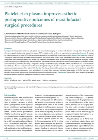
Fig. 1. Appearance of scars in patients of the control group on day 10 after
surgery.
Fig. 2. Appearance of scars in patients of the control group on day 30 after
surgery.
proLékaře.cz | 22.2.2022 | login: menchisheva.y@kaznmu.kz

PLATELET-RICH PLASMA IMPROVES ESTHETIC POSTOPERATIVE OUTCOMES
Acta Chir Plast 2021; 63(3): 118– 126
122
Me = 80.1 pixels (P
25
= 47.0; P
75
= 113.4)
which is less than the median of the scar
width in patients of the control group
Me = 99.3 pixels (P
25
= 71.1; P
75
= 130.4),
P < 0.05 (Graph 3).
Evaluation of scars
POSAS observer scale
We used the POSAS questionnaire
30 and 90 days after surgery to evaluate
the quality of postoperative scars. The
POSAS questionnaire (observer part) in-
cludes six indicators (vascularity, pig-
mentation, thickness, relief, pliability,
surface area) which were assessed by
physicians using a 1–10 scoring scale.
Thirty days after surgery, the mean
score value of all six indicators of the
scale was 5.8 ± 0.14 in the control group,
which was about 2.3× greater than the
treatment group mean. The mean value
in the treatment group was 2.5 ± 0.14
(P < 0.05). Ninety days after surgery, the
mean score value of the control group
was 3.7 ± 0.23 and the mean value in the
treatment group was 1.6 ± 0.07 (P < 0.05)
(Tab. 3).
POSAS patient scale
The POSAS questionnaire (patient part)
included six questions which were as-
sessed by patients using a 1–10 scoring
scale.
Thirty days after surgery, the mean pa-
tient POSAS score of the control group
was 5.0 ± 0.75, which was about 1.9times
greater than the treatment group mean.
The mean value in the treatment group
was 2.7 ± 0.35 (P < 0.05).
1.0 mm (Р
25
= 1.0; Р
75
= 2.5) and 2.0 mm
(Р
25
= 1.0; Р
75
= 3.0) in the treatment and
control groups, respectively (P < 0.05).
On the 30
th
day, this indicator was
3.0 mm (Р
25
= 2.0; Р
75
= 4.0) in the control
group, which is greater than the median
value of the treatment group – 1.5 mm
(Р
25
= 1.0; Р
75
= 3.5) (Graph 2).
Using the Image J program, we meas-
ured the width of the scars in pix-
els on the 10
th
and 30
th
days. The me-
dian widths of the postoperative scar
were 57.6 pixels (P
25
= 44.0; P
75
= 92.7)
and 62.8 pixels (P
25
= 46.7; P
75
= 120.1)
in the treatment and control groups
on 10
th
day after surgery, respectively
(P < 0.05). One month after the surgical
procedure, the postoperative scar width
in patients of the treatment group was
than the median width in the treatment
group 1.0 mm (Р
25
= 1.0; Р
75
= 1.5) on
3.5 days after surgery (P < 0.05). On the
7
th
day after surgical procedure, the me-
dian widths of postoperative wounds
were 2.0 mm (Р
25
= 1.0; Р
75
= 3.0) and
1,0 mm (Р
25
= 1.0; Р
75
= 2.0) in the con-
trol and treatment groups, respectively
(P < 0.05).
The most noticeable changes were
on the 10
th
and 30
th
days after opera-
tion. The scars of the patients in the
control group (Fig. 1, 2) were distin-
guishable from the normal surround-
ing skin on the 10
th
and 30
th
days after
operation as opposed to the treatment
group patients (Fig. 3, 4), who received
PRP injections. So, on the 10
th
day after
surgery, the median scar widths were
Fig. 4. Appearance of scars in patients
of the treatment group on day 30
after surgery.
Fig. 3. Appearance of scars in patients
of the treatment group on day 10
after surgery.
Graph 2. The expansion of postoperative scars (mm) at: A) 3 days; B) 5 days; C) 7 days; D) 10 days; E) 30 days after surgery.
0
1
2
3
4
5
6
7
8
9
A)
treatment group
0
1
2
3
4
5
6
7
8
9
B)
0
1
2
3
4
5
6
7
8
9
C)
0
1
2
3
4
5
6
7
8
9
D)
control group
0
1
2
3
4
5
6
7
8
9
E)
proLékaře.cz | 22.2.2022 | login: menchisheva.y@kaznmu.kz

PLATELET-RICH PLASMA IMPROVES ESTHETIC POSTOPERATIVE OUTCOMES
Acta Chir Plast 2021; 63(3): 118– 126
123
terpretation in the control group, the
treatment results had a slightly nega-
tive effect, while in the treatment group,
they did not have a negative effect. The
differences between the mean values of
DQLI on the 30
th
and 90
th
days after sur-
gery in the two groups are displayed in
Graph 4.
Discussion
The main results of the study are the
2-fold reduction of the scar width on
the 90
th
day after surgery and higher
patient satisfaction obtained from
questionnaires.
There is a number of studies offering
different speed and time of centrifuga-
tion to obtain PRP. The methods of prep-
aration of PRP are different in many ways
[36–39], which explains the lack of stand-
ardized methods of obtainment and ap-
plication of PRP. The therapeutic effect
of PRP could be achieved by increas-
ing the concentration of platelets twice
[40]. We were guided by the method of
Akhmerov et al [41] in choosing the time
and speed of rotation to PRP (specifically
5 min at a speed of 3,000 rpm). Regard-
ing the choice of the frequency of in-
prox. 4times more than in the treatment
group, i.e. 3.1 ± 4.25 (P < 0.05). Accord-
ing to the interpretation of DQLI in the
control group, the postoperative out-
comes of maxillofacial surgical proce-
dures had a strong negative impact on
the patients’ lives, while in the treatment
group, these outcomes had a slight
negative impact. Ninety days after sur-
gery, the mean values of scores were
4.3 ± 2.91 and 1.7 ± 1.82 points in the
control and treatment groups, respec-
tively (P < 0.05). According to the in-
Ninety days after surgery, the mean
score value of the control group was
2.7 ± 0.48, which was about 1.8times
more than the treatment group mean.
The mean value in the treatment group
was 1.5 ± 0.14 (P < 0.05). The differences
between the mean values in the two
groups are displayed in Tab. 4.
Results of the DQLI
Thirty days after surgical procedures,
the mean score value in the control
group was 12.7 ± 6.7, which was ap-
Graph 3. The expansion of postoperative scars (pixels) at: A) 10 and B) 30 days
after surgery.
treatment group
control group
0
50
100
150
200
250
300
350
400
A)
0
50
100
150
200
250
300
350
400
B)
Tab. 3. The mean observer POSAS scores in the control ad treatment groups 30 and 90 days after surgical procedures.
N Indicators
30 days after surgery
90 days after surgery
control group
N = 50
treatment group
N = 50
P-value
control group
N = 50
treatment group
N = 50
P-value
mean ± SE median
(Р
25
–Р
75
) mean ± SE
median
(Р
25
–Р
75
)
mean ± SE median
(Р
25
–Р
75
) mean ± SE
median
(Р
25
–Р
75
)
1. vascularity
5.6 ± 0.14
7.0
(6.0–7.0) 2.2 ± 0.14
2.0
(1.75–3.0) Р < 0.05 3.0 ± 0.14
3.0
(2.0–4.0) 1.6 ± 0.18
1.0
(1.0–2.0)
Р < 0.05
2. pigmentation 6.4 ± 0.16
7.0
(6.0–7.0) 3.1 ± 0.1
3.0
(2.0–4.0) Р < 0.05 3.8 ± 0.12
4.0
(3.0–4.0) 1.8 ± 0.13
2.0
(1.0–2.0)
Р < 0.05
3. thickness
5.8 ± 0.16
7.0
(6.0–7.25) 2.2 ± 0.14
2.0
(1.75–3.0) Р < 0.05 4.6 ± 0.15
5.0
(4.0–5.0) 1.7 ± 0.18
2.0
(1.0–2.0)
Р < 0.05
4. relief
6.0 ± 0.16
6.5
(6.0–7.0) 2.3 ± 0.13
2.0
(2.0–3.0) Р < 0.05 3.2 ± 0.19
4.0
(3.0–4.0) 1.7 ± 0.16
2.0
(1.0–2.0)
Р < 0.05
5. pliability
6.1 ± 0.16
6.0
(5.75–7.0) 2.5 ± 0.13
2.0
(1.0–2.0) Р < 0.05 4.0 ± 0.16
4.0
(3.0–5.0) 1.6 ± 0.17
2.0
(1.0–2.0)
Р < 0.05
6. surface area
5.4 ± 0.16
6.0
(5.75–7.0) 2.6 ± 0.11
1.5
(1.0–2.0) Р < 0.05 3.6 ± 0.17
4.0
(3.0–5.0) 1.3 ± 0.14
1.0
(1.0–2.0)
Р < 0.05
POSAS – patient and observer scar assessment scale
proLékaře.cz | 22.2.2022 | login: menchisheva.y@kaznmu.kz

PLATELET-RICH PLASMA IMPROVES ESTHETIC POSTOPERATIVE OUTCOMES
Acta Chir Plast 2021; 63(3): 118– 126
124
jection of PRP several studies reported
about multiplicity of PRP application
[43–45] or injection [46] during or after
surgery.
We used a single plasma injection,
since we believe that a single plasma in-
jection as a stimulator of regeneration is
sufficient to start the process of normal
wound healing. A single PRP injection
has also been suggested in the studies
of Eichler et al [47].
Considering the anatomical features
of blood supply and innervation on
Graph 4. The mean values of the dermatological quality of life index in patients
of the control and the treatment groups at 1 and 3 months after surgical
procedures.
0
5
10
15
20
25
1 month
3 months
treatment group
control group
Tab. 4. The mean patient POSAS scores in the control and treatment groups 30 and 90 days after surgical procedures.
N Indicators
30 days after surgery
90 days after surgery
control group
N = 50
treatment group
N = 50
P-value
control group
N = 50
treatment group
N = 50
P-value
mean ± SE median
(Р
25
–Р
75
) mean ± SE
median
(Р
25
–Р
75
)
mean ± SE median
(Р
25
–Р
75
) mean ± SE
median
(Р
25
–Р
75
)
1.
Has the scar
been painful
the past few
weeks?
2.9 ± 0.13
3.0
(2.0–3.25)
1.4 ± 0.07
1.0
(1.0–2.0)
Р < 0.05 1.1 ± 0.05
1.0
(1.0–1.0)
1.0 ± 0.02
1.0
(1.0–1.0)
Р < 0.05
2.
Has the scar
been itching
the past few
weeks?
2.5 ± 0.27
3.0
(2.0–4.0)
1.9 ± 0.18
2.0
(1.0–2.0)
Р < 0.05 1.3 ± 0.07
1.0
(1.0–2.0)
1.3 ± 0.07
1.0
(1.0–2.0)
Р < 0.05
3.
Is the scar
color differ-
ent from the
color of your
normal skin
at present?
6.6 ± 0.23
5.0
(3.0–8.0)
3.3 ± 0.14
2.0
(2.0–3.0)
Р < 0.05 3.2 ± 0.14
3.0
(2.0–4.0)
1.9 ± 0.14
2.0
(2.0–3.0)
Р < 0.05
4.
Is the stiff-
ness of the
scar differ-
ent from your
normal skin
at present?
6.0 ± 0.27
6.0
(2.0–8.25)
2.9 ± 0.21
1.0
(1.0–2.25)
Р < 0.05 3.2 ± 1.33
3.0
(2.0–4.0)
1.7 ± 0.14
1.0
(1.0–2.25)
Р < 0.05
5.
Is the thick-
ness of the
scar differ-
ent from your
normal skin
at present?
6.6 ± 0.21
6.0
(2.0–7.75)
3.5 ± 0.25
2.0
(1.0–3.25)
Р < 0.05 3.7 ± 1.43
4.0
(3.0–5.0)
1.8 ± 0.15
2.0
(1.0–3.25)
Р < 0.05
6.
Is the scar
more irregu-
lar than your
normal skin
at present?
5.7 ± 0.31
5.0
(2.0–6.0)
3.4 ± 0.23
2.0
(1.0–2.25)
Р < 0.05 3.7 ± 1.51
4.0
(2.75–5.0)
1.7 ± 0.12
2.0
(1.0–2.25)
Р < 0.05
POSAS – patient and observer scar assessment scale
proLékaře.cz | 22.2.2022 | login: menchisheva.y@kaznmu.kz

PLATELET-RICH PLASMA IMPROVES ESTHETIC POSTOPERATIVE OUTCOMES
Acta Chir Plast 2021; 63(3): 118– 126
125
study more objective than those con-
ducted previously.
Conclusion
The current study demonstrates two
findings: the first was that the use of PRP
improves postoperative wound healing
and results in better esthetic outcomes
in the postoperative period; the second
finding was that the patient satisfaction
with the results and quality of life was
higher in the treatment group where
PRP was used.
Role of authors:
Yuliya Menchisheva: originate con-
cept and design of the study, operation of the patients,
measurement of the scars, PRP preparation, acqui-
sition, analysis and interpretation of the data, critical
revision of the manuscript, crafting of the manuscript.
Ulmeken Mirzakulova:
operation of the patients,
analysis and interpretation of the data, crafting of the
manuscript, statistical analysis.
Dildora Usupova:
operation of the patients, measure-
ment of scars of patients, data analysis.
Gulzhan Yermukhanova:
review of the literature,
critical revision of the manuscript, crafting of the
manuscript.
Zhanagul Rysbayeva:
review of the literature, crafting
of the manuscript, statistical analysis.
Ethical considerations:
All participating patients sig-
ned informed consent forms to be eligible for research.
Ethics approval was obtained from the local ethics Co-
mmittee. The study protocol conformed to the ethical
guidelines of the 1975 Declaration of Helsinki.
Conflict of interest:
We are declaring that no com-
peting interests exist. We declare that we received no
financial support for the research, authorship or publi-
cation of this article.
References
1.
Borrione P., Gianfrancesco AD., Pereira MT.,
et al. Platelet-rich plasma in muscle healing.
Am J Phys Med Rehabil.
2010, 89(10): 854–861.
2.
Chicharro-Alcántara D., Rubio-Zaragoza M.,
Damiá-Giménez E., et al. Platelet rich plasma:
new insights for cutaneous wound healing man-
agement.
J Funct Biomater.
2018, 9(1): 10.
3.
Sclafani AP., Azzi J. Platelet preparations for
use in facial rejuvenation and wound healing:
a critical review of current literature.
Aesth Plast
Surg
. 2015; 39(4): 495–505.
4.
Hall MP., Band PA., Meislin RT., et al. Platelet-
-rich plasma: current concepts and application
in sports medicine.
J Am Acad Orthop Surg
. 2009,
17(10): 602–608.
5.
Li X., Xu C., Hou YL., et al. Are Platelet concen-
trates an ideal biomaterial for arthroscopic ro-
tator cuff repair? A meta-analysis of randomi-
zed controlled trials.
Arthroscopy
2014, 30(11):
1483–1490.
spective on their scarring is ultimately
the most important in relation to pa-
tients’ quality of life.
Many studies evaluate the use of PRP
in combination with other treatment
methods. These studies have had very
similar designs, often being presented
in a couple of groups, where researchers
compare results of treatment methods,
one of which includes the use of PRP.
Majani et al write about the treatment of
patients with traumatic scars [49]. In this
study which used a small sample size,
and the Manchester Scar Scale, it was
found that PRP was associated with bet-
ter treatment results.
It was our aim to find studies not lim-
ited to only facial surgeries or dermatol-
ogy, where authors describe the esthetic
results after surgeries. The most exten-
sive study in our search was made by
Balbo et al, where a five-year analysis of
the results of 115 patients with the am-
putations or wounds of fingers treated
with platelet gel was presented [50]. The
difference of this study from the study
conducted at our hospital is the applica-
tion of the platelet rich gel, not the in-
jection of PRP directly into the soft tis-
sues as it was done in our case. Balbo et
al reported that the recovery of soft tis-
sues of all patients ranged from 80 to
100% (median time 3 weeks) and the es-
thetic results were satisfactory in nearly
all cases that were shown in numerous
photos after surgeries. According to the
article, patients who have undergone
surgeries were satisfied with the results
of the treatment afterwards, but these
claims were not supported by objective
quantitative data.
None of the studies found in the liter-
ature were directly comparable to our
study in terms of design or methodol-
ogy. However, all studies found simi-
lar results. A strength of our study was
that we measured outcomes from dif-
ferent perspectives (both that of the pa-
tient and the surgeon) and triangulated
results from different methods of meas-
urement. We feel that this makes our
the face and neck, we propose to in-
ject PRP leaving 0.5 cm from the edge
of the wound. We were guided by the
regulation of the Republic of Kazakh-
stan (No. 666, November, 2009) on the
procurement and processing of blood
components, choosing the amount of
blood taken from the patient. According
to the recommendations of Akhmerov
et al [41], in the treatment of various dis-
eases (in such fields of medicine as sur-
gery, traumatology, gynecology), it is
necessary to inject 3–9 mL of PRP, de-
pending on the clinical case. A total of
9 mL of blood was required to obtain
3 mL of PRP. In current study, one tube of
9 mL was required for wounds < 10 cm
in length (3 mL of PRP), two tubes for
10–20 cm wounds (6 mL of PRP), and
three tubes for wounds > 20 cm (9 mL
of PRP).
The goal of PRP is to minimize wound
complications and attain better esthetic
outcomes. Previous studies have shown
the efficiency of PRP in different wound
healing processes, but few have pro-
vided an assessment of the influence of
PRP on skin quality or assessed patient
satisfaction with treatment results.
Several studies have evaluated the po-
tential of platelet rich plasma to treat
scar tissues. Willemsen et al reported
that platelet rich plasma reduced re-
covery time and improved esthetic out-
comes in facial rejuvenation [48]. They
observed when the patients could re-
turn to work or restart their social activi-
ties after surgery. The authors conducted
questionnaires about the appearance
of 82 patients’ faces after 4 weeks. They
used three questions with a scale 1–10
and surveyed only surgeons. Although
they used a different scale than the one
used in our study, the results were simi-
lar: both studies found that scarring was
less pronounced in the treatment group
relative to the control group. Our study
also surveyed patients. We consider this
a strength of our study because a sur-
geon’s perspective alone may not be
objective, and because the patient per-
proLékaře.cz | 22.2.2022 | login: menchisheva.y@kaznmu.kz

PLATELET-RICH PLASMA IMPROVES ESTHETIC POSTOPERATIVE OUTCOMES
Acta Chir Plast 2021; 63(3): 118– 126
126
on periimplant bone regeneration.
Bone
. 2004,
34(4): 665–671.
41.
Akhmerov RR., Korotkova OI., Ovechkina
MV., et al. Use of the autoplazma containing
thrombocytes in a dermatokosmetologiya and
an odontology. Plasmolifting™ technology. Plas-
tic surgery and cosmetology.
Russian Journal
(Plasticheskaya khirurgiya i kosmetologiya)
2013,
1: 94.
42.
Hom DB., Linzie BM., Huang TC. The hea-
ling effects of autologous platelet gel on acute
human skin wounds.
Arch Facial Plast Surg
. 2007,
9(3): 174–183.
43.
Khalafi RS., Bradford DW., Wilson MG. Topi-
cal application of autologous blood products
during surgical closure following a coronary ar-
tery bypass graft.
Eur J Cardiothorac Surg
. 2008,
34(2): 360–364.
44.
Kazakos K., Lyras DN., Verettas D., et al. The
use of autologous PRP gel as an aid in the man-
agement of acute trauma wounds.
Injury
2009,
40(8): 801–805.
45.
Yoo J., Roth K., Hughes B., et al. Evaluation of
postoperative drainage with application of pla-
telet-rich and platelet-poor plasma following
hemithyroidectomy: a randomized controlled
clinical trial.
Head Neck
. 2008, 30(12): 1552–1558.
46.
Guo Y., Qiu J., Zhang C. Follow-up study on
platelet-rich plasma in repairing chronic wound
nonunion of lower limbs in 47 cases.
Zhongguo
Xiu Fu Chong Jian Wai Ke Za Zhi
. 2008, 22(11):
1301–1305.
47.
Eichler C., Najafpour M., Sauerwald A., et al.
Platelet-rich plasma in the treatment of subcu-
taneous venous access device scars: a head-to-
-head patient survey.
Biomed Res Int
. 2015, 2015:
630601.
48.
Willemsen JC., van der Lei B., Vermeulen KM.,
et al. The effects of platelet-rich plasma on reco-
very time and aesthetic outcome in facial rejuve-
nation: preliminary retrospective observations.
Aesthetic Plastic Surg
. 2014, 38(5): 1057–1063.
49.
Majani U., Majani A. Correction of scars by
autologous fat graft and platelet rich plasma
(PRP).
Acta Medica Mediterranea
2012, 28(2):
99–100.
50.
Balbo R., Avonto I., Marenchino D., et al. Pla-
telet gel for the treatment of traumatic loss of
finger substance.
Blood Transfus.
2010, 8(4):
255–259.
Yuliya Menchisheva, MD, PhD
S. D. Asfendiyarov Kazakh National
Medical University
Tole bi 94
Almaty, 050000
Kazakhstan
e-mail: menchisheva.y@kaznmu.kz
Submitted: 30. 4. 2021
Accepted: 24. 7. 2021
24.
Lacci KM., Dardik A. Platelet-rich plasma:
support for its use in wound healing.
Yale J Biol
Med
. 2010, 83(1): 1–9.
25.
Chicharro-Alcántara D., Rubio-Zaragoza M.,
Damiá-Giménez E., et al. Platelet rich plasma:
new insights for cutaneous wound healing ma-
nagement.
J Funct Biomater
. 2018, 9(1): 10.
26.
Wang L., Gu Z., Gao C. Platelet-rich plasma for
treating acute wounds: a meta-analysis.
Zhon-
ghua Yi Xue Za Zhi.
2014, 94(28): 2169–2174.
27.
Prabhu R., Vijayakumar C., Bosco Chan-
dra AA., et al. Efficacy of homologous, platelet-
-rich plasma dressing in chronic non-healing ul-
cers: an observational study.
Cureus
. 2018, 10(2):
e2145.
28.
Malay S., Chung KC. How to use outcomes
questionnaires: pearls and pitfalls.
Clin Plast
Surg
. 2013, 40(2): 261–269.
29.
Rezaei F., Rezaei F., Abbasi H., et al. A compa-
rison of doctor/patient satisfaction with aesthe-
tic outcomes of rhinoplasty: a prospective study.
J Med Life
. 2019, 12(4): 374–380.
30.
Fearmonti R., Bond J., Erdmann D., et al. A re-
view of scar scales and scar measuring devices.
Eplasty
. 2010, 10: e43.
31.
Commander SJ., Chamata E., Cox J., et al. Up-
date on postsurgical scar management.
Semin
Plast Surg
. 2016, 30(3): 122–128.
32.
Marshall CD., Hu MS., Leavitt T., et al. Cutane-
ous scarring: basic science, current treatments,
and future directions.
Adv Wound Care
(New Ro-
chelle)
2018, 7(2): 29–45.
33.
Garg S., Dahiya N., Gupta S. Surgical scar re-
vision: an overview.
J Cutan Aesthet Surg
. 2014,
7(1): 3–13.
34.
Jourdan M., Madfes DC., Lima E., et al. Skin
care management for medical and aesthetic
procedures to prevent scarring.
Clin Cosmet In-
vestig Dermatol
. 2019, 12: 799–804.
35.
Moher D., Schulz KF., Altman DG. The CON-
SORT statement: revised recommendations for
improving the quality of reports of parallel-
-group randomized trials.
Ann Intern Med
. 2001,
134(8): 657–662.
36.
Breddin HK. Can platelet aggregometry be
standardized?
Platelets
2005, 16(3–4): 151–158.
37.
Araki J., Jona M., Eto H., et al. Optimized
preparation method of platelet-concentrated
plasma and noncoagulating platelet-derived
factor concentrates: maximization of platelet
concentration and removal of fibrinogen.
Tissue
Eng Part C Methods
. 2012, 18(3): 176–185.
38.
Dugrillon A., Eichler H., Kern S., et al. Autolo-
gous concentrated platelet-rich plasma (cPRP)
for local application in bone regeneration.
Int J
Oral Maxillofac Surg
. 2002, 31(6): 615–619.
39.
Franco D., Franco T., Schettino AM., et al. Pro-
tocol for obtaining platelet-rich plasma (PRP),
platelet-poor plasma (PPP), and thrombin for
autologous use.
Aesth Plast Surg
. 2012, 36(5):
1254–1259.
40.
Weibrich G., Hansen T., Kleis W., et al. Effect
of platelet concentration in platelet-rich plasma
6.
Sanchez M., Delgado D., Sґanchez P., et al. Pla-
telet rich plasma and knee surgery.
Biomed Res
Int
. 2014, 2014: 890630.
7.
Creaney L., Hamilton B. Growth factor deli-
very methods in the management of sports in-
juries: the state of play.
Br J Sports Med
. 2008,
42(5): 314–320.
8.
Dawood AS., Salem HA. Current clinical appli-
cations of platelet-rich plasma in various gyne-
cological disorders: An appraisal of theory and
practice.
Clin Exp Reprod Med
. 2018, 45(2): 67–74.
9.
Galal M., Khalifa A., Abd El Hafez M., et al. Pla-
telet-rich plasma (PRP) in obstetrics and gyne-
cology. Egypt J Hosp Med. 2021, 83(1): 889–894.
10.
de Vos RJ., van Veldhoven PL., Moen MH.,
et al. Autologous growth factor injections in
chronic tendinopathy: a systematic review.
Br
Med Bull
. 2010, 95: 63–77.
11.
Ahmed M., Reffat SA., Hassan A., et al. Plate-
let-rich plasma for the treatment of clean diabe-
tic foot ulcers.
Ann Vasc Surg
. 2017, 38: 206–211.
12.
Ji-Jun H., Hui-Hui S., Qing L., et al. Efficacy
of using platelet-rich plasma in spinal fusion
surgery-a preferred reporting items for sys-
tematic reviews and meta-analyses-compli-
ant meta-analysis.
World Neurosurg
. 2020, 139:
e517–e525.
13.
Kaplan N. A study of platelet rich plasma co-
mmonly used in neurosurgery practice.
Merit
Res J Med Sci.
2019, 7(1): 1–7.
14.
Ronci C., Ferraro AS., Lanti A., et al. Platelet-
-rich plasma as treatment for persistent ocular
epithelial defects.
Transfus Apher Sci
. 2015; 52(3):
300–304.
15.
Alio JL., Arnalich-Montiel F., Rodriguez AE.
The role of ”eye platelet rich plasma” (E-PRP) for
wound healing in ophthalmology.
Curr Pharm
Biotechnol
. 2012, 13(7): 1257–1265.
16.
Albanese A., Licata ME., Polizzi B., et al. Pla-
telet-rich plasma (PRP) in dental and oral sur-
gery: from the wound healing to bone regenera-
tion. Immun Ageing. 2013, 10(1): 23.17. Carlson
NE., Roach RB. Jr. Platelet-rich plasma: clinical
applications in dentistry.
J Am Dent Assoc
. 2002,
133(10): 1383–1386.
18.
Khatu SS., More YE., Gokhale NR., et al. Pla-
telet-rich plasma in androgenic alopecia: myth
or an effective tool.
J Cutan Aesthet Surg
. 2014,
7(2): 107–110.
19.
Oryan A., Alidadi S., Moshiri A. Platelet-rich
plasma for bone healing and regeneration.
Ex-
pert Opin Biol Ther
. 2016, 16(2): 213–232.
20.
De Sousa A. Psychological issues in oral and
maxillofacial reconstructive surgery.
Br J Oral
Maxillofac Surg
. 2008, 46(8): 661–664.
21.
De Sousa A. Psychological issues in acqui-
red facial trauma.
Indian J Plast Surg
. 2010, 43(2):
200–205.
22.
Gasparyan AY., Ayvazyan L., Pretorius E.,
et al. Platelets in rheumatic diseases: friend or
foe?
Curr Pharm Des
. 2014, 20(4): 552–566.
23.
Cole BJ., Seroyer ST., Filardo G., et al. Platelet-
-rich plasma: where are we now and where are
we going?
Sports Health
. 2010, 2(3): 203–210.
proLékaře.cz | 22.2.2022 | login: menchisheva.y@kaznmu.kz


