Library search
Search Results
-
 Optical and biometric indicators of the eye in children with juvenile glaucoma combined with myopia
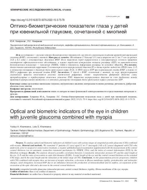
in LibraryPurpose. To analyze optical and biometric indicators of children's eyes with various stages of congenital juvenile glaucoma (CJG) combined with myopia. Material and methods. We examined 17 patients (31 eyes) aged 11 to 17 (averagely 14.0 ± 0.2 years) diagnosed with CYG who underwent, prior to surgical or conservative treatment, a regular ophthalmological examination supplemented with the measurement of the central corneal thickness (on an automatic contactless tonometer-pachymeter by NIDEK, USA), and the index of corneal deformation (ICD) by the Shkrebets technique. Results. The analysis showed a possible correlation between 1) tonometric intraocular pressure (P) and the axial length of the eye, 2) P t and the ratio of excavation to optic disk diameter (E/ON), 3) axial length of the eye and the central corneal thickness at the terminal CYG stage, and 4) the axial length and the refraction at the initial stage of CYG. Conclusion. As the glaucomatous process progresses, children with CYG combined with myopia show an increase of myopic refraction, a decrease in fibrous membrane rigidity, pretrabecular and trabecular changes, axial elongation, increased IOP due to an imbalance between the production of intraocularfluid and its outflow, an expansion of the excavation of the optic disk and a decrease in the central corneal thickness.
-
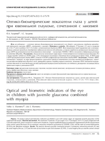 Optical and biometric indicators of the eye in children with juvenile glaucoma combined with myopiaPurpose. To analyze optical and biometric indicators of children's eyes with various stages of congenital juvenile glaucoma (CJG) combined with myopia. Material and methods. We examined 17 patients (31 eyes) aged 11 to 17 (averagely 14.0 ± 0.2 years) diagnosed with CYG who underwent, prior to surgical or conservative treatment, a regular ophthalmological examination supplemented with the measurement of the central corneal thickness (on an automatic contactless tonometer-pachymeter by NIDEK, USA), and the index of corneal deformation (ICD) by the Shkrebets technique. Results. The analysis showed a possible correlation between 1) tonometric intraocular pressure (Pt) and the axial length of the eye, 2) Pt and the ratio of excavation to optic disk diameter (E/ON), 3) axial length of the eye and the central corneal
in Library -
 Неврологические проявления остеохондроза позвоночника и грыжи межпозвонкового диска после микродискэктомии
Scientific works of gifted youth and medicine of the XXI centuryНеврологические проявления остеохондроза позвоночника являются чрезвычайно распространенной патологией, которая ведет к длительной утрате трудоспособности и к социальной дезадаптации лиц трудоспособного возраста. По данным эпидемиологических исследований последних лет, дегенеративные изменения в позвоночнике в возрасте 20-25 лет имеют 53% людей в общей популяции Земли. В возрастной группе от 22 до 40 лет, боль по поводу заболеваний позвоночника составляет 15-20%, а в возрасте 45-65 лет достигает 23%. Частота быстро повышается с возрастом и составляет у лиц 40-50 лет 93%, а у людей старше 60 лет — 100%. Доля дегенеративно-дистрофических поражений позвоночника в поясничном отделе составляет более 80%, среди которых 60% обусловлены грыжей межпозвонкового диска
-
 Osteochondrosis of the spine, characterized by degenerative changes in the intervertebral disc and adjacent vertebral bodies, is one of the most common diseases of the musculoskeletal system [10]
 -
 DIAGNOSTIC ROLE OF OPTICAL COHERENT TOMOGRAPHY OF ANGIOGRAPHY IN PRIMARY OPEN-ANGLE GLAUCOMAThe review presents data from foreign literature on the diagnostic value of optical coherence tomography angiography (OCT-A) as a method for studying the microvasculaturc of the eye in early diagnosis and monitoring of glaucoma. Numerous foreign researchers have studied the OCT-A parameters, which provide information on the anatomy and physiology of the microcirculation of the retina and the optic nerve head. OCT-A can provide an assessment of the vasculature within the optic nerve, peripapillary superficial retina, macula, and parapapillary choroid in glaucoma. The density of the peripapillary superficial retinal vessels allows the diagnosis and detection of the progression of glaucoma, as well as the thickness of the peripapillary RNFL. A decrease in the density of the peripapillary vessels of the intact field or unaffected glaucomatous eyes indicates that vascular changes may occur before noticeable damage to the visual field.
Medicine and innovations -
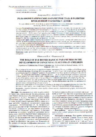
 Purpose: to study the interrelation between the biomechanical and the biometric parameters of the eye in different stages of infantile glaucoma. Material and methods. A total of 19 patients (37 eyes) aged from 3 to 10 years with a non-operated primary infantile glaucoma were examined. Of these 6 patients (9 eyes) had initial stage, 7 patients (9 eyes) had advanced stage), 6patients (9 eyes) hadfar advanced stage, and 7patients (10 eyes) had terminal stage, respectively. A combination of different stages of the disease was found in 11 children, while 8 children had the same stage of the disease in both eyes. Examination methods included visometry, ophthalmoscopy, determining the excavation of the optic disc, A -scan recording the anterior-posterior axis (APA) of the eye, Filatov — Kalfa elastotonometry with weights of 5g, 10g, 15g. Results. The gradient of elastic curve rise was noted in all stages, but it was the highest in the terminal stage, where APA was greater according to the severity of the disease. APA and elastic curve raise was found to correlate in the far-advanced and the terminal stages of glaucoma. Conclusion. The changes in the biomechanical properties of the fibrous membrane of the eye in children with infantile glaucoma depend on the severity of the disease, which is manifested in changes in the biometric parameters. Sclera rigidity is reduced in the far-advanced and terminal stages of infantile glaucoma, which can lead to an underestimation of the true level oflOPand the risk of glaucoma development in children.
 -
 Early diagnostics of glaucoma with low oftalmotonus in the conditions of the Samarkand areaIn some cases it is difficult to make a diagnosis of low pressure glaucoma. 79 patients had low pressure glaucoma. 64 (81%) patients made up 80/60-100/60 mm Hg, but in 15 (19%) it was 100-130/85-90 mm Hg and if taking into consideration their age they were refered to hypotonics too. A careful clinical ophthalmologic examination was performed to all patients. Analysis of intraocular pressure revealed in 52 (65,7%) patients ophthalmotonus being at 16-19 mm Hg, in 27 (34,3%) ranging 20-22 mm Hg. At an early stage of the process there had been noted single absolute scotomas in central vision which may be increased if intraocular pressure is high. There was marked an early appearance of some changes in the optic nerve, to be specific and to be an evident informative sings in diagnosis of low pressure glaucoma. A direct correlation between the state of peripheric vision and the changes in the optic disc has been established. In therapy of patients with local hypotensive drugs it is advisable to prescribe medicine for increasing arterial pressure, improving microcirculation and angioprotectors. Indication to surgical procedure is recommended in case of progressing disturbance of visual function
Journal problems of biology and medicine -
 The role of eye biomechanical parameters in the development of congenital glaucoma in children
in LibraryThe objective of this review was to study biomechanical parameters of the eye in children with different stages of congenital glaucoma. 20 patients (40 eyes) aged from 1 month to 3 years with simple primary congenital not operated glaucoma were examined. 7 patients (14 eyes) entered in the 1st group (developed stage), 8 patients (16 eyes) — the 2nd group (passed stage), 5 patients (10 eyes) — the 3rd group (end stage) respectively. Research methods included visiometry, refractometry, ophthalmoscopy, definition of the optic disc excavation, ultrasound А-scan of the anterior posterior size of the eye, Filatov’s — Kalfa elastotonometry with weights 5g, 10g, 15g.
The analysis of the data shows that gradient of the elastotonometry graphs elevation registered in all groups, but the highest was in group of children with terminal stage. Thus the anteroposterior size of the eye and intraocular pressure increase corresponding to disease severity. Biomechanical parameters of the eye increase according to the stages of a congenital glaucoma first form. The decrease of sclera rigidity shows morphological changes of the eye tissue and severity of glaucoma process. -
 The effect on the human body of solar radiation in the conditions of the samarkand regionThe problem of interaction between man and his environment is especially acute in regions with a dry hot climate, which include Uzbekistan. Solar radiation is the totality of solar matter and energy entering the Earth. Energy propagates in the form of electromagnetic waves at a speed of 300 thousand kilometers per second, passes through the atmosphere and reaches the Earth in 8 minutes. earthly the surface is under the influence of both direct and scattered by the earth's atmosphere, the sun's rays. It is the scattering of blue-blue rays in the atmosphere that explains the blueness of the sky on a clear day. Yellow-orange color The finite disk is due to the fact that the waves corresponding to it pass almost without dispersion. The electromagnetic spectrum of solar radiation consists of infrared (50%), visible (41%) and ultraviolet (9%) parts. Because they quanta have different energies, they have a variety of effects on a person. The hygienic significance of solar radiation is also extremely high. Its regulation is carried out in accordance with SNiP, which for solar radiation are compiled taking into account light climatic features of various geographical zones and are taken into account in the design and construction of various facilities
Journal problems of biology and medicine -
 Evaluation of the 3 mm thickness splint therapy on temporomandibular joint disorders (TMDS)
Actual problems of dentistry and maxillofacial surgery 4Temporomandibular disorders (TMDs) encompass internal derangements of the temporomandibular joint (TMJ), abnormalities of masticatory muscles and the neighboring structure of the TMJ, and TMJ-related headache conditions. In all manifestations of TMDs, the major negative effects the patients experience include jaw movement limitations and of course slight to severe pain in the head and neck regions. TMDs include TMJ and facial pain, including tenderness to touch the facial region muscle (particularly masticatory muscles and the TMJ), uncoordinated jaw movements, and the presence of joint noise .While many research studies have evaluated diet intake problems during postop patient follow-ups of TMDrelated surgeries, some studies have also considered diet intake before and after treatment of both nonsurgical and surgical evaluations of TMD patients in the context of jaw movement and the level of pain the patient experienced.
-
 Results of treatment after the operational spondilodistsit at injuries of a backboneThe research is based on studying of a tunelization at diagnostics and treatment of the post-operational spondilodistsit. At the time of inspection and treatment the Samarkand regional hospital of an orthopedics and a consequence of a trauma were in unit of a vertebrologiya 31 patients. From the total number of patients was 9 (29%) women, there were 22 men (71%). The age of patients was intensified from 18 to 63 years with lesions of lumbar vertebrae and disks, adjacent to them, - spondilodistsity. And with the medical purpose we carried out by the purpose of confirmation of the diagnosis low-invasive interventions - a tunnelization of a body of a vertebra and a disk through an arch root with a biopsy and the subsequent sanation of the center (A way of treatment of the spondilodistsit No. IAP 05393). Good results are received at 22 (71%) patients - full retrogress of a pain syndrome at rest and turn of a bed, decrease of feeling of fatigue at vertical position, improvement of clinical laboratory indicators. Satisfactory results are received at 8 (25,8%) patients, the unsatisfactory result is noted at 1 patient (3,2%). Thus, the tunnelization and sanations of the center antibiotics at a limited or initial spondilodistsit is a low-traumatic and effective method of single-step pressure decrease in a zone of pathology and providing local etiologies and pathogenetic treatment. The material intake for a bacteriological and histological research allows to reduce time of verification of process and to quickly provide positive clinical effect
Journal problems of biology and medicine -
 Параметры жевательных мышц у детей с зубочелюстными аномалиями при мышечно- суставных дисфункциях ВНЧС.
Actual problems of dentistry and maxillofacial surgery 4Асимметричное сокращение жевательных мышц приводит к несогласованному движению обеих нижнечелюстных головок в суставных ямках, что вызывает повреждение суставных поверхностей, сдавление отдельных участков внутрисуставного диска, дистальное смещение головок нижней челюсти.
-
 Complex treatment of glaucomatous optic neuropathy by endonasal electrophoresis in combination with electrostimulation (revie
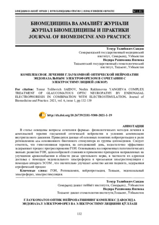
Journal of Biomedicine and PracticeThe article points out the issues associate with combination of pharmaco-physiologic methods of treatment in complex treatment of glaucomatous optic neuropathy in condition with compensate intraocular pressure. There was informationabout main conceptions of neuroprotection and the role of retinalamin as a main biogenic stimulator from the citomedin group. It should be noted that, these days, hypotensive therapy is not enough effectively retain process of the progression of GON. It is more reasonable to use drugs directed to improve a blood supply in the optic nerve area, mainly via targeted delivery with a help of endonasal electrophoresis and percutaneous electrostimulation with ESOM apparatus that considerably improve a quality of patient`s life by retaining atrophic process
-
 Clinical manifestations and results of surgical treatment of intervertebral disc herniations of lumbosacral regionIt has been studied 83 patients with a diagnosis of "Osteochondrosis of the lumbosacral region, inter-vertebral disc herniations" operated in the Clinic of Neurosurgery of Samarkand Medical Institute. Among 83 operated patients more often observed extravertebral (monoradiculary pain, positive symptom of La-segue) and vertebral (flattening of lumbar lordosis and limited mobility in the lumbar region of spine and the varying degrees of scoliosis) syndromes. Advanced interlaminary and interlaminary techniques are the most effective among the existing sur-gical treatment of intervertebral disc herniations and they allow achieving full recovery approximately of 70.0% of the patients, the relative recovery on every fourth patient after surgery. Determination of the regularities of the clinical course, the timely application of modern methods of surgical treatment of patients with disc herniations of lumbosacral region contribute to improving the results of treatment, prevention of various complications and disability
Journal problems of biology and medicine -
 Complex treatment of glaucomatous optic neuropathy by endonasal electrophoresis in combination with electrical stimulation (review)
in LibraryThe article points out the issues associate with combination of pharmaco-physiologic methods of treatment in complex treatment of glaucomatous optic neuropathy in condition with compensate intraocular pressure. There was informationabout main conceptions of neuroprotection and the role of retinalamin as a main biogenic stimulator from the citomedin group. It should be noted that, these days, hypotensive therapy is not enough effectively retain process of the progression of GON. It is more reasonable to use drugs directed to improve a blood supply in the optic nerve area, mainly via targeted delivery with a help of endonasal electrophoresis and percutaneous electrostimulation with ESOM apparatus that considerably improve a quality of patient`s life by retaining atrophic process
-
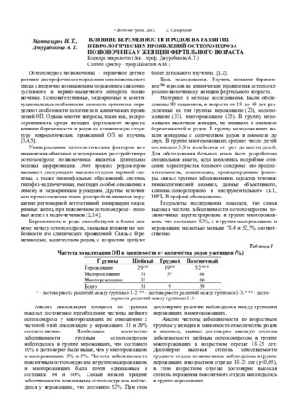 Влияние беременности и родов на развитие неврологических проявлений остеохондроза позвоночника у женщин фертильного возрастаОстеохондроз позвоночника - первичное дегенеративно-дистрофическое поражение межпозвонкового диска с вторично возникающим поражением связочносуставного и нервно-мышечного аппарата позвоночника. Психовегетативные, эндокринные и конституциональные особенности женского организма определяют особенности патогенеза и клинических проявлений ОП. Однако многие вопросы, такие как, распространенность среди женщин фертильного возраста, влияние беременности и родов на клиническую структуру неврологических проявлений ОП не изучены
Doctor's Herald -
 The role of eye biomechanical parameters in the development of congenital glaucoma in children
in LibraryTug'ma glaukoma bilan og'rigan bolalarda ko'zning biomexanik ko'rsatkichlari uning turli bosqichlari bilan o'rganildi. Oddiy birlamchi tug'ma operatsiyasiz glaukoma bilan og'rigan 1 oydan 3 yoshgacha bo'lgan 20 bemor (40 ko'z) tekshirildi. Ulardan 1-guruhga (ilg'or bosqich) mos ravishda 7 (14 ko'z), 2-chi (ilg'or bosqich) - 8 (16 ko'z), 3-chi (terminal bosqich) 5 bemor (10 ko'z) kiradi. Tadqiqot usullari orasida visometriya, refraktometriya, oftalmoskopiya, optik diskni qazib olishni aniqlash, ko'zning old-orqa o'lchamini ro'yxatga olish bilan A-skanerlash, 5 g og'irlikdagi elastotonometriya; janubiy; 15d, shuningdek, Filatov-Kalf usuli bilan. Olingan ma'lumotlarning tahlili shuni ko'rsatdiki, elastokrivning ko'tarilish gradienti barcha guruhlarda qayd etilgan, ammo eng yuqori ko'rsatkich terminal bosqichi bo'lgan bolalar guruhida bo'lgan, ko'zning anteroposterior hajmi esa mos ravishda kattalashgan. kasallikning og'irligi bilan. Ko'z ichi bosimining oshishi kasallikning og'irligiga mutanosib ravishda qayd etilgan. Konjenital glaukomaning oddiy shakli rivojlanishi bilan ko'zning biomexanik ko'rsatkichlari bosqichlarga ko'ra ortadi. Shu bilan birga, tolali membrananing qattiqligining pasayishi ko'zning to'qimalarida morfologik o'zgarishlarni va glaukoma jarayonining zo'ravonligini ko'rsatadi.
-
 The relationship between functional and anatomical-optical parameters of the eye in congenital glaucoma in children
in LibraryAim of research. To study the dynamics of the FBS of the eyes in children with congenital glaucoma by age. The studies were carried out in the clinic of TashPMI, on 132 eyes with congenital glaucoma. By age, patients were distributed according to the classification of E.S. Avetisova 2003. At the advanced stage of FBS, the eye exceeded the average statistical norm by 2.9 mm, 2.3 mm, 2.3 mm, at the advanced stage by 4.7 mm, 4.8 mm, 6.3 mm, and at the terminal stage by 7.5 mm, respectively, in each age group of eyes. An increase in the size of the anterior-posterior axis of the eye in congenital glaucoma depends not only on the violation of the hemo hydrodynamic processes of the eye with the accumulation of intraocular fluid, but also depends on the age-related dynamics of eye growth.
-
 Our experience of surgical treatment of idiopathic scoliosis of thoracic localizationTo analyze results of surgical treatment of patients with thoracic idiopathic scoliosis. Materi- al and methods. Fifty-two patients with Lenke type 1 idiopathic scoliosis were operated on. Follow-up periods ranged from 2 weeks to 8 years (mean 1.8 years). Surgical treatment included four types of operation: spine deformity correction with CD instrumentation; supramalleolar-and-skull traction and CDI correction; discec-tomy and interbody fusion with bone autograft and CDI correction; supramalleolar-and-skull traction, discec- tomy and underbody fusion with bone auto graft, and CDI correction. Patients were interrogated with pre-and postoperative SRS-24 questionnaires. Results. Scoliosis was corrected from a mean of 67.7° to 26.6°, with a mean deformity value being 30.3° at the last follow-up. Thus, postoperative progression of the thoracic curve with a mean follow-up 1.8 years was 3.7° (9 % from the achieved correction). Anterior fusion provided a three- fold decrease in postoperative progression. Sagittal shape of the thoracic and lumbar spine remained within norm limits. The location of the lowest instrumented vertebra (LIV) relative to a neutral vertebra, lower stable vertebra and neutralized disc did not reliably influence on the postoperative course. Postoperative deformity progression was associated only with increase in LIV tilt. SRS-24 data showed a high rate of patients' satisfac-tion with the obtained effect of treatment, the rate growing with the extension of follow-up terms. Severe com-plications were not observed. Conclusion. Modern 3rd generation segmental instrumentation allows to obtain stable and high results of treatment for single curve thoracic idiopathic deformities, while all regularities of postoperative course are not fully understood yet
Journal problems of biology and medicine