Library search
Search Results
-
 Использование интерактивных приемов в обучении говорению на иностранном языкеПоявление интерактивных методов обучения примерно в 1990-х годах позволило развиваться творческой стороне личности, раскрыть ранее не поддерживаемые грани мыслительной деятельности обучающихся. Интерактивные методы сделали обучение более интересным, обучающиеся стали больше вовлечены в процесс, а также стали взаимодействовать не только между собой, но и с педагогом в целом.
Modern innovative research current problems and development trends: solutions and prospects -
 Эволюция понятия творческое мышлениеВ данной стате раскрывается понятие творческое мышление. Выделены основные научные направления, занимавшиеся вопросами творчества, креативности и творческого мышления.
Modern innovative research current problems and development trends: solutions and prospects -
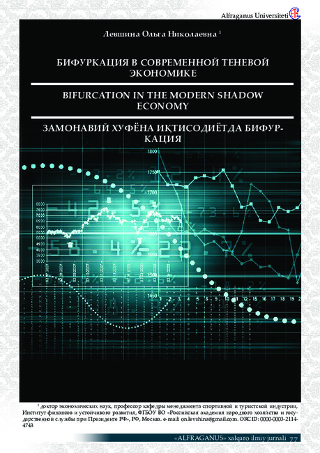
In the world scientific literature, it is traditionally customary to highlight a significant tax burden, excessive regulation of organizational and economic activities and a significant share of the state in the total volume of turnover in the economy as the main reasons for the shadow economy’s emergence and development. The key prerequisites for the formation and growth of the shadow economy are the emergence and development of a structural and economic crisis in the country’s economy, significant government intervention in the economy, and the build-up and intensification of competition. And, as a result, typical features of the shadow economy include the avoidance of taxes by economic entities, the presence of hidden unemployment, a significant level of corruption, the existence of barter trade, the withdrawal of domestic capital abroad, and the presence of a «double» accounting system. Thus, as statistics and practice of economic indicators of recent decades have shown, shadow activities in the micro-, meso- and macromarkets pose a threat to the economic security of the country and the region and destroy the existing structure of socioeconomic relationships.
-
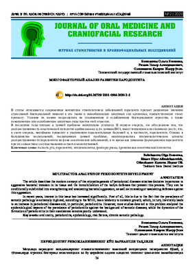 MULTIFACTOR ANALYSIS OF PERIODONTITIS DEVELOPMENTThe article describes the modern concept of the etiopathogenesis of periodontal diseases attaches decisive importance to aggressive bacterial invasion in its tissue and the immobilization of the body's defenses that prevent this process. They can be conditionally subdivided into strengthening and weakening bacterial aggression, as well as increasing or weakening defenses against this aggression. In recent years, interest in this issue has increased significantly. First of all, this is due to the fact that the prevalence of somatic pathology is extremely high and, according to the WHO, has a tendency to constant growth, which, in turn, inevitably leads to an increase in periodontal diseases and, in particular, periodontitis. However, most studies devoted to this problem analyzed the epidemiological aspects of the prevalence of periodontitis against the background of somatic diseases, while the dynamics of the formation of periodontitis in their coexistence remains poorly understood.
Journal of oral medicine and craniofacial research -
 Актуализация содержательного компонента учебных программ повышения квалификации преподавателей в области цифровизации образования
Цифровизация современного образования: проблема и решениеВ статье представлен содержательный компонент учебных программ повышения квалификации в области цифровизации образования, разработанный с целью формирования профессиональных компетенций преподавателей естественнонаучных и социогуманитарных дисциплин.
-
 QUALITY OF LIFE IN PATIENTS WITH CHRONIC RECURRENT LIP FISSUREBased on the results of the study, it was found that patients with chronic recurrent lip crack (CRLC) have a more severe dental status and need a comprehensive dental examination in order to optimize the treatment and rehabilitation process and a differentiated individual approach to providing specialized dental care.
Journal of oral medicine and craniofacial research -
 Polyalkylene guanidine chloride as a preservative for carbohydrate feed in the top dressing for beesCarbohydrate feeding for bees is a must in the autumn-winter period. With long-term presence of liquid feed in the form of sugar syrup, they become sour, microflora changes, which is not permissible when feeding insects. For long-term storage of cooked carbohydrate feed, preservatives must be used. This work presents the results of studying the use of polyalkylene guanidines as a preservative for longterm storage of carbohydrate feed. Studies have been conducted on the toxicity of this drug when using it.
Prospects for the development of veterinary science and its role in ensuring food safety -
 Peculiarities of the state of cerebral hemodynamics in patients with vertebro-basilar insufficiency
Journal of Biomedicine and PracticeThe paper presents the results of our own studies of the cerebral (arterial and venous) hemodynamic state with duplex scanning of the neck vessels, transcranial dopplerography of the cerebral vessels in patients with vertebro-basilar insufficiency. The study of venous blood flow characteristic of patients with venous encephalopathy revealed increased diameter of the internal jugular vein, increased blood flow rate and decreased pulsatility index of the basal Rosenthal vein, increased LVBF (linear velocity of blood flow) and diameter of the vertebral vein
-
 Slaughter and meat qualities of agricultural poultry when using the anti-stress drug in their dietsThis article discusses information on the use of an anti-stress drug in feeding broiler chickens, on its effect on the slaughter and meat qualities of poultry. Studies to study the effect of the anti-stress additive Fid-Food Magic Antistress Mix were carried out on young farm poultry meat cross “Ross – 308” in the conditions of the research center for the safety and efficiency of the use of feed and additives of the Volgograd State Agrarian University in Volgograd
Prospects for the development of veterinary science and its role in ensuring food safety -
 ASSESSMENT OF THE PREVALENCE AND SEVERITY OF CARIES IN PATIENTS WITH GASTROESOPHAGEAL REFLUX DISEASEThe study is devoted to the study of the features of the clinical course of care in patients with various stages of gastroesophageal reflux disease. The results of a comprehensive clinical examination confirm the aggravating effect of GERD on the course of dental caries. Moreover, with the progression of gastroesophageal reflux disease and its transition from endoscopically "negative" to the catarrhal and metaplastic stages, the severity of the course and the intensity of carious lesions arc aggravated.
Medicine and innovations -
 Обеспечение прослеживаемости сырья и животноводческой продукции в рамках систем HACCPСогласно законодательству Украины, внедрение систем безопасности продукции HACCP является обязательным для всех предприятий пищевой промышленности. Важной составляющей систем HACCP является обеспечение надлежащей сырьевой/продуктовой прослеживаемости «от поля до стола». В животноводческом секторе основой эффективной прослеживаемости является полная идентификация животных, в мясой и молочной промышленности – всеобъемлющие системы внутренней и внешней прослеживаемости
Prospects for the development of veterinary science and its role in ensuring food safety -
 Качество и безопасность пищевых продуктов на основе принципов системы хасспВ статье приведены результаты анализа управления системой безопасности мясных паштетов на основе принципов ХАССП.
Prospects for the development of veterinary science and its role in ensuring food safety -
 The role of magnetic resonance imaging in the comprehensive radial diagnosis of volumetric masses of the eye organ
Catalog of abstractsRelevance of the problem. The difficulties of diagnostics of orbital diseases are well known. Especially difficult is intraspecies differentiation among the multitude of tumour, pseudotumour, inflammatory, vascular, endocrine and other diseases occurring here, manifested by the symptom complex of unilateral exophthalmos [Beradze I.N., 1978; Brovkina A.F., 1993].
Malignant intraocular neoplasms are the main cause of death of patients with diseases of the organ of vision, with 45-48% of patients dying from metastases in the first 5 years after enucleation [Alekseeva I.B., 1990, Barkhash S.A.1978, Brovkina A.F..1991, 1997; Keizer R.W.. Viclvoyc G.L.,1986],
Retinoblastoma is the most frequent malignant neoplasm in children. According to different authors, the frequency of its occurrence is 1 case per 14000 - 35000 newborns. [Bobrova N.F. and Vit V.V., 1993; Brovkina A.F., 1997; Provenzale J.M., et al., 1995; Skulski M., et al., 1997; Weber A.L., Mafee M.F, 1992; Wilms G., et al., 1989]. The frequency of patients with the most malignant intraocular tumour in adults - uveal melanoma has recently reached 7-9 people per 1 million population [Brovkina A.F., 1997; Kotslyansky E.O., 1989; Yushko N.A., Peskova L.I., Kalenich L.A., 1989; Peyster R.G., Augsburger J..I., Shields J.A., 1988; Romani A.. Baldeschi L., ct al 1998; Scott I.U., 1998].
The fundamental difference in treatment tactics, depending on the stage of development, size and topography of the tumour, as well as the seriousness of the prognosis in retinoblastomas and melanomas sharply increase the requirements for the accuracy of their differential diagnosis. At the same time, the number of diagnostic errors in ocular tumours continues to be 10-30% even when complex clinical and instrumental examination is applied in specialised ophthalmological centres [Ternovoy S.K., Panfilova G.V., Rogozhin V.A., 1979; Friedman F.E., Malyuta G.D., Kodzov M.V., 1995; Song G.X., 1991].
Widely used in ophthalmological practice traditional diagnostic methods (ophthalmoscopy, gonioscopy, diaphanoscopy, fluorescence angiography, laboratory tests) are insufficient to obtain comprehensive information about the localisation, nature of growth and prevalence of volumetric pathological formations of the eye and orbit. This circumstance and not quite satisfactory results of surgical treatment are the causes of high mortality of patients [Muratova T.T., Nigmanova N.H., Kozlovskaya G.M.. 1989, Naches A.I., 1980; Cheremisin V.M., Trufanov G.E., Kholin A.V., 1991]. Untimely or erroneous recognition of pathological processes of the orbit leads to a sharp deterioration of visual functions, up to blindness, and in some cases to the death of the patient [Yuzhakov A.M., Travkin A.G., Kiseleva O.A., 1991]. All this determines the importance of timely and accurate diagnosis of diseases of the orbit, on the one hand, and the difficulty of such diagnosis - on the other [Gabunia R.I., Kolesnikova E.K., Tumanov L.B., 1982].
The fact that the orbit is closed from direct inspection and palpation by bone walls and the eyeball, indicates the advantage of radial diagnostics in comparison with other methods of examination. In the arsenal of clinicians there is a great variety of methods of clinical-radial diagnostics of orbital pathology, however, at present the information in the literature about their resolving capabilities and significance in comparative aspect is incomplete and not fully studied. The priority of using one or another instrumental investigation, their sequence and expedient combination have not been determined yet. This makes it difficult to choose the optimal standardised approach for diagnosis and adequate treatment [Cheremisin V.M., Trufanov G.E., 1993, Weber A.L., Sabates N.R., 1996; Wenig V.M., Mafee M.F., 1998].
Thus, the study of these and other questions, contributing to the improvement of diagnostics and treatment of patients with neoplasms of the eye and ocular cavity, should be recognised as urgent urgent.
Purpose of the study. Comparative evaluation of magnetic resonance tomography capabilities and development of algorithms for complex radial diagnostics of volumetric formations of the visual organ. To solve this goal we set the following tasks.
1. To study the normal picture of the magnetic resonance image of the visual organ in comparison with other methods of visualisation.
2. To find out the possibilities of magnetic resonance tomography, ultrasound and computed tomography in detection and evaluation of intraocular neoplasms.
3. To determine the role and place of magnetic resonance tomography in differential diagnostics of volumetric pathological formations of the eye cavity in comparison with other radial methods of research.
4. To determine the indications and to develop an algorithm for the complex application of radiography, ultrasound, computer and magnetic resonance tomography for diagnostics of volumetric formations of the eye organ.
Scientific novelty.
The present work is the first to give a detailed and detailed description of the complex clinical and radiation examination, with generalisation and standardisation of magnetic resonance, computer and ultrasound semiotics of volumetric pathological formations of the eye and eye cavity. The conducted clinical and instrumental investigations allowed to determine the diagnostic value and resolving capabilities of each of the applied methods. The ultrasound, CT and MRI signs of volumetric formations of the eye organ were studied, clarified and supplemented taking into account the use of low-field magnetic field and general-purpose ultrasound apparatus. The developed standardised diagnostic algorithm of examination of patients with this pathology is new, thanks to which the pre-oppositional diagnosis of tumour and other diseases of the visual organ is improved and the total radiation load on the patient is reduced.
Conclusions
1. MPT will provide an opportunity to study the weight of the soft tissue and anatomical components of the ocular cavity, up to the optic nerve sheath and perineural liquor space, the orbital apex and chiasmal-sellar region, as well as to assess the condition of adjacent structures of the brain and facial skull. The method is limited in the evaluation of changes in the bony walls of the orbital cavity.
2. MRI is inferior in detecting characteristic signs of retinoblastoma (presence of calcification). The sensitivity of MRI was 66.6%, while for ultrasound and CT these values were 96.1 and 100%, respectively. But when the tumour spreads rstrobulbarly outside the eyeball (at 3-4 stages) the informativeness of MRI increases significantly. In uveal melanoma the sensitivity and specificity of MRI reaches 100%.
3. Both MRI and CT have a high detection rate (98.1% and 95.8% respectively) of benign orbital tumours of both primary and secondary origin. However, MRI is the preferred method of investigation. MRI is especially informative when a cranioorbital tumour and pseudotumour are suspected. The sensitivity of the method is 90.9% and 91.6%, respectively
4. In some cases ultrasound can be used to differentiate between encapsulated and diffuse neoplasms, which facilitates the diagnosis. However, when the pathological process is localised near the orbital apex, the diagnostic value of ultrasound decreases. In such cases it is advisable to use MRI.
5. In detection of primary and secondary malignant tumours of the orbital cavity both MRI and CT are quite informative (sensitivity 97,2% and 95,4% respectively), but the most comprehensive information about the state of bone walls will be provided by CT. When the process spreads intracranially, the value of MRI increases significantly, especially with the use of contrast enhancement.
6. The developed algorithm of complex clinical and radiation examination of patients with the use of ultrasound, CT and MRI is the most effective in the diagnosis of volumetric pathological formations of the eye and eye cavity, allowing to reduce to an adequate minimum the total radiation load on the patient and diagnostic period, excluding duplication of research techniques and choosing the most informative in each case, which in turn allows to develop appropriate treatment tactics and reduce the level of disability of the patient. -
 THE ROLE OF COMORBID PATHOLOGY IN THE DEVELOPMENT AND SEVERITY OF PERIODONTAL DISEASESIn the course of the study, the necessity and degree of participation of narrow-profile specialists in the treatment of periodontal diseases were determined for the timely detection of comorbid pathology and the appointment of adequate therapy. 225 patients with generalized periodontitis of various severity were studied, 40 patients without GP and pathology of COPD formed a control group. Statistically significant differences in the frequency of registered pathology in patients of the compared groups with the control indicators were established, as well as correlation relationships between the frequency of detected diseases and the severity of periodontal pathology. Thus, the total frequency of previously existing diseases increased in the series of HPLT, GPST and GPTS from 32.47 ± 5.34% in HPLS; to 51.25 ± 3.83 % in GPST to 86.96 ± 3.14 % in patients with GPTS ( linear correlation coefficient χ2 = 96.167; P ≤ 0.001); the corresponding dynamics of the newly detected pathology was 42.86 ± 5.63 % ; 47,65 ± 3,83 % and 13.04 ± 3,14 % (χ2 =65,087 ; P ≤ 0.001); and the corresponding frequency of all somatic pathology, requiring systematic medical correction made 75,32±4,93%; 99,41±0.52% and 100,00 ± 0,00 % (χ2 = 235,351; P ≤ 0.001). On the basis of the research, define the tactics of local therapy for the following pathogenetic mechanisms of development of periodontal disease: disorders of microcirculation, the prevalence of lipid peroxidation, increased cytokine aggression and increasing bone resorption. When making a diagnosis, it is necessary to individualize the volume and methods of therapy as much as possible based on the assessment of individual clinical and laboratory parameters of the patient, by identifying markers that determine the priority mechanisms of the development of the disease.
Journal of oral medicine and craniofacial research -
 Features of the organization of veterinary events for cattle leukemiaThe article presents an analysis of anti-leukemia measures in the farms of the Chelyabinsk region, an economic assessment of veterinary measures, identifies the main disadvantages and gives recommendations to improve the effectiveness of health measures.
Prospects for the development of veterinary science and its role in ensuring food safety -
 Determination of water quality by the indicators of zoobenthos (by the example of the chirchik river)Water resources of the Tashkent region are intensively usedfor water supply, meeting the needs of industries, agriculture, energy, fish farming and recreation. In the thesis, on the example of the Chirchik River, the issues of the impact of anthropogenic activities and the accumulation of environmental damage are considered. The material for the work was the modern data of the author and thirdparty researchers, which made it possible to characterize the prospects for the development of the water economy of the Tashkent region.
Prospects for the development of veterinary science and its role in ensuring food safety -
 International arbitration in switzerland: recent developments
Prospects of development of international commercial arbitration in UzbekistanAs an Uzbek lawyer practicing international arbitration in Switzerland, I have been closely monitoring the recent developments in Switzerland’s arbitration landscape. Today, I am delighted to share my observations and insights on these changes with you.
The 2021 has been a turning point for the Swiss arbitration community because of two major developments: the revised Swiss arbitration law entered into force in January 2021 and the revised Swiss Rules of arbitration entered into force in June 2021. -
 The multifactorial nature of the etiology of periodontal diseases dictates the need to consider the probability of their development and features of the clinical course using multifactorial and risk assessment models. Models for predicting variants of the course of pathology are the basis for making decisions about methods and means of prevention and prescribing individual therapy. This was the basis for the assessment of the "total risk of generalized periodontal disease".
 -
 Role of comorbid pathology in the development and severity of parodontal diseases
Journal of Biomedicine and PracticeIn the course of the study, the need and degree of participation of narrow-profile specialists in the treatment ofparadontal diseases was determined for the timely detection of comorbid pathology and the appointment of adequate therapy.
We studied 225 patients with generalized parodontitis of varying severity. 40 patients without generalized parodontitis (GP) and pathology of the oral mucosa (POM) formed a control group.
Statistically significant differences in the frequency of registered pathology in patients of the compared groups with control indicators, as well as correlations between the frequency of detected diseases and the severity ofparadontal pathology, were established. Thus, the total incidence of previously existing diseases increased in the series of mild generalized parodontitis (MGP), moderate generalized parodontitis (MODERGP) and severe generalized parodontitis (SGP) from 32.47 ± 5.34% with MGP; up to 51.25 ± 3.83% with MODERGP up to 86.96 ± 3.14% in patients with SGP (linear correlation coefficient χ² = 96.167; P ≤ 0.001); the corresponding dynamics of the newly discovered pathology was 42.86 ± 5.63%; 47.65 ± 3.83% and 13.04 ± 3.14% (χ² = 65.087; P ≤ 0.001); and the corresponding frequency of all somatic pathology requiring systematic drug correction was 75.32 ± 4.93%; 99.41 ± 0.52% and 100.00 ± 0.00% (χ² = 235.351; P ≤ 0.001).
Based on the studies performed, local therapy tactics should be determined for the following pathogenetic mechanisms of the development of parodontitis: microcirculation disorders, the prevalence of lipid peroxidation processes, an increase in cytokine aggression and an increase in bone resorption.
When making a diagnosis, the scope and methods of therapy should be maximally individualized based on an assessment of the patient's individual clinical and laboratory parameters, by identifying markers that determine the priority mechanisms of the development of the disease. -
 POTENTIAL RISK FACTORS IN THE DEVELOPMENT AND PROGRESSION OF NONSPECIFIC INTERSTITIAL PNEUMONIACurrently, the interest of a number of domestic and foreign researchers in the problem of timely diagnosis and treatment of respiratory diseases has sharply increased. Nonspecific interstitial pneumonia (NPSI) was considered an orphan disease. Currently, there is a significant increase in this pathological condition, in particular, it is associated with the coronovirus pandemic, where interstitial pneumonia is a serious complication. The prevalence of NSAIDs is approximately the same as that of ILF. The prevalence of NSIPs is about 40 patients per 100 thousand people. Potential risk factors include viruses, smoking, diabetes mellitus, gastroesophageal reflux, organic and inorganic dust, heredity, etc. With NSAIDs, chronic inflammation and persistent viral infections can synergistically support autoimmune lesions that were previously described as ILF / idiopathic fibrosing alveolitis. However, causative antigens in NSIP remain unknown.
Journal of Cardiorespiratory Research -
 Teaching and methodological manual as an effective preparation tool for arabic language testing (level A1-A2)Educational and methodological manuals developed by teachers of the Department of Arabic Philology in the Institute of Asian and African Countries at Moscow State University are an effective tool for diagnosing and systematizing knowledge of Arabic grammar in its basic part, as well as for testing and developing practical speaking and listening skills within certain topics in the volume required for testing in Arabic (basic level).
Arabic language in the era of globalization: innovative approaches and teaching methods -
 DIFFERENTIAL DIAGNOSTICS OF NON-SPECIFIC INTERSTITIAL PNEUMONIAA retrospective analysis of case histories of 82 patients with nonspecific interstitial pneumonia, 24 patients with idiopathic pulmonary fibrosis, 8 with exogenous allergic alveolitis, 12 with systemic scleroderma and 6 with drug pneumonia was carried out in hospital treatment in the pulmonology department of the Samarkand city medical hospital in 2010-2019 years. It has been established that in the differential diagnosis of nonspecific interstitial pneumonia and other lung diseases, particular attention should be paid to the anamnesis, clinical presentation and CT signs.
Journal of Cardiorespiratory Research -
 On the issue of modern methods of monitoring the degree of knowledge in the Russian language
Traditions and innovations in language research and teachingВ статье описываются разные приемы установления уровня знаний по русскому языку необходимых для получения национальных и международных сертификатов обучающимися. Задания, описанные в статье позволяют обучающимся определить круг недостаточно усвоенных вопросов, а своевременная и тщательная работа по устранению пробелов в знаниях учащихся – залог высоких результатов обучения.
-
 Современные компетенции педагога в современном образовании
Science, society, education in modern realitiesВ статье исследуется проблема формирования и развития информационно-коммуникативных компетенций будущего учителя в условиях цифровой трансформации образовательного процесса. Актуальность исследования обусловлена задачами цифровизации образования и направленностью на развитие всех видов цифровой деятельности педагога.
-
 Interactive Training Model When Observing The Transformed Phraseologists In Literary Text
Traditions and innovations in language research and teachingThe article provides material from a practical lesson in a student classroom. The authors show how a subjective-subjective environment is created when carrying out work on the analysis of transformed verbal phraseological units using examples from the ironic detective story by D. Dontsova “This Bitter Sweet Revenge”. The result of using an interactive teaching model in increasing students’ motivation to study the Russian language and research activities.