Library search
Search Results
-
 От мудрого правления к стратегическому руководству: что не так с переводами документов ООНС возникновением международных организаций в 1940-х гг. в сфере развития широко распространился так называемый Developmentspeak, или язык развития, представляющий собой специализированный вариант английского языка со своим особым лексиконом. Основные понятия, термины и жаргонизмы языка развития представляют собой громкие слова (buzzwords) и броские фразы (catchphrases), которые весьма сложны как для понимания, так и для перевода.
Translation studies: problems, solutions and prospects -
 ABSTRACT: The high prevalence of tuberculosis among the population determines the relevance of information on timely diagnosis and treatment, this pathology. The clinical manifestations of tuberculosis in the oral cavity depending on the form, modem methods of diagnosis and differential diagnosis of the disease, as well as approaches to conservative treatment of specific lesions of the oral mucosa arc described.
 -
 Methodological approach to ensuring the safety of consumer goods in economic integrationThe article analyzes methodological approaches that determine the importance of food security in the context of consumer goods security. Improved food security concept by economic criteria.
Economics And Education -
 Results of epidemiological studies prevalence of iodine deficiency diseases in the republic of uzbekistanIn spite of the works performed against IDD, it still remains severe in Uzbekistan. Epidemiological studies performed in 1998 and 2004 demonstrated that endemic goiter prevalence in children constituted 72,8% and 58,8 %, respectively. Progress to lower iodine deficiency was due to the activities to liquidation of iodine deficiency disorders (IDD) including supply with iodizing equipment and supplementation with potassium iodate. The law of Republic of Uzbekistan “Prevention of iodine deficiency disorders” was adopted in May 2007. Current publication demonstrates iodine deficiency level, in accordance with WHO recommendations with sentinel method, in Uzbekistan. It has been revealed that goiter prevalence decreased to 40,2%. Population consuming iodized salt and population having normal ioduria values increased 62,4 % и 63,7 %. Thus we demonstrated that iodine deficiency disorders in Uzbekistan decreases although remains on severe level.
Doctor's Herald -
 An approach to prosthetics of teeth in children with multiple adentities encountered in practice
StomatologiyaTo improve the quality of dentures in children with multiple edentulism.
-
 Improving the tactics of treating patients with combined craniocerebral and maxillofacial injuriesIt is investigated 156 patients with combined cranio-maxillofacial trauma. Ways of improvement of tactics of treatment of patients with combined craniocereberal and maxillofacial damages are developed and defined. Modern techniques of cranial-maxillofacial surgery allowing in shorter terms and in fuller volume to spend rehabilitation of patients with heavy combined by a cranial-maxillofacial trauma.
Stomatologiya -
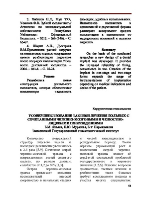
 Methods to assessment pain syndrome using scales and questionnaires in patients with trigemI- NAL neuralgia
NeurologyThis article presents the results of a study of pain syndrome in patients with severe stenosis of the carotid and vertebral arteries (group 1, 50 patients) and patients without stenosis (group 2, 50 patients) using scales and questionnaires. When assessing the sensory, emotional, and evaluative components of the McGill questionnaire, it was found that the pain syndrome in the first group was more pronounced than in the second. A significant correlation was found between the results on the Beck Depression Scale and VAS, which shows that patients of older age groups with stenosis have a significantly stronger pain syndrome.
-
 This article is devoted to the study of the role of chronic brain ischemia as a possible additional risk factor for inflammation of the oral mucosa (IOM). Patients with disorders of cerebral circulation caused by atherosclerosis of cerebral vessels were studied. It was revealed that the systemic immune inflammation observed in brain ischemia play an important role in the Genesis of inflammation in the SOPR. This is evidenced by an increase in the level of proinflammatory cytokines along with an increase in neuro-specific protein and enzyme, which is one of the triggers of inflammation of the IOM. against the background of chronic brain ischemia
 -
We conducted a retrospective study of 75 cases of pregnancy and childbirth in women with obesity, which were divided into 2 groups: group 1 - 45 pregnant women with obesity and group 2 - 30 pregnant women with overweight. The control group consisted of 16 healthy pregnant women with normal weight growth rates. The effect of overweight and obesity in women on the course and outcome of pregnancy and childbirth was studied. In obese pregnant women, the risk of developing hypertensive conditions increases 3.5 times. Gestational hypertension was detected in 15.5%, and preeclampsia in 26.6% of obese pregnant women. Gestational pyelonephritis was detected in 20% of group 1 and in 10% of pregnant women in group 2. In women of the 1st group, in 22% of cases, the pregnancy was terminated before 12 weeks. Physiological childbirth in patients of group 1 occurred in 68.5% of cases, and in group 2, it was 80%. The study of anthropometric data of newborns showed that 28.5% had an increased body weight, and 25.7% of newborns had indicators of a large fetus.
-
Изучить качество жизни у пациентов с невралгией тройничного нерва на фоне консервативного и хирургического лечения.
-
 USE OF OSTEOPLASTIC MATERIALS BASED ON HYDROXYAPATITE AND COLLAGEN IN RESTORATION OF DEFECTS OF THE JAW BONESA review of studies on the use of osteoplastic materials based on hydroxyapatite and collagen in the restoration of defects in the jaw bones is reviewed. A special place among bioactive ceramics is occupied by hydroxyapatite, which has a high affinity for bone tissue. It has been shown that biocomposite materials based on collagen stimulate osteogenesis and without contraindications, with the exception of individual intolerance. Need to research and improve osteoplastic materials based on the natural components of the bone.
Stomatologiya -
 The role of magnetic resonance imaging in the comprehensive radial diagnosis of volumetric masses of the eye organ
Catalog of abstractsRelevance of the problem. The difficulties of diagnostics of orbital diseases are well known. Especially difficult is intraspecies differentiation among the multitude of tumour, pseudotumour, inflammatory, vascular, endocrine and other diseases occurring here, manifested by the symptom complex of unilateral exophthalmos [Beradze I.N., 1978; Brovkina A.F., 1993].
Malignant intraocular neoplasms are the main cause of death of patients with diseases of the organ of vision, with 45-48% of patients dying from metastases in the first 5 years after enucleation [Alekseeva I.B., 1990, Barkhash S.A.1978, Brovkina A.F..1991, 1997; Keizer R.W.. Viclvoyc G.L.,1986],
Retinoblastoma is the most frequent malignant neoplasm in children. According to different authors, the frequency of its occurrence is 1 case per 14000 - 35000 newborns. [Bobrova N.F. and Vit V.V., 1993; Brovkina A.F., 1997; Provenzale J.M., et al., 1995; Skulski M., et al., 1997; Weber A.L., Mafee M.F, 1992; Wilms G., et al., 1989]. The frequency of patients with the most malignant intraocular tumour in adults - uveal melanoma has recently reached 7-9 people per 1 million population [Brovkina A.F., 1997; Kotslyansky E.O., 1989; Yushko N.A., Peskova L.I., Kalenich L.A., 1989; Peyster R.G., Augsburger J..I., Shields J.A., 1988; Romani A.. Baldeschi L., ct al 1998; Scott I.U., 1998].
The fundamental difference in treatment tactics, depending on the stage of development, size and topography of the tumour, as well as the seriousness of the prognosis in retinoblastomas and melanomas sharply increase the requirements for the accuracy of their differential diagnosis. At the same time, the number of diagnostic errors in ocular tumours continues to be 10-30% even when complex clinical and instrumental examination is applied in specialised ophthalmological centres [Ternovoy S.K., Panfilova G.V., Rogozhin V.A., 1979; Friedman F.E., Malyuta G.D., Kodzov M.V., 1995; Song G.X., 1991].
Widely used in ophthalmological practice traditional diagnostic methods (ophthalmoscopy, gonioscopy, diaphanoscopy, fluorescence angiography, laboratory tests) are insufficient to obtain comprehensive information about the localisation, nature of growth and prevalence of volumetric pathological formations of the eye and orbit. This circumstance and not quite satisfactory results of surgical treatment are the causes of high mortality of patients [Muratova T.T., Nigmanova N.H., Kozlovskaya G.M.. 1989, Naches A.I., 1980; Cheremisin V.M., Trufanov G.E., Kholin A.V., 1991]. Untimely or erroneous recognition of pathological processes of the orbit leads to a sharp deterioration of visual functions, up to blindness, and in some cases to the death of the patient [Yuzhakov A.M., Travkin A.G., Kiseleva O.A., 1991]. All this determines the importance of timely and accurate diagnosis of diseases of the orbit, on the one hand, and the difficulty of such diagnosis - on the other [Gabunia R.I., Kolesnikova E.K., Tumanov L.B., 1982].
The fact that the orbit is closed from direct inspection and palpation by bone walls and the eyeball, indicates the advantage of radial diagnostics in comparison with other methods of examination. In the arsenal of clinicians there is a great variety of methods of clinical-radial diagnostics of orbital pathology, however, at present the information in the literature about their resolving capabilities and significance in comparative aspect is incomplete and not fully studied. The priority of using one or another instrumental investigation, their sequence and expedient combination have not been determined yet. This makes it difficult to choose the optimal standardised approach for diagnosis and adequate treatment [Cheremisin V.M., Trufanov G.E., 1993, Weber A.L., Sabates N.R., 1996; Wenig V.M., Mafee M.F., 1998].
Thus, the study of these and other questions, contributing to the improvement of diagnostics and treatment of patients with neoplasms of the eye and ocular cavity, should be recognised as urgent urgent.
Purpose of the study. Comparative evaluation of magnetic resonance tomography capabilities and development of algorithms for complex radial diagnostics of volumetric formations of the visual organ. To solve this goal we set the following tasks.
1. To study the normal picture of the magnetic resonance image of the visual organ in comparison with other methods of visualisation.
2. To find out the possibilities of magnetic resonance tomography, ultrasound and computed tomography in detection and evaluation of intraocular neoplasms.
3. To determine the role and place of magnetic resonance tomography in differential diagnostics of volumetric pathological formations of the eye cavity in comparison with other radial methods of research.
4. To determine the indications and to develop an algorithm for the complex application of radiography, ultrasound, computer and magnetic resonance tomography for diagnostics of volumetric formations of the eye organ.
Scientific novelty.
The present work is the first to give a detailed and detailed description of the complex clinical and radiation examination, with generalisation and standardisation of magnetic resonance, computer and ultrasound semiotics of volumetric pathological formations of the eye and eye cavity. The conducted clinical and instrumental investigations allowed to determine the diagnostic value and resolving capabilities of each of the applied methods. The ultrasound, CT and MRI signs of volumetric formations of the eye organ were studied, clarified and supplemented taking into account the use of low-field magnetic field and general-purpose ultrasound apparatus. The developed standardised diagnostic algorithm of examination of patients with this pathology is new, thanks to which the pre-oppositional diagnosis of tumour and other diseases of the visual organ is improved and the total radiation load on the patient is reduced.
Conclusions
1. MPT will provide an opportunity to study the weight of the soft tissue and anatomical components of the ocular cavity, up to the optic nerve sheath and perineural liquor space, the orbital apex and chiasmal-sellar region, as well as to assess the condition of adjacent structures of the brain and facial skull. The method is limited in the evaluation of changes in the bony walls of the orbital cavity.
2. MRI is inferior in detecting characteristic signs of retinoblastoma (presence of calcification). The sensitivity of MRI was 66.6%, while for ultrasound and CT these values were 96.1 and 100%, respectively. But when the tumour spreads rstrobulbarly outside the eyeball (at 3-4 stages) the informativeness of MRI increases significantly. In uveal melanoma the sensitivity and specificity of MRI reaches 100%.
3. Both MRI and CT have a high detection rate (98.1% and 95.8% respectively) of benign orbital tumours of both primary and secondary origin. However, MRI is the preferred method of investigation. MRI is especially informative when a cranioorbital tumour and pseudotumour are suspected. The sensitivity of the method is 90.9% and 91.6%, respectively
4. In some cases ultrasound can be used to differentiate between encapsulated and diffuse neoplasms, which facilitates the diagnosis. However, when the pathological process is localised near the orbital apex, the diagnostic value of ultrasound decreases. In such cases it is advisable to use MRI.
5. In detection of primary and secondary malignant tumours of the orbital cavity both MRI and CT are quite informative (sensitivity 97,2% and 95,4% respectively), but the most comprehensive information about the state of bone walls will be provided by CT. When the process spreads intracranially, the value of MRI increases significantly, especially with the use of contrast enhancement.
6. The developed algorithm of complex clinical and radiation examination of patients with the use of ultrasound, CT and MRI is the most effective in the diagnosis of volumetric pathological formations of the eye and eye cavity, allowing to reduce to an adequate minimum the total radiation load on the patient and diagnostic period, excluding duplication of research techniques and choosing the most informative in each case, which in turn allows to develop appropriate treatment tactics and reduce the level of disability of the patient. -
 Cross-cultural communication challenges in translation/interpretationThe dynamics of the scientific and cultural contacts between independent Uzbekistan and foreign countries arouse high demands on the quality of translation and require the study of the methods of translation used in the transmission of the content of various texts. The role of any language in translation is the same as that which it always plays in the life of society; it acts as the most important means of human communication, reflects any changes in the political and economic life of the society and forms one of the incredible parts of human behavior
Translation studies: problems, solutions and prospects -
 Comparative analysis of the results of corneal diameters by different methods of measurement in children with primary congenital glaucom
Journal of Biomedicine and PracticeThe article provides a comparative analysis of the study of the diameter of the cornea in children with primary congenital glaucoma. Three methods, which are used in pediatric ophthalmology, are described. The first method is measuring the diameter of the cornea using a school ruler, the second method is using a surgical compass and the third is a new method developed with the use of a gauge in the form of glasses and a computer program. All the positive and negative aspects of methods for studying the diameter of the cornea are described in detail.
-
 The use of integrated scales in purulent - inflammatory processes of the maxillofacial regionThe article provides an overview of the literature on the problem of determining the severity and prognosis of pyoinflammatory diseases of the maxillofacial region. The use of integrated point scales is a widespread and accessible method that has not been sufficiently introduced into clinical practice. Integrated point scales can be used both to assess the severity and prognosis of pyoinflammatory diseases, and to determine the effectiveness of therapy and its correction, as well as to prevent complications.
Medicine and innovations -
 In this article, the history of Surkhan oasis dance, which is a part of Uzbek national dance art, and local aspects related to it are expressed.
 -
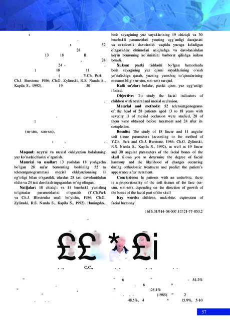
 Сохта прогеник прикусли беморларни турли ортодонтик аппаратлар ёрдамида даволаш
Actual problems of dentistry and maxillofacial surgery 4Прогения сагиттал сатх йўналишдаги прикус аномалияси. Барча аномалиялар ичида сохта прогения 13-14% ташкил этиб ,унинг мажбурий ва фронтал турлари фарқланади. (Ф.Я.Хорошилкина). Шунга асосан, аномалияни тарқалиш даражаси ,уни аниқлаш ва даволаш долзарб хисобланади.
-
 The study of environmental certification as one of the factors for quality management and economic securityThe article analyzes the essence of the concept of "ecological certification" as one of the factors ensuring economic security. It also provides a methodological approach to studying the stages of environmental certification, which serves as a means of controlling the entry of environmentally harmful products into the country. The certification system has been improved, not limiting certification and contributing to the integration of the country's economy into the world market and meeting international obligations.
Economics and innovative technologies -
 Contemporary views on the role of genital papillomaviral infection in the development of precanceral diseases and cervical cancer, ways of their prevention
in LibraryHuman papillomaviruses (HPV) are sexually transmitted and often occur in young people. They are usually neutralized by the immune system. However, the continued presence of high-risk HPV (HR) can lead to the development of abnormal cervical cells; this condition is considered precancerous if at least two-thirds of the surface layer of the cervix is affected. Human papillomavirus (HPV) is a common virus that can cause about 6 different types of cancer in later life. Cancer can develop years, and sometimes decades, after a person is infected with HPV.
-
 Review of early orthodontic treatment for class III malocclusion
Actual problems of dentistry and maxillofacial surgery 4Evaluate the effectiveness of orthodontic/orthopedic methods used in the early treatment of Class III malocclusion in the short and long terms.
-
 Клинико-неврологические и нейро-физилогические особенности невралгии тройничного нерва
Scientific works of gifted youth and medicine of the XXI centuryРаспространенность невралгии тройничного нерва (НТН) достаточно велика и составляет до 30-50 больных на 100 000 населения, а заболеваемость по данным ВОЗ находится в пределах 2-4 человек на 100 000 населения. Заболевание чаще возникает после 40 лет и преобладает у женщин. Проблема болей в лице, связанных с поражением тройничного нерва на разных уровнях, изучается длительное время. В то же время сегодня не существует четко определенной концепции патогенеза этой нозологии, и методы патогенетического лечения, соответственно, так же разнообразны. В последние годы проведены новые исследования, выполненные на современном техническом уровне, которые расширили представления о патогенезе НТН.
-
 Роль заместительной гормональной терапии при ортопедическом стоматологическом лечении женщин в постменопаузе
Actual problems of dentistry and maxillofacial surgery 4Исследованиями последних лет установлено влияние системного остеопороза на состояние зубочелюстной системы. У женщин в постменопаузе наблюдается быстрое снижение минеральной плотности кости (МПК) из-за дефицита гормона (эстрогена).
-
 Вросший ноготь (unguis incarnates, ingrown toenail или onychocryptosis) - сложный патологический комплекс, возникающий вследствие ряда причин и сопровождающийся совокупностью морфологических и функциональных изменений со стороны ногтей пальцев стоп, их матриксов и мягких тканей. Эта патология яатяется одной из наиболее частых причин обращения к хирургам в амбулаторных условиях - от 0,5 до 10% больных [1,2,3]. По данным Д.И. Муратова (1971), удельный вес больных среди обратившихся за хирургической помощью составляет 0,71%, а среди общего числа первичных амбулаторных больных — от 0,5 — 1,1%
 -
 Анализ репродуктивных нарушений у женщин с миомой матки и/или аденомиозом и методы коррекции
in LibraryСреди структурных аномалий в гинекологии миома матки и аденомиоз представляют собой две различные, хотя часто сосуществующие патологии с заметной распространенностью у женщин репродуктивного возраста. До настоящего времени были предложены различные механизмы, чтобы объяснить влияние каждого из них на репродуктивный результат [3].
-
 Применение гидроксиапатита и коллагена при эндопротезировании нижней челюсти
Actual problems of dentistry and maxillofacial surgery 4Повышение эффективности хирургического лечения костных дефектов нижней челюсти с использованием комбинированного костнопластического материала на основе гидроксиапатита и коллагена.