

Доктор ахборотномаси № 1 (98)—2021
108
Today, severe traumatic brain injury attracts the attention of many researchers around the
world. The huge scale of today's traumatism has made it not only a medical, but also an acute so-
cial problem for the whole country [1,6,8,9].
The main percentage of injuries is traumatic brain injury. Traumatic brain injury averages 30
-40% in terms of injury ratio. It ranks first among the causes of disability in the world population.
Traumatic brain injury affects the temporary disability of the population. Among the causes of
death of the population of working age, traumatic brain injury is even ahead of cardiovascular and
oncological diseases [11].
Severe traumatic brain injury often causes damage to the basal structures of the brain. In this
case, central reflex and humoral changes occur throughout the div. As a result, for the first time
after a traumatic brain injury, microcirculation disorders occur throughout the div, the same
changes lead to systemic damage to all internal organs, ultimately causing multiple organ failure.
In connection with this, changes in the kidneys are observed more often, and are manifested by the
corresponding clinical picture [7].
The reasons for the high mortality rate in patients with traumatic brain injury are largely as-
sociated with the development of intra- and extracranial complications. Clinical studies prove a
change in the kidneys (cystopyelitis, renal failure) most often, and complications in this organ are
DOI: 10.38095/2181-466X-2021981-108-111 UDK 611-018.61:616.831.001(756):616-092.2
MACROSCOPIC AND MICROSCOPIC CHARACTERISTICS OF KIDNEYS OF WHITE
UNBORED RATS AFTER SEVERE CRANIAL INJURY
G. Kh. Khuseynova
Bukhara state medical institute, Bukhara, Uzbekistan
Key words:
traumatic brain injury, kidney, macroscopic features, microscopic features, topography, abdominal cavity.
Таянч сўзлар:
бош мия жароҳати, буйрак, макроскопик хусусиятлари, микроскопик хусусиятлари ,
топография, қорин бўшлиғи.
Ключевые слова:
черепно-мозговая травма, почка, макроскопические особенности, микроскопические осо-
бенности, топография, брюшная полость.
This article provides information on the results of scientific studies that allow to evaluate and study the features
of macroscopic and microscopic characteristics of the kidneys of 3-month-old white outbred rats with severe traumatic
brain injury. Traumatic brain injury was performed using the Traffic Accident model, and rat kidneys were isolated
and macroscopic and microscopic examinations were performed in the order in which anatomical parameters were
established.
ОҒИР БОШ МИЯ ЖАРОҲАТЛАНИШИДАН КЕЙИНГИ ОҚ ЗОТСИЗ КАЛАМУШЛАР
БУЙРАКЛАРИНИНГ МАКРОСКОПИК ВА МИКРОСКОПИК ХУСУСИЯТЛАРИ
Г. Х. Хусейнова
Бухоро давлат тиббиѐт институти, Бухоро, Ўзбекистон
Ушбу мақолада оғир травматик мия жароҳати олган 3 ойлик оқ каламуш буйракларининг макроскопик
ва микроскопик хусусиятларини баҳолаш ва ўрганиш имконини берадиган илмий тадқиқот натижалари
ҳақида маълумот берилган. Травматик мия шикастланиши ―Йўл-транспорт ҳодисаси‖ модели ѐрдамида амалга
оширилди анатомик параметрларини аниқлаш мақсадида макроскопик ва микроскопик текширишлар
ўтказилди.
МАКРОСКОПИЧЕСКАЯ И МИКРОСКОПИЧЕСКАЯ ХАРАКТЕРИСТИКА ПОЧЕК БЕЛЫХ
БЕСПОРОДНЫХ КРЫС ПОСЛЕ ТЯЖЕЛОЙ ЧЕРЕПНО-МОЗГОВОЙ ТРАВМЫ
Г. Х. Хусейнова
Бухарский государственный медицинский институт, Бухара, Узбекистан
В данной статье представлена информация о результатах научных исследований, позволяющих оценить
и изучить особенности макроскопической и микроскопической характеристики почек 3-месячных белых бес-
породных крыс с тяжелой черепно-мозговой травмой. Черепно-мозговая травма была выполнена с использо-
ванием модели «Дорожно-транспортное происшествие», и почки крысы были изолированы и провели макро-
скопическое и микроскопическое исследования в том порядке, в котором были установлены анатомические
параметры.
Оригинальная статья

Доктор ахборотномаси № 1 (98)—2021
109
mainly infectious and inflammatory [16].
Signs of damage to the parenchyma in the kidneys are observed more often. After traumatic
brain injury in the kidneys, dyscirculatory changes initially develop in dynamics, especially at the
border of the kidney layers. After 12 hours in the cortical layer of the kidney due to ischemic cir-
culatory disorders, metabolic-dystrophic and necrotic changes develop. The main pathomorpho-
logical signs of changes in the renal tissue after traumatic brain injury are ischemic necrosis of the
epithelium of the convoluted tubules, interstitial edema and disorganization of the connective tis-
sue, as well as acute venous congestion and diapedesic hemorrhages at the border of the layers and
in the medulla of the kidney [2].
In the majority of patients with traumatic brain injury in the acute stage, there are signs of
damage to the renal parenchyma, that is, an increase in the number of red blood cells, protein se-
cretion and the presence of casts in the urine. In many patients with acute injuries, these deviations
last more than 2-3 weeks. In the kidneys, for the first time hours after an acute traumatic brain in-
jury, pronounced discirculatory and hemorrhagic changes are observed, and after 24 hours ischem-
ic metabolic-dystrophic and necrotic phenomena of the epithelium of the convoluted tubules in-
crease. 3-4 days after traumatic brain injury, the morphological picture of shock cortical necrosis
of the kidney develops [4].
To date, there have been no detailed studies of the effects of traumatic brain injury on the
kidney structure. Now, at the Department of Clinical Anatomy of the Bukhara State Medical Insti-
tute, we continue research on the effect of traumatic brain injury on changes in the morphofunc-
tional parameters of the kidneys. This will allow us to deepen our understanding of the mecha-
nisms of an adequate response of the kidneys to the effect of exogenous factors, that is, traumatic
brain injury and the order of possible sequential changes in the morphometric parameters of this
organ.
Morphological examination of kidney biopsy specimens is an important method for studying
the state of the renal tissue in traumatic brain injury and early prediction of the course of the dis-
ease. Analysis of structural changes in the kidneys in various parts of the nephrons in the experi-
mental modeling of traumatic brain injury is one of the most important tasks of modern modern
nephrology [3,5,10,12,13,14,15].
Object:
To study the morphological features of the kidneys of 3-month-old white outbred
rats after severe traumatic brain injury.
Materials and methods:
For the study, laboratory white outbred rats were used: 8 males
and 8 females of three months of age, whose average weight was 100.6 ± 10.3 g. Work with labor-
atory animals was carried out in compliance with the basic regulatory and ethical requirements for
laboratory animals. All animals were anesthetized under light ether anesthesia. The experimental
rats were fixed on a vehicle with wheels, which were made by hand to avoid a fracture of the jaw,
the head of the animal was fixed on a soft pillow. The restrained laboratory rats were dispersed in
the vehicle and hit a wooden barrier with their frontal head. As a result of a traumatic brain injury
experiment, 16 white rats died on the spot. The closed craniocerebral trauma was reproduced using
the "Traffic Accident" method. As a result of this experiment, 16 white rats died on the spot. All
rats that died during the experiment were decapitated on the spot by instant decapitation of the ani-
mals. After opening the abdominal cavity, the kidney was removed for further study.
Results and discussion:
The rat kidney has a smooth one papillary, bean-shaped and red-
dish-brown color. The weight of each kidney is approximately 0.45-0.5 g. The sizes of the kidneys
are variable, as the length of the kidneys of a 3-month-old rat is shown below (Fig. 2.) they aver-
aged 14-16 mm, the width is 5-8 mm (Fig. 1) and the thickness is 3-5 mm. The kidneys are located
in the lumbar region, in front of the twelfth thoracic - second lumbar segment. The lobulation of
the kidneys in these laboratory rats is poorly expressed, the cranial and caudal edges of the kidneys
are dull. In the kidney, a convex lateral and somewhat concave medial edge are distinguished. And
also outside, the kidney is covered with dense fibrous connective tissue and weakly expressed fatty
membranes, a serous membrane lying on the ventral surface of the organ. Kidney preparations
G. Kh. Khuseynova

Доктор ахборотномаси № 1 (98)—2021
110
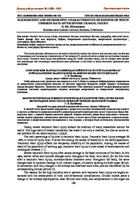
Fig. 2. hemorrhage in the interstitial kidneys.
Coloring G-E.
were prepared using standard histological techniques. The preparations are stained with hematoxy-
lin and eosin. Microscopy of preparations in transmitted light was carried out using a trinocular
microscope with a microscope magnification × 60, × 80. Histological images were obtained using
a microscope camera and the analysis of the images obtained was carried out using specialized
software for medicine.
Conclusions:
It is possible to draw such a conclusion from all this, studies that were carried
out with laboratory rats in severe cases of craniocerebral trauma using the "Road Traffic Accident"
method ends in the death of animals in the experiment of white outbred rats and practically does
not lead to visible macroscopic changes in the kidneys and organs nearby. The study of histologi-
cal preparations of rat kidneys after severe traumatic brain injury revealed pronounced changes in
blood flow and structural changes in the renal parenchyma, in particular, expansion of capillaries
and veins, perivascular hemorrhages in the initial period.
Оригинальная статья
Fig. 1. Hemorrhage of the proximal tubules is
noted space with blood vessels. Coloring G-E.
Fig. 3. Macroscopic picture of kidneys 3 a month
-old rat after TBI (top view).
Fig. 4. The location of the kidneys in the lumbar
region.

Доктор ахборотномаси № 1 (98)—2021
111
References:
1. Babayan E., V. Zelman., Yu.S. Polushin, A.B. Shchegolev // Anesthesiology and Reanimatology. 2005. - No. 4. -
S. 4-14.
2. Badmaeva L.N. Laboratory methods for establishing the prescription of traumatic brain injury in forensic medi-
cine // Sud.-med. expertise. - 2003. No. 1. - S. 37-39.
3. Barinov E.F. The role of eNOS in the pathomorphism of the vascular glomeruli of the kidneys of rats in diabetes
mellitus / E.F. Barinov, Kh.V. Grigoryan, O. N. Sulaeva // Morphology. - 2008. - T. II, No. 1 - S. 29-32.
4. Chelnokov V.S., Ilyina E.V. Pathomorphological changes in traumatic brain injury // Sud.-med. expertise. - 2001. -
No. 1. - S. 7-9
5. Chereshnev V.A. Morphological and hematological criteria for the effectiveness of treatment of experimental pye-
lonephritis with a complex of natural cytokines and antibacterial peptides / V.A. Chereshnev, P. Kosareva, N.
Averyanova, N.A. Zimushkina, E.I. Samodelkin, I. Loginova // Perm Medical Journal. - 2008. - No. 2. - S. 5-13.
6. Fayziev Kh.B., Khuseynova G. Kh. // Macroscopic topography of the spleen of white outbred rats after a severe
traumatic brain injury caused by an accident // Problems of Biology and Medicine 2020, no. 6 (124). S. 185-188.
7. Fursov I.V., V.V. Grave, 2013. " Extracranial complications of severe traumatic brain injury "Tavricheskiy medico
-biological bulletin 2013, volume 16, No. 3, part 3 (63)
8. Khuseynova G.Kh. / "Modeling of traumatic brain injury under the influence of nucleotides." // Khuseynova
G.Kh., New day in medicine. - No. 4 (32) -2020.- P.598-600.
9. Khuseynova G.Kh., Teshaev Sh.Zh. // "Comparative characteristics of the morphometric parameters of the kidneys
in different phases of traumatic brain injury." // New day in medicine. - 2020, 2/1 (30/1) pp. 101-103.
10. Kireeva E.P. The relationship between the initial kidney damage and the environmental toxic load on the div
with lead and cadmium and its prevention (epidemiological and experimental studies): Author's abstract. dis. Hon-
ey candidate. science. - Yekaterinburg, 2007 .- 24 p.
11. Kondakov E.N., Krivetsky V.V. Traumatic brain injury: a guide for doctors in non-specialized hospitals. - SPb .:
SpetsLit, 2002.-271S.
12. Kropachev A.Yu. Model development and morphological characteristics of the kidneys in incomplete (variable)
occlusion of the urinary tract / A.Yu. Kropachev, D.A. Sosnin, G.A. Sklyarenko, V. Novochadov // Bul. Volgo-
grad Scientific Center of the Russian Academy of Medical Sciences. - 2008. - No. 1. - pp. 24–26.
13. Sosnin D.A. Model development and morphological characteristics of the kidneys with incomplete (varying) oc-
clusion of the urinary tract // Bulletin of the Volgograd Scientific Center of the Russian Academy of Medical Sci-
ences. Morphology. Pathology. - 2008. - T. I. - S. 24–26.
14. Veselova M.V. Antioxidant activity of polyphenols in yew of the Far East / M.V. Veselova, S.A. Fedoreev, N.A.
Vasilevskaya, V.A. Denisenko, A. Gerasimenko // Chemical and pharmaceutical journal. - 2007. - T. 41, No. 2. -
S. 29-34.
15. Zakharova S.G. Features of the nephroprotective action of furosemide and some mitochondrial substrates in kid-
ney damage with mercury dichloride: Author's abstract. dis. Honey candidate. science. - Ufa, 2008 .- 22 p.
16. Zhanaspaev A.T., Ishmukhamedov R.Sh. Optimization of treatment of inflammatory complications of traumatic
brain injury // Polytrauma. - 2008. - No. 1. - P.87-91
G. Kh. Khuseynova


