Поиск по библиотеке
Результаты поиска
-
 Случай из практики: Лечения радикулярной кисты нижней челюсти путем создание декомпрессионного окна
in LibraryЭффективная декомпрессия кисты, которая достигается путем создания «окна» в костной полости позволяет не просто санировать полость кисты растворами антисептиков, но и получить необходимый гистологический материал (костного материала и оболочки кисты) для дальнейших исследований. Последующее заполнение дефекта костной полости новообразованной костной тканью происходит вследствие вторичного остеогенеза.
-
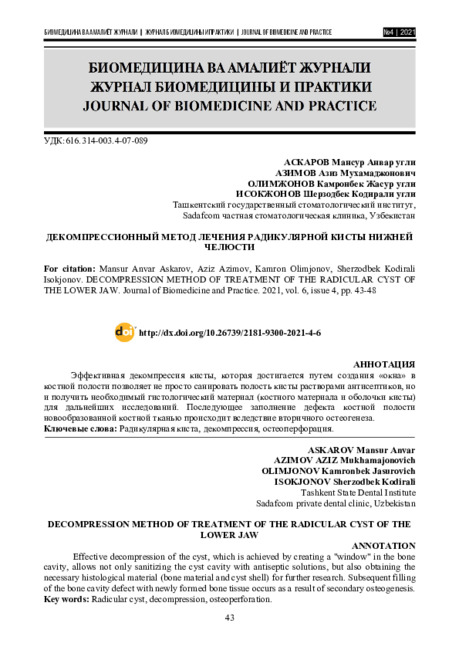
Эффективная декомпрессия кисты, которая достигается путем создания «окна» в костной полости позволяет не просто санировать полость кисты растворами антисептиков, но и получить необходимый гистологический материал (костного материала и оболочки кисты) для дальнейших исследований. Последующее заполнение дефекта костной полости новообразованной костной тканью происходит вследствие вторичного остеогенеза.
-
 Абстракт. Дифференциальная диагностика воспалительных заболеваний, солидных опухолей и кистозных образований в области головы и шеи, к сожалению, до настоящего времени представляет собой сложную задачу для специалистов. В структуре кистозных образований мягких тканей шеи и их осложнений стойко занимают первое место тирсоглоссальные кисты: 24% взрослых и 50% детей, госпитализируемых в стационары, страдают этими заболеваниями Тщательный анализ современной литера гуры свидетельствует о настоятельной необходимости разработки алгоритма дифференциально-диагностических признаков сонографии при кистозных образованиях шеи и определение эффективности метода.
 -
This article presents a clinical case of the treatment of a mandibular radicular cyst from 3.6 teeth by creating a decompression "window", which allows the cyst volume to be reduced up to complete restoration of the bone structure as well as ensuring the integrity of the surrounding anatomical structures. The subsequent filling of the cavity with newly formed bone is due to secondary osteogenesis. This operation can be performed on an outpatient basis in a dental surgery room under local anesthesia
-
Application Of Decompression Treatment For Mandibular Radicular Cysts
The American Journal of Medical Sciences and Pharmaceutical ResearchThis article presents a clinical case of the treatment of a mandibular radicular cyst from 3.6 teeth by creating a decompression "window", which allows the cyst volume to be reduced up to complete restoration of the bone structure as well as ensuring the integrity of the surrounding anatomical structures. The subsequent filling of the cavity with newly formed bone is due to secondary osteogenesis. This operation can be performed on an outpatient basis in a dental surgery room under local anesthesia
-
 В нашем исследовании больных выявлены следующие варианты клинических проявлений лямблиоза: выделяют латентную, субклиническую и клиническую формы лямблиоза. Когда у латентных детей не было жалоб секреция кисты колебалась в среднем на 0,6 кисты в поле зрения. При субклинической и клинической форме наблюдаются: боли в животе, кишечный синдром и желудочная диспепсия. Секреция кисты при субклинической форме кисты была 1,5 в поле зрения. При лямблиозе клиническая форма клинической картины была наиболее выражена, кисты секреции в среднем составляли 2,5 кисты в поле зрения. Разделяя латентную, субклиническую и клиническую формы лямблиоза, назначаются соответствующие диетотерапия и медикаментозная противолямблиозная терапия.
 -
В этой статье предоставляется информация о распространении цисты эхнококоса у овец
-
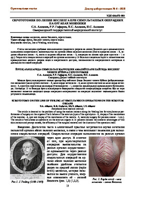 Статья посвящена проблеме использования срединного разреза по линии Веслинга для одномоментного выполнения оперативного вмешательства на органах обеих половин мошонки (Киста придатка яичка - 8, водянка оболочек яичка - 4, киста и водянка оболочек яичка - 3, операции на яичках при раке простате - 1 и др.). Результаты симультанных операций на органах мошонки у 16 больных свидетельствуют о косметических преимуществах данного разреза кожи и оперативного доступа, экономичности операционного материала и длительности самой операций.
 -
 О ЩАДЯЩЕМ ЭНДОНАЗАЛЬНОМ УДАЛЕНИИ КИСТ ВЕРХНЕЧЕЛЮСТНЫХ ПАЗУХКисты верхнечелюстной пазухи чаще всего возникают в результате закупорки выводного отверстия железы, выделяющей слизь, и отёчности слизистой оболочки. Методом выбора при лечении больных с кистой верхнечелюстного синуса остается хирургический. В ринологии все шире используются щадящие методы хирургических вмешательств, атравматический характер которых практически не влияет на дальнейшее формирование анатомических структур носа, околоносовых пазух и их физиологические функции. Мы применили методику щадящего эндоназального удаления кисты верхнечелюстного синуса через нижний носовой ход, при интактных структурах остиомеатального комплекса, с использованием порт- проводника. Предложенное нами щадящее хирургическое вмешательство переносится легче, чем радикальная операция на этом синусе. Срок пребывания больных в стационаре составил от 2 до 5 дней.
Журнал стоматологии и краниофациальных исследований -
 Актуальность. Кисты яичников - это доброкачественные опухоли, среди гинекологических заболеваний кисты яичников у женщин в возрасте 18-48 лет составляют 14-19%. Кисты яичников могут быть различных размеров, гормонально активные или неактивные. Кисты яичников могут осложняться перекрутом ножки кисты, нагноением, разрывом, кровотечением в брюшную полость.
 -
 Декомпрессионный метод при лечении кист челюстей
Актуальные вопросы хирургической стоматологии и дентальной имплантологииВ структуре стоматологических заболеваний пациенты с одонтогенными кистами челюстей занимают важное место. Радикулярные кисты составляют 94-96% среди одонтогенных кист челюстей, выявляющихся у взрослых. Наиболее частая локализация радикулярных кист на верхней челюсти, реже – на нижней [1,3,5]. Несмотря на современные консервативные методики лечения, нуждаемость в хирургическом лечении одонтогенных кист не уменьшается. Основным хирургическим методом лечения одонтогенных кист челюстей является операция цистэктомия, реже – цистотомия
-
Improvement Of Treatment Of Odontogenic Cysts Of The Jawbone In Children
The American Journal of Medical Sciences and Pharmaceutical ResearchThe improvement of methods of treatment of odontogenic cysts of the jaw remains the actual problem of surgical stomatology. This is caused by the widespread of the disease, the possibility of such complications as cyst abscesses, osteomyelitis development, jaw deformities, tooth loss, pathological fracture occurrence and even the so-called central jaw cancer from the epithelium of cyst walls, and also rather frequent relapses after surgical treatment carried out.
-
 В данной статье нами рассмотрен собственный опыт диагностики и лечения кист подколенной ямки. Исследование включало материалы и данные по 96 пациентам с кистой Бейкера. В статье рассмотрены преимущества, недостатки, а также некоторые особенности различных методов клинико-инструментального обследования пациентов с данной патологией.
 -
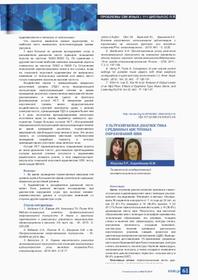 Изучение диагностических признаков тиреог-лоссальных и дермоидных кист шеи с помощью ультразвукового исследования. Материал и методы: обследованы 36 пациентов в возрасте от 1 -го года до 32 лет. из низ 22 (61,1%) женщины и 14 (38,9%) мужчин. У 22 (61,1%) были тиреоглоссальные кисты шеи, у 14 (38,9%) дермоидные кисты шеи. Результаты: при кистозных образованиях шеи с помощью сонографии оценивались локализация образования, его размеры, толщина стенок и наличие септ (перегородок), контуры стенок, внутренняя эхогенность, солидный компонент, эхотекстура, наличие артефакта дистального акустического усиления, свищей, кровотока при цветовом допплеровском картировании (ЦДК). Выводы: основными достоверными УЗ-признаками срединных кистозных образований шеи были локализация, контуры стенок, эхогенность, эхотекстура. Наличие перегородок, неправильных контуров, а также солидного компонента позволило диагностировать тиреоглос-сальную кисту в 86,4% (оценка SIST).
 -
 Кисты верхнечелюстной пазухи чаще всего возникают в результате закупорки выводного отверстия железы, выделяющей слизь, и отёчности слизистой оболочки. Методом выбора при лечении больных с кистой верхнечелюстного синуса остается хирургический. В ринологии все шире используются щадящие методы хирургических вмешательств, атравматический характер которых практически не влияет на дальнейшее формирование анатомических структур носа, околоносовых пазух и их физиологические функции. Мы применили методику щадящего эндоназального удаления кисты верхнечелюстного синуса через нижний носовой ход. при интактных структурах остиомеаталыюго комплекса, с использованием порт-проводника. Предложенное нами щадящее хирургическое вмешательство переносится легче, чем радикальная операция на этом синусе. Срок пребывания больных в стационаре составил от 2 до 5 дней.
 -
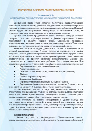 Киста зубов. Важность своевременного лечения
Актуальные вопросы профилактики стоматологических заболеваний и детской стоматологииДентальная киста зубов является достаточно распространенной патологией в особенности у детей так, как дети меньше соблюдают правила гигиены и больше подвержены инфицированию ротовой полости. В данной работе будет рассматриваться информация о дентальной кисте, её профилактике и последствиях при несвоевременном лечении.
-
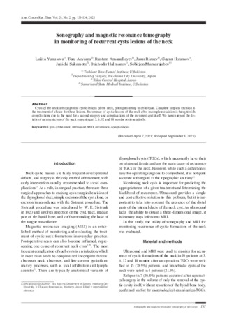 Sonography and magnetic resonance tomography in monitoring of recurrent cysts lesions of the neck
in LibraryСysts of the neck are congenital cystic lesions of the neck, often presenting in childhood. Complete surgical excision is the treatment of choice for these lesions. Recurrence of cystic lesions of the neck after incomplete excision is fraught with complications due to the need for a second surgery and complications of the recurrent cyst itself. We herein report the de-tails of recurrent cysts of the neck presenting at 3, 6, 12 and 18 months postoperatively.
-
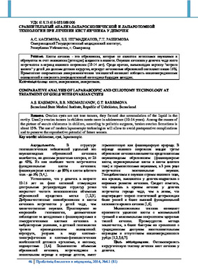 Сравнительный анализ лапароскопической и лапаротомной технологии при лечении кист яичника у девочекКисты яичника - это образования, которые не являются истинными опухолями и образуются за счет накопления (ретенции) жидкости в полости. Опухоли яичников у девочек чаще всего встречаются в период полового созревания (10-14 лет). Среди причин, вызывающих картину "острого живота" у детей ,по данным детских хирургов, перекрут яичниковых образований составляет около 15%. Применение современных лапароскопических технологий позволит избежать послеоперационных осложнений и сохранить репродуктивный потенциал будущих женщин
Журнал проблемы биологии и медицины -
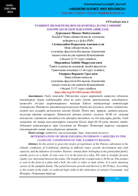 ОПРЕДЕЛЕНИЕ ВЫСОКОУРОЖАЙНЫХ СОРТОВ ПОТИССОНА В КЛИМАТИЧЕСКИХ УСЛОВИЯХ ТАШКЕНТСКОЙ ОБЛАСТИ.В этой статье переведены результаты исследования выращивания Патисона в климатических условиях Узбекистана, посадка в разных временах, рост, развитие и урожайность, а также влияние погоды. Патисон - однолетние ростение относящиеся роду тыквенных ростений, обычно растёт как кустарник, в редких случаях бывает в форме хлыста. Стебель прямой, гранёный, жёстко кистовой, сокращённый между узлами. Длина кривого стебля состовляет 30-60 см. Продукт овальный в форме пластинки и колокольчика, окраска белая или тёмно-жёлтая. Период современни пророста до сбора урожая составляет 40-50 дней. По результатам исследования добились высоко урожайности в выращивании сортов Солнышко и Фонарик, Летающая тарелка.
Современная наука и исследования -
 Экспериментальное обоснование эффективной терапевтической дозы альбендазола для профилактики рецидива эхинококкозаДля изучения влияния альбендазола в различных дозировках на образование эхинококковой кисты (мочевого пузыря) были проведены опыты на овцах (8 овец), искусственно зараженных эхинококкозом. Для определения действия альбендазола исследовали ткани печени овец, подвергавшихся и не подвергавшихся химиотерапии. Сравнительное изучение формирующихся эмбриональных пузырей у овец, получавших и не получавших альбендазол, показало достоверную разницу в динамике формирования эмбриональных пузырей. Альбендазол в дозе 5 мг/кг массы оказывает фатальное действие на эмбриональную форму эхинококка и является эффективной лечебной дозой без токсического действия в профилактике рецидивов эхинококкоза.
Журнал проблемы биологии и медицины -
Background: The present study attempted to clarify the typical anatomical variants of Thyroglossal cysts (TGC). Patients and methods: Clinically and epidemiologically 67 previously non-experienced patients with TGC 1.5 to 73.0 years old were examined. Results: Based on clinical and ultrasound examinations of 121 patients with 67 thyroglossal cysts, the most typical cyst of anatomical variations was specified. It was noted that, concerning the hyoid bone, thyroglossal cysts may be suprahyoid (located at the root of the tongue), parahyoid (broadly adjoining the hyoid), prelingual (located in the front of the hyoid in the hypo lingual region), postlingual (located behind the hyoid bone in the prenatal and peri-laryngeal spaces), or sublingual (located the book from the hyoid bone). An ultrasound examination facilitated the identification of thyroglossal cysts with-out clinical manifestations (23 sublingual cysts among 37 [62.2%] were incidentally revealed by the ultrasound examination), which is important when selecting the most appropriate surgical treatment. Conclusion: Ultrasound studies facilitate the identification of TGCs located at the root of the tongue without any clinical manifestations, which is important when determining the degree of surgical treatment to perform.
-
 Морфологические и рентгенологические исследования эхинококкоза и пециломикоза легкихНаблюдения и исследования проведены на 189-ти больных эхинококкозом легких (БЭЛ), возраст которых от 16 до 63 лет и 213 больных пециломикозом. Предварительные рентгенологические исследования позволили выполнить видеоторакоскопическую эхинококэктомию с применением минидоступа. Рентгенологически у больных легких в возрасте от 18 до 25 лет с выставленным диагнозом эхинококкоз выявлялись в легких кисты до 5 см в диаметре и они диагностировались как начинающийся эхинококкоз. Трижды проведенный курс лечения таких больных альбендазолом не дал положительных результатов. Новым иммунологическим методом реакцией антигенсвязывания лимфоцитов (АСЛ) на пециломикоз у этих больных была установлена тяжелая форма пециломикоза легких. Инфицирование грибами рода Paecilomyces, относящихся к группе условно-патогенных микроорганизмов, перестало быть редким явлением и стало реальной опасностью для жизни человека. Появилась необходимость изменить подход к заболеваниям, вызываемым грибами рода Paecilomyces, не как к узкой и редкой патологии, а рассматривать её как генерализованную инфекцию с возможным поражением различных органов и систем, но чаще всего бронхо-легочной системы при паразитарных заболеваниях
Журнал проблемы биологии и медицины