


Scientific
Journal «ScienceRise
:
Medical Science» №6(45)2021
34
UDC 616
DOI: 10.15587/2519-4798.2021.250239
PEDIATRIC SURGICAL SEPSIS: DIAGNOSTICS AND INTENSIVE THERAPY
Elmira Satvaldieva, Gulchehra Ashurova, Otabek Fayziev, Abdumalik Djalilov
The aim:
Optimization of diagnostics and schemes of pathogenetic intensive therapy of surgical sepsis in chil-
dren based on clinical and laboratory criteria and bacteriological monitoring.
Materials and methods:
The research period is 2018-2020. The object of the study (n=73) – children with surgi-
cal pathology (widespread peritonitis, bacterial destruction of the lungs, post-traumatic brain hematomas, ab-
dominal trauma, etc.). Research methods: microbiological monitoring to determine the sensitivity of the micro-
organism to antibiotics was carried out before and at the stages of treatment (sputum, urine, wound, bron-
choalveolar lavage, tracheal aspirate, blood, contents from drainages, wound surface). Determination of the
sensitivity of the isolated strains to antibiotics was carried out by the disk-diffusion method. To determine pre-
dictors of sepsis in surgical patients, clinical (mean arterial pressure (mAP), heart rate (HR), respiratory rate
(RR), SpO
2
, etc. and laboratory parameters on days 1–2 (up to 48 hours) of sepsis identification, days 4 and 8 of
intensive therapy. Procalcitonin was determined by immunofluorescence on a Triage® MeterPro analyzer (Bi-
osite Diagnostics, USA). Blood gases and electrolytes were analyzed using a Stat Profile CCX analyzer (Nova
Biomedical, USA).
Results:
studies have shown the effectiveness of complex intensive care in 86.3 % of cases. Mortality was found
in 13.7 % of cases. Patients with severe surgical pathology died: widespread peritonitis, severe TBI + coma with
irreversible neurological disorders, urosepsis against the background of chronic renal failure, after repeated
surgical interventions, due to the development of refractory septic shock (SS).
Conclusions.
Early diagnosis of sepsis, rational early ABT under the control of microbiological monitoring,
non-aggressive infusion therapy with early prescription of vasopressors (SS) with constant monitoring of the
child's main life support organs contribute to an improvement in sepsis outcomes and a decrease in mortality
Keywords:
pediatric sepsis, balanced crystalloids, respiratory support, septic shock
How to cite:
Satvaldieva, E., Ashurova, G., Fayziev, O., Djalilov, A. (2021). Pediatric surgical sepsis: diagnostics and intensive therapy. ScienceRise: Medical
Science, 6 (45), 34–42. doi: http://doi.org/10.15587/2519-4798.2021.250144
© The Author(s) 2021
This is an open access article under the Creative Commons CC BY license hydrate
1. Introduction
Sepsis, as a life-threatening problem in modern
medicine, has been repeatedly revised by the internation-
al medical community over the past 3 decades (Sepsis-1-
Sepsis-2-Sepsis-3), definitions, approaches to early diag-
nosis and intensive care have changed, also changed rat-
ing scales of severity and prognosis of sepsis. The results
of recent studies show that the information value of the
criteria for systemic inflammatory response syndrome
(IRS) is very low [1, 2]. It is proved that the very process
of interaction of micro- and macroorganisms is more
complex and is characterized by the versatility of the
reaction of the latter to microbial invasion, the manifesta-
tions of which determine gender, age, race, genetic fac-
tors and concomitant pathology [1, 2]. As a result, sepsis
has been defined as life-threatening organ dysfunction
(OD) resulting from dysregulation of the host's response
to infection.
All changes in the diagnosis and treatment of sep-
sis affected mainly adult patients and, to a lesser extent,
children. It is important that among the highlighted pedi-
atric aspects of sepsis treatment there are no recommen-
dations that are not classified according to the degree of
evidence [3].
A multicenter study of sepsis in children
(n=6925, SPROUT, 2014), conducted in 26 countries (in
128 pediatric intensive care units), revealed a significant
variability in the incidence of sepsis from 6.2 % in Eu-
rope to 23.1 % in Africa, in the United States on ave-
rage – 8.2 % [4]. On average, mortality from sepsis was
24 %. The most frequent foci of infection were the res-
piratory system (40 %) and blood flow (19 %) [5–7]. A
detailed review of the epidemiology and geography of
sepsis (2019) showed that in countries with a high level
of economy, the incidence of sepsis varied widely from
1.4 % (Japan) to 7.7 % (USA), mortality from sepsis was
7–17 %. %, from the septic shock (SS) - 51 %. In small
economies, the incidence of severe sepsis in children was
1–26 %, and the mortality rate was 12–35 %. The authors
associate these significant fluctuations with various diag-
nostic criteria for sepsis and economic factors [8, 9]. Al-
so, after discharge from the hospital, a fifth of the surviv-

Scientific
Journal «ScienceRise
:
Medical Science» №6(45)2021
35
ing children with sepsis were found to have moderate
functional disability [10].
Thus, the need for early diagnosis and treatment of
sepsis in children is confirmed by the continuing high rates
of morbidity and mortality. To facilitate the diagnosis of
sepsis in children, the pSOFA and PELOD-2 children's
scales have been developed in recent years. They do not
have 100 % specificity, but their use will help in the early
diagnosis of sepsis [10, 11]. The authors of [12, 13] ob-
served a very high predictive accuracy of these scales.
As a result of the selection of literary sources
collected in the Pub Med, Science Direct, Cochrane
Library databases, with a search depth of 10 years
(2009–2019), 36 articles were selected on the problem
under study. The number of controlled clinical trials of
childhood sepsis is very small (with the exception of
neonatal sepsis), and they all reflect an unsolved prob-
lem, a lack of a single concept and protocols for diag-
nosis and treatment.
The aim of the research
. Optimization of diag-
nostics and schemes of pathogenetic intensive therapy of
surgical sepsis in children based on clinical and laborato-
ry criteria and bacteriological monitoring.
2. Materials and methods
A prospective, non-randomized case-control
study. Research period 2018-2020 The inclusion criteria
for patients in the study were signs of organ dysfunction
(2+), procalcitonin >0.5 ng/ml, pSOFA>3 points, age –
children under 18 years of age and the presence of the
required examination volume. The exclusion criterion is
the disagreement of the patient or his relatives to partici-
pate in the study. The study included 73 patients who
underwent interventions for intestinal obstruction – 14,
generalized peritonitis – 15, traumatic rupture of the in-
testine – 8 and esophagus – 1, bacterial destruction of the
lungs – 6, wound infection – 3; craniocerebral trauma
(subdural, intracerebral hematomas) – 11; congenital
anomalies of the urinary tract, ureterohydronephrosis (2–
4 degrees, chronic renal failure, urosepsis) – 15. The age
composition of patients: preschoolers 2–5 years – 11,
schoolchildren 6–12 years old – 44, adolescents 13–18
years old – 18 Schoolchildren prevailed and accounted
for 60.3 % in the general structure of patients.
During the study, permission was obtained from
all parents of patients in accordance with the code of
ethics (2013). Approved by the conclusion of the expert
commission of the ethical committee of the clinic of the
Tashkent Pediatric Medical Institute of the Ministry of
Health of the Republic of Uzbekistan (No. 475 of
21.10.2021). Artificial lung ventilation (ventilators SAV-
INA, SULLA) lasting more than 48 hours was performed
in 27 patients (36.9 %), of which nosocomial pneumonia
was detected in 19 children (70.3 %). The length of stay
in the intensive care unit averaged 19.3±5.6 days.
Microbiological monitoring to determine the sensi-
tivity of the microorganism to antibiotics was carried out
before and at the stages of treatment (sputum, urine, wound,
bronchoalveolar lavage, tracheal aspirate, blood, contents
from drains, wound surface). Determination of the sensitivi-
ty of the isolated strains to antibiotics was carried out by the
disk-diffusion method. The results of microbiological moni-
toring are presented in diagrams 1,2,3,4.
To determine predictors of sepsis in surgical
patients, we analyzed the clinical (mean arterial pres-
sure (mAP), heart rate (HR), respiratory rate (RR),
blood oxygen saturation (SpO
2
), and laboratory pa-
rameters on day 1–2 (up to 48 hours) detection of sep-
sis, 4 and 8 days of intensive care. Thrombocytopenia
was diagnosed with a platelet count <120,000/μL of
blood, immunoglobulinemia G – with a serum level <7
g/L. Assessment of the state of the immune system
was carried out based on a quantitative determination
of the concentration of serum immunoglobulins IgG
by flow cytometry. A Mindray BA-88A automatic
biochemical analyzer was used to study AST, total
protein, albumin, creatinine, and blood sugar. Procal-
citonin was determined by the immunofluorescence
method using a Triage® MeterPro analyzer (Biosite
Diagnostics, USA). Blood gases and electrolytes were
analyzed using a Stat Profile CCX analyzer (Nova
Biomedical, USA). The results of clinical and labora-
tory studies are presented in Table 1. At all stages of
intensive care, monitoring of the parameters RR, HR,
BP, SpO
2
, T div (Nihon Kohden) was carried out.
Statistical data processing was performed using the
Statistica 6.1 statistical software package (StatSoft,
USA, 2003). Comparison of independent groups for
quantitative characteristics was carried out using the
Mann-Whitney U-test, qualitative comparison of inde-
pendent groups - by analyzing contingency tables us-
ing the two-sided Fisher's exact test for unrelated
groups or the χ
2
method with Yates' correction de-
pending on the expected frequencies of the function.
3. Results
The diagnosis of sepsis was based on clinical and
laboratory data and confirmed by identification of the
pathogen culture in blood and / or other biosubstrates.
Sowing of the same culture of the pathogen in 2 or
more loci was bacteriologically confirmed by sepsis and
was etiologically proven. Objective indicators of organ
dysfunction were taken into account (100 % of cases).
As noted above, sepsis is a heterogeneous process with
pronounced individual variability, which complicates
its diagnosis and treatment [14–17]. When making a
diagnosis, the most important thing is the clinical pic-
ture of the disease. However, it is no less important for
practicing physicians to monitor indicators of metabo-
lism, hemodynamics, blood circulation and biomarkers
of sepsis [18].
Patients who developed sepsis had severe hy-
permetabolic syndrome, which was manifested by
tachycardia and tachypnea, hyperthermia, low levels
of albumin and total protein in the blood. Among
them, on the 2nd day (stage 1), hypoglobulinemia G
and thrombocytopenia were more common (Table 1).
Protein catabolism in patients was accompanied by a
decrease in the synthesis of globulins (IgG) and the
development of a secondary immunodeficiency state.
There was a moderate increase in the level of fibrino-
gen, which characterizes the severity of the syndrome
of disseminated intravascular coagulation, against the
background of an inflammatory reaction with damage
to microvessels, hemoconcentration, endothelial dis-
orders, etc.

Scientific
Journal «ScienceRise
:
Medical Science» №6(45)2021
36
Table 1
Clinical, biochemical and special markers of sepsis in children (n=73, M
±m
)
Index
1–2
nd
day (48 h)
4
th
day
8
th
day
MAP, mm Hg
84.5±4.3
80±4.8
72±3.5*^
Heart rate, min
129.4±7.2
118.6±5.7
107±5.1*^
Breathing rate, min
-1
34.2±3.4
29.1±3.2
25.3±2.7*
Body temperature,
о
С
37.9±2.0
37.5±1.8
37.0±1.4
SpO
2
, %
96±4.9
97±4.7
98±3.9
Leukocytes, 10
9
/ l
15.8±5.3
12.8±2.4
9.05±1.7**
Neutrophils, %
81.6±2.9
78.9±2.8
70.6±2.4**
Hemoglobin, g / l
105±5.6
114±4.3
117±3.8*
Platelets, 10
9
/ l
120.5±6.1
124.3±7.5
140.2±5.5*
Fibrinogen, g / l
5.1±1.2
4.8±1.3
4.0±1.4
Bicarbonate, mmol / l
23.2±2.5
22.8±2.1
22.1±2.4
AST, u / l
1.9±0.40
1.0±0.36
0.8±0.32*
Total protein, g / l
48.4±7.5
49.9±6.7
58.0±6.8*
Albumin, g / l
27.2±3.9
28.9±4.5
31.0±4.2
Creatinine, μmol / l
97.5±5.5
89.9±4.9
87.5±3.8*
Glucose, mmol / l
7.1±0.2
7.0±0.1
6.5±0.2
Ig G, g / l
6.01±1.7
6.58±1.9
7.0±1.8
C-reactive protein, mg / l
34.0±3.9
27.0±3.7
15.0±1.9**^
Procalcitonin, ng / ml
2.60±0.3
2.10±0.7
1.8±0.2*
pSOFA, points
9 ±2.5
7±1.6
4±0.9
Note: reliability of data on indicators for 1–2 days; * – p<0.05; ** – p<0.01; ^ – reliability of data to indicators on the 4th day,
^ – p<0.05
Blood culture – a specific and affordable method –
has always been considered the “gold standard” for diag-
nosing infection, but its sensitivity does not exceed 25–
42 %. In addition, due to the use of antibiotics before
blood sampling, blood cultures are often false negative.
The causative agent remains unknown in 30–75 % of
children with sepsis [20].
Blood sampling for bacteriological examination
was carried out before the start of antimicrobial treat-
ment. In most patients, blood samples and biomaterials
from other loci were taken for bacteriological examina-
tion 2–3 times during their stay in the intensive care unit.
The largest number of isolates was isolated from tracheal
aspirate (sputum) – 39.7 %, surgical drains – 32.8 %,
urine – 27.3 %, and blood – 26 %.
We followed the standard of testing blood for ste-
rility from two peripheral veins at intervals of up to
30 minutes in two vials. Blood sampling from a central
venous catheter was performed on the condition that it
had just been inserted. To diagnose or exclude catheter-
associated sepsis, blood sampling from a previously in-
serted catheter was allowed.
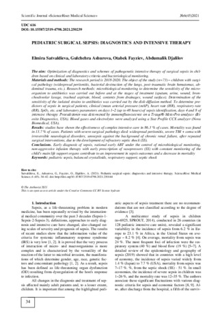
Bacteriological examination from different loci
revealed the following data: from blood (Fig. 1) – staphy-
lococcus, coagulase-negative – 42.5 % (8), St Aureus –
26.3 % (5), Streptococcus viridans et pneumoniae –
10.5 % (2), Enterococcus faecium – 5.4 %, Kl pneu-
moniae – 10.5 %, Pseudomonas spp. – 5.4 %. Gram-
positive bacteria predominated: Staphylococcus, coagu-
lase negative, and St aureus.
Fig.
1. Microbiological monitoring of blood culture
42.5
26.3
10.5
10.5
5.4
5.4
Hemoculture, n=19
Staphylococcus,
coagulase negative
St Аureus
Streptococcus viridans
et pneumoniae-
Kl pneumoniae
Enterococcus faecium
Pseudomonas spp.-

Scientific
Journal «ScienceRise
:
Medical Science» №6(45)2021
37
In turn, from surgical drains, peritoneal fluid, cere-
brospinal fluid (Fig. 2) was sown in 29.1 % of cases (7) –
Kl pneumoniae
, in 25 % (6) –
Pseudomonas
Aeruginosa
,
in 16.6 % (4) –
St. Aureus
, in 20.2 % (5) –
Acinetobacter
,
in 8.3 % (2) –
Enterobacteriaceae
.
From bronchoalveolar aspirate (Fig. 3) –
Kl.
pneumoniae
– 27.5 % (8),
Ps. Aeruginosa
– 24.1 % (7),
St. Aureus
– 20.6 % (6),
Pneumococcus
– 17.2 % (5),
Acinetobacter
– 10.3 % (4) was obtained.
All over the world, multi-resistant superbugs, rep-
resentatives of the ESCAPE group (
Enterococcus Faeci-
um,
St.
aureus. Kl.
Pneumonia, Acinetobacter,
Ps.aeruginosa, Enterobacter spp
.), Pose a particular
problem. Local monitoring confirmed the dominant posi-
tion in the structure of the studied isolates of bacteria
such as
St. aureus et epidermidis, Ps. aeruginosa, Kl.
pneumoniae
and
Acinetobacter
. In our study,
Kl. pneu-
moniae exceeded Ps.aeruginosa
.
Thus, the analysis of changes in the bacteriologi-
cal landscape showed that the proportion of gram-
negative microflora among the studied isolates remains
consistently high. Fungi of the genus Candida were sown
in 12.5 % of cases and were part of the polymicrobial
flora. In general, when summarizing the results of other
biological media of the patient, representatives of gram-
negative
flora
(
Enterobacteriaceae,
Pseudomonas,
Kl.pneumoniae
) were the main causative agents of sepsis
in 47.6 % of cases, gram-positive (
St. Aureus et epider-
midis, Enterococcus, Pneumococcus
) – in 30 %, polymi-
crobial – in 21.8 % (Fig. 4).
Fig. 2.
Microbiological monitoring of peritoneal fluid and drains
Fig. 3. Microbiological monitoring of broncho-alveolar aspirate
29 %
25 %
17 %
20 %
9 %
Discharge of drainage, peritoneal fluid, etc., n=24
Kl.Pneumonia
P. Аeruginosa
St.Aureus
Acinetobacter spp.
Enterobacteriacea
28 %
24 %
21 %
17 %
10 %
Broncho-alveolar aspirate,n=29
Kl.Pneumonia
P. Aeruginosa
St aureus
Pneumococcus
Acinetobacter

Scientific
Journal «ScienceRise
:
Medical Science» №6(45)2021
38
Fig. 4. The main causative agents of sepsis
Multicomponent intensive therapy for sepsis in-
cluded detoxification therapy, respiratory support (if nec-
essary, mechanical ventilation), correction of water-
electrolyte, hemodynamic disorders, inotropic support,
nutritional and immunotherapy.
Intensive therapy for sepsis / SS (the main provi-
sions of the local protocol. Syndromic therapy of organ
dysfunction was performed in all patients with sepsis / SS:
1. Respiratory support, mechanical ventilation (in
36.9 % of cases). Ventilation was carried out in con-
trolled pressure (CP) mode with a quick transition to
auxiliary ventilation modes. Gas exchange was moni-
tored by KOS and blood gases, SpO
2
– 90–95 %.
CP, PRESSURE CONTROL SYSTEM. The lead-
ing controlled variable is inspiratory pressure (Pinsp).
Optional-f, PEEP. Initial installation parameters: Pinsp
<28 cm of water gauge; PEEP – 5–8 cm of water gauge
(prevention of electrical injury); inspiration time 0.8 s
(physiological); BH (f) – 20 (children >5 years old);
FiO
2
– 0.8 (ideally 0.5–0.6)
2. Infusion-transfusion therapy. The calculation of
infusion
therapy
for
sepsis
averaged
4–6
(4+2) ml/kg/hour with compensation for current losses.
The qualitative composition of IT was represented by
balanced crystalloids (Ringer's lactate solution), less of-
ten 0.9 % sodium chloride solution, as well as colloids
(albumin)
until
mBP
reached
≥60
mmHg,
CVP – 8 mm Hg. In liquid refractory shock, when after
intravenous administration of 2 boluses of 20 ml/kg of
fluid (40 ml/kg) for 1 hour, A/D remains below the age
norm, vasopressor support (dopamine, dobutamine,
adrenaline, norepinephrine) was started, which depended
on the type of septic shock. In hyperdynamic shock,
norepinephrine was administered at a dose of 0.05–
0.1 μg/kg/min. Epinephrine (0.05–0.2 μg/kg/min) re-
placed dopamine in children with hypodynamic shock.
Dobutamine was prescribed to patients with low cardiac
output and high vascular resistance (cold extremities,
delayed capillary filling, decreased urine output after IT
at normal blood pressure). Later, when the condition sta-
bilized, the child received a physiological daily need for
fluid, if necessary, against the background of diuretic
therapy. Transfused with hemoglobin 70–90 g/l, erythro-
cyte mass. For fibrinolytic bleeding, fresh frozen plasma
was transfused at a dose of 15 ml/kg.
3. Hormone therapy SS. Steroids for the treatment
of refractory shock to infusion and vasopressor therapy.
Children with resistance to catecholamines, with suspect-
ed adrenal insufficiency, were treated with hydrocorti-
sone 1–2 mg/kg/day intravenously, then 150–250 mg for
3–4 injections.
4. Antibiotic therapy. Broad-spectrum antibiotics
were prescribed within 2–3 hours of the diagnosis of
sepsis. Considering the severity of the patient's condition
caused by the septic process, the initial antibiotic therapy
included 2 broad-spectrum antibiotics (3rd and 4th gen-
eration cephalosporins, 3rd generation aminoglycosides),
often in conjunction with metronidazole. Protected beta-
lactam antibiotics took precedence. The revision of the
ABT scheme was carried out after receiving the results of
a microbiological study (after 48–72 hours) and evaluat-
ing the clinical data in order to narrow the antibacterial
spectrum to an adequate one (the principle of de-
escalation).
So, in gram-negative sepsis, a deescalation mode
of etiotropic ABT with protected CPs of 3-4 generations
in combination with AH of the 3rd generation was used,
then, if necessary, and according to microbiological mon-
itoring data, the course of ABT was changed in extreme-
ly severe cases – carbapenems (KB, imipenem, mero-
penem), fosfomycin, fluoroquinolones 3–4 generations
(reserve) in combination with other antibacterial drugs.
The reasons for transferring patients to fluoroquinolones
were: lack of effect from the previous ABT; high sensi-
tivity of pathogens to them [20–22]. As a result of the
study, a high resistance of Kl was revealed. pneumoniae
to cephalosporins 3–4, KB and even fluoroquinolones.
For multi-resistant gram-negative flora, Polymyxin E (so-
dium colistimethate) was prescribed. It is used at a dose of
3–5 mg/kg/s every 8 hours intravenously (1 mg –
12500 IU) to patients without renal pathology (2/3 of the
unchanged state is excreted by the kidneys during the day).
In the case of gram-positive sepsis, the emphasis
was placed on the use of antibiotics from the groups of
oxazolidinones and glycopeptides. In the presence of
methicillin-resistant S. aureus (MRSA), coagulase-
30 %
48 %
22 %
Gram+
Gram-
Polimicroby

Scientific
Journal «ScienceRise
:
Medical Science» №6(45)2021
39
negative staphylococcus, glycopeptides (vancomycin,
teicoplanin) were used, and in the case of vancomycin-
resistant strains – linolid. According to indications, anti-
fungal drugs (fluconazole) were included in the ABT
regimen for no more than 5 days.
The duration of antimicrobial therapy for sepsis
averaged 16±4.5 days. Patients with sepsis received from
1 to 5 courses of ABT, one course for 8–10 days.
5. Nutritional support (NS). The choice of NS
method depended on the severity of nutritional status and
gastrointestinal dysfunction. Patients with sepsis were
given parenteral nutrition (PN) when full enteral nutrition
was not available. The regime of round-the-clock admin-
istration of nutrients was observed, which is associated
with better tolerance and metabolism. An early meal was
prescribed – within 48 hours. Nutritional support: energy
value of food -25-30 kcal / kg of div weight per day;
protein – 1.5–2.0 g/kg/day; glucose – 30–70 % of non-
protein calories while maintaining glycemic levels below
6.1 mmol/l; lipids – 15–20 % of non-protein calories.
Glutamine 0.5 ml/min for 2 hours, 1.5–2 ml/kg/day for
5 days, infusion rate: 0.5 ml/min. Priority of enteral nu-
trition (glucose+IV). Contraindications to any nutritional
support were refractory shock (arterial hypotension on
the background of infusion of epinephrine or norepineph-
rine at a dose of more than 0.1 μg/kg/min); decompen-
sated metabolic acidosis.
6. Immune replacement therapy. The results of the
use of intravenous immunoglobulin G (IVIG, Biovena) at
a dose of 0.4 g/kg/day from the 4th day of illness showed a
relative stabilization of the clinical and laboratory manifes-
tations of sepsis and the cessation of the decrease in globu-
lins (IgG) by the beginning of the 2nd week of illness.
IVIG was administered for 5 days against the background
of complex pathogenetic intensive therapy for sepsis.
As can be seen from Table 1, against the back-
ground of the application of this protocol, there was a
relative stabilization of clinical and biochemical parame-
ters by 4 days of intensive care, HR and RR decreased by
8.4 % and 15 %, blood leukocytes by 19 %, procalcitonin
and CR-protein by 19.3 % and 21 % respectively. After 2
weeks of complex intensive therapy, a significant stabili-
zation of many of the studied parameters of homeostasis
was noted. Procalcitonin and CR-protein at the 3rd stage
of the study decreased by 30.8 and 55.9 % in relation to
the initial data of the 1st stage. According to the results
of pSOFA assessment in patients with sepsis, a tendency
towards a decrease in signs of organo-systemic damage
from stages 1 to 3 was revealed: 9 points – 7 points –
4 points, respectively. Procalcitonin correlated with the
severity of the patient's condition on the pSOFA scale. In
10 patients, despite the ongoing complex treatment, the
PCT index remained stable.
Clinical case
Patient – girl A., 1 year 2 months. (Fig. 5). Date
of entry 12.08.19, Complaints (according to the mother):
hyperthermia, lack of appetite, anxiety, shortness of
breath, groaning breathing. Anamnesis: Illness for
10 days. In September 2019, she underwent inpatient
treatment for pneumonia. In November she received a
prophylactic vaccination against pneumococcal infection +
against measles, rubella and mumps. From 01.12.19 the
condition worsened. Anxiety, fever, shortness of breath,
refusal to eat, weakness, and abdominal pain appeared.
Objectively: the general condition is severe, multiple
organ dysfunction: acute respiratory failure of the 2nd
degree, acute cardiovascular failure of stage 2B, acute
cerebral failure, toxic encephalopathy. The child is le-
thargic. The skin and visible mucous membranes are
sharply pale, bluish, dry. Breathing moans rapidly with
the participation of accessory muscles. In the lungs on
the right, hard wire breathing with dry wheezing. On the
left, breathing is weakened. Deaf heart sounds, tachycar-
dia. The abdomen is enlarged, swollen. Liver + 3.5 cm.
There was no defecation for 2 days. Oliguria.
Ultrasound of the heart from 12/08/2019 – Peri-
cardial effusion: an increase in the amount of fluid in the
pericardium over the entire surface by 21-23 mm. Fibrin
deposits. Ultrasound of the pleural cavity from
08.12.2019: free fluid is determined in the pleural cavi-
ties: On the right, 20.0 ml. There is still 80.0 ml. Clinical
and biochemical analyzes (selectively): Нв 77 g/l; Leu-
kocytosis – 11,8 ×10
9
/l, Neutrophils 86 %, ESR –
18 mm/h. Medium molecules – 0.758 units. Total protein –
47.8 g/l. Urea – 18.2 mmol/l; AST – 4.8. Procalcitonin –
17 ng/ml, CRP – 42 mg/l. In urine, blood and throat:
St.
aureus, Ps. Aeruginosa.
Fig. 5. X-ray picture lung -12.08.2019. Hydrothorax
from the left. Pericarditis
The clinical diagnosis was made:
Main: Bacterial destruction of the lungs, pulmo-
nary-pleural-mediastinal form.
Complications: Hospital sepsis (Gr + and Gr-).
Multiple organ dysfunction. Pyothorax on the left, puru-
lent pericarditis. Toxic hepatitis-nephritis (hepato-renal
syndrome). DIC syndrome. Encephalopathy.
On 10.12.19 the operation was performed for
health reasons: sternotomy, pericardiotomy. Anterior
pericardiectomy. Sanitation and drainage of the cavity.
Purulent effusion in a volume of 100.0 ml.
Thoracocentesis 12/10/19: from the left pleural
cavity through the drainage allocated 110.0 ml of puru-
lent effusion. The drain is connected to active aspiration.
Intensive care: 1 course of ABT Cefoperazone +
sulbactam + Vancomycin, 2 course – Meropenem +
Anzolid (Oxazolidinone group). Infusion-transfusion
therapy (Ringer, saline, albumin, washed erythrocytes).

Scientific
Journal «ScienceRise
:
Medical Science» №6(45)2021
40
Immunosubstitution therapy (Intravenous native immu-
noglobulins G (IVIG, Bioven) 0.4 g/kg/s for 7 days).
Correction of the main organs of life support with respir-
atory support. Mixed parenteral-enteral nutrition (with
the pharmacological nutrient glutamine).
Dynamics of the state: The child was on mechani-
cal ventilation for 17 days. For 2 weeks, purulent effu-
sion from the left pleural cavity and pericardium is daily
45–50.0 ml and 10 ml, respectively. Flushing of the peri-
cardium and left pleural cavity with antibiotics was car-
ried out for 2 weeks. Functioning of the cutaneous-
mediastinal fistula. Constant subfebrile condition. Peri-
cardial and pleural effusion – St. aureus
By the beginning of 3 weeks, the child's condition
began to stabilize, according to the general condition and
according to the results of the dynamic examination,
there was a clear positive trend. Decrease in signs of or-
gano-systemic damage. Clinical and biochemical analyz-
es: Нв 117 g/l; Blood leukocytes – 9.8 ×10
9
/l, ESR –
13 mm/h. Total protein – 58.8 g/l; Urea – 7.2 mmol/l;
AST – 1.0; Procalcitonin – 1.7 ng/ml. After 4 weeks, the
child was transferred to a specialized surgical depart-
ment, where an operation was performed on adhesions
(decortication of the lungs).
4. Discussion of research results
According to international protocols [23–31], con-
firmed sepsis / SS requires rapid provision of venous
access and initiation of infusion (vasopressors, if neces-
sary), administration of antibiotics 1-3 hours before sam-
pling for microbiological examination. Studies have
shown a correlation between increased mortality and
delayed ABT prescription after sepsis / SS is identified.
In children, a 1 hour delay in antibiotic treatment is inde-
pendently associated with increased mortality [9, 33].
In our work, against the background of antibacte-
rial therapy, according to the data of local microbiologi-
cal monitoring, by the 4th day of illness, a decrease in
blood leukocytes by 19 %, procalcitonin and CR protein
by 19.3 % and 21 %, respectively, was revealed. By the
8th day of intensive therapy, there was a significant sta-
bilization of many of the studied homeostasis indicators.
Procalcitonin and CR-protein at the 3rd stage of the study
decreased by 30.8 and 55.9 % in relation to the initial
data of the 1st stage. The results of the pSOFA assess-
ment in septic patients showed a tendency towards a de-
crease in signs of organ-systemic damage from stage 1 to
stage 3: 9 points – 7 points – 4 points, respectively. Pro-
calcitonin correlated with the severity of the patient's
condition on the pSOFA scale.
In addition, the inclusion of glutamine in nutri-
tional support for patients with surgical sepsis contribut-
ed to a decrease in intoxication, a decrease in hyperca-
tabolism and the restoration of nutritional status at the
study stages, which confirms the studies of other authors
on the need to include glutamine in the parenteral-enteral
nutrition program to prevent mucosal atrophy, stimulate
the immune the function of the intestinal lymphoid appa-
ratus and the reduction of bacterial translocation [34].
An integral part of our treatment was early immuno-
corrective therapy with intravenous immunoglobulins G. In
sepsis, the state of immunosuppression leads to the devel-
opment of secondary immunodeficiency and worsens the
prognosis, therefore today IVIG is positioned as second-line
drugs in demand in patients with an unfavourable course of
the disease, resistance of pathogens to antimicrobial drugs
and a high-risk death [35]. In patients on the background of
intensive therapy with IVIG (Bioven), an increase in immu-
noglobulins G was observed by the 4th and 8th days of in-
tensive therapy by 9.5 % and 16.5 %, respectively.
Stabilization of the patient's condition was noted in
86 % of cases. The transfer of patients from the ICU was
decided individually based on a comprehensive assessment
of the dynamics of the patient's condition. The main crite-
ria for the transfer of the patient to the surgical department
were: positive dynamics of the course of the pyoinflamma-
tory process (sanitation of the focus of infection), no signs
of a systemic inflammatory reaction, decreased leukocyto-
sis, procalcitonin value ≤ 0.5 ng / mg, and the sum of
pSOFA points ≤3. The study of procalcitonin at the stages
of the study showed that with timely sanitation of the pyo-
inflammatory focus and adequate etiotropic antibiotic
therapy, this biomarker tends to decrease.
5. Conclusions
Thus, both gram-positive and gram-negative mi-
croorganisms are involved in the development of surgical
sepsis in children, and the proportion of the latter is in-
creasing. The most common pathogens of blood cultures
were Staphylococcus, coagulase-negative and Staphylo-
coccus aureus (68.4 %); in other studied loci
Ps.aeruginosa, Kl. pneumoniae and Acinetobacter (surgi-
cal drains, peritoneal fluid 76 %, bronchoalveolar aspi-
rate 64 %). Given the high proportion of multi-resistant
flora, empirical combined deescalation of ABT with
broad-spectrum antibiotics was prescribed, followed by
its revision based on microbiological monitoring and
clinical and laboratory data of a patient with sepsis. De-
spite the fact that the developed protocol for intensive
therapy of sepsis adheres to the basic principles of ABT
(immediate initiation after detection of sepsis, empirical
antibiotic therapy, its correction after a positive bacterio-
logical analysis, the use of the evidence base in the
treatment of gram-positive and gram-negative bacteria),
the mortality rate in surgical sepsis was 13 % (10 patients
with widespread peritonitis, severe concomitant traumat-
ic brain injury, cerebral coma with irreversible neurolog-
ical disorders; urosepsis, chronic renal failure after re-
peated surgical interventions due to the development of
refractory shock against the background of gram-
negative surgical sepsis).
In 86.3 % of cases, the effectiveness of complex in-
tensive therapy for surgical sepsis was noted. Early diag-
nosis of sepsis, rational early antibiotic therapy under the
control of microbiological monitoring, non-aggressive
infusion therapy with early prescription of vasopressors
(SS) with constant monitoring of the main organs of the
child's life support – contribute to an improvement in sep-
sis outcomes and a decrease in mortality.
Conflict of interests
The authors declare that they have no conflicts of
interest.
Financing
The study was performed without financial support.

Scientific
Journal «ScienceRise
:
Medical Science» №6(45)2021
41
References
1. Rudnov, V. A., Kulabukhov, V. V. (2015). Sepsis and teragnostics on the way to personalized medicine. Bulletin of Anes-
thesiology and Reanimatology, 6, 60–67.
2. Vincent, J.-L., Martin, G. S., Levy, M. M. (2016). qSOFA does not replace SIRS in the definition of sepsis. Critical Care,
20 (1). doi: http://doi.org/10.1186/s13054-016-1389-z
3. Mironov, P. I., Lekmanov, A. U. (2013). Diagnostic and therapeutic aspects of sepsis in pediatrics from the point surviving
Sepsis Campa. Russian Bulletin of Pediatric Surgery, Anesthesiology and Reanimatology, 3 (2), 38–47.
4. Weiss, S. L., Fitzgerald, J. C., Pappachan, J., Wheeler, D., Jaramillo-Bustamante, J. C., Salloo, A. et. al. (2015). Global
Epidemiology of Pediatric Severe Sepsis: The Sepsis Prevalence, Outcomes, and Therapies Study. American Journal of Respiratory
and Critical Care Medicine, 191 (10), 1147–1157. doi: http://doi.org/10.1164/rccm.201412-2323oc
5. Dugani, S., Kissoon, N. (2017). Global advocacy needed for sepsis in children. Journal of Infection, 74, S61–S65. doi:
http://doi.org/10.1016/s0163-4453(17)30193-7
6. Plunkett, A., Tong, J. (2015). Sepsis in children. BMJ, 350 (10), h3017. doi: http://doi.org/10.1136/bmj.h3017
7. Souza, D. C. de, Brandão, M. B., Piva, J. P. (2018). From the International Pediatric Sepsis Conference 2005 to the Sepsis-
3 Consensus. Revista Brasileira de Terapia Intensiva, 30 (1). doi: http://doi.org/10.5935/0103-507x.20180005
8. Machado, F., de Souza, D. (2018). Epidemiology of Pediatric Septic Shock. Journal of Pediatric Intensive Care, 8 (1), 3–
10. doi: http://doi.org/10.1055/s-0038-1676634
9. Tan, B., Wong, J. J.-M., Sultana, R., Koh, J. C. J. W., Jit, M., Mok, Y. H., Lee, J. H. (2019). Global Case-Fatality Rates in
Pediatric Severe Sepsis and Septic Shock. JAMA Pediatrics, 173 (4), 352–261. doi: http://doi.org/10.1001/jamapediatrics.2018.4839
10. Lekmаnov, А. U., Mironov, P. I., Rudnov, V. А., Kulаbukhov, V. V. (2018). modern definitions and principles of inten-
sive care of sepsis in children. Messenger of anesthesiology and resuscitation, 15 (4), 61–69. doi: http://doi.org/10.21292/2078-5658-
2018-15-4-61-69
11. Singer, M., Deutschman, C. S., Seymour, C. W., Shankar-Hari, M., Annane, D., Bauer, M. et. al. (2016). The Third International
Consensus Definitions for Sepsis and Septic Shock (Sepsis-3). JAMA, 315 (8), 801–810. doi: http://doi.org/10.1001/jama.2016.0287
12. Matics, T. J., Pinto, N. P., Sanchez-Pinto, L. N. (2019). Association of Organ Dysfunction Scores and Functional Outcomes Fol-
lowing Pediatric Critical Illness*. Pediatric Critical Care Medicine, 20 (8), 722–727. doi: http://doi.org/10.1097/pcc.0000000000001999
13. Schlapbach, L. J., Straney, L., Bellomo, R., MacLaren, G., Pilcher, D. (2017). Prognostic accuracy of age-adapted SOFA,
SIRS, PELOD-2, and qSOFA for in-hospital mortality among children with suspected infection admitted to the intensive care unit.
Intensive Care Medicine, 44 (2), 179–188. doi: http://doi.org/10.1007/s00134-017-5021-8
14. Dellinger, R. P., Levy, M. M., Rhodes, A., Annane, D., Gerlach, H. et. al. (2013). Surviving Sepsis Campaign: Interna-
tional Guidelines for Management of Severe Sepsis and Septic Shock, 2012. Intensive Care Medicine, 39 (2), 165–228. doi:
http://doi.org/10.1007/s00134-012-2769-8
15. Emr, B. M., Alcamo, A. M., Carcillo, J. A., Aneja, R. K., Mollen, K. P. (2018). Pediatric Sepsis Update: How Are Chil-
dren Different? Surgical Infections, 19 (2), 176–183. doi: http://doi.org/10.1089/sur.2017.316
16. Wheeler, D. S., Wong, H. R., Zingarelli, B. (2011). Pediatric Sepsis – Part I: “Children are not small adults”. The Open
Inflammation Journal, 4, 4–15. doi: http://doi.org/10.2174/1875041901104010004
17. Wheeler, D. S. (2011). Pediatric Sepsis: Markers, Mechanisms, and Management. The Open Inflammation Journal, 4 (1),
1–3. doi: http://doi.org/10.2174/1875041901104010001
18. Velkov, V. V. (2012). Presepsin – the new highly effective biomarker of sepsis. Clinical and laboratory consultation,
3 (41), 64–70.
19. Dewi, R., Somasetia, D. H., Risan, N. A. (2016). Procalcitonin, C-Reactive Protein and its Correlation with Severity
Based on Pediatric Logistic Organ Dysfunction-2 (PELOD-2) Score in Pediatric Sepsis. American Journal of Epidemiology and In-
fectious Disease, 4 (3), 64–67.
20. Agyeman, P. K. A., Schlapbach, L. J., Giannoni, E., Stocker, M., Posfay-Barbe, K. M., Heininger, U. et. al. (2017). Epi-
demiology of blood culture-proven bacterial sepsis in children in Switzerland: a population-based cohort study. The Lancet Child &
Adolescent Health, 1 (2), 124–133. doi: http://doi.org/10.1016/s2352-4642(17)30010-x
21. Sabirov, D. M., Satvaldieva, E. A. (2013). Prophylactic and therapeutic application of fluoroquinolones in surgery infec-
tion. Bulletin of emergency medicine, 2, 91–94. Available at: https://cyberleninka.ru/article/n/primenenie-ftorhinolonov-v-
profilaktike-i-lechenii-hirurgicheskoy-infektsii
22. Kuo, K.-C., Yeh, Y.-C., Chiu, I.-M., Tang, K.-S., Su, C.-M., Huang, Y.-H. (2020). The clinical features and therapy of
community-acquired gram negative bacteremia in children less than three years old. Pediatrics & Neonatology, 61 (1), 51–57. doi:
http://doi.org/10.1016/j.pedneo.2019.05.009
23. Boeddha, N. P., Schlapbach, L. J., Driessen, G. J., Herberg, J. A., Rivero-Calle, I. et. al. (2018). Mortality and morbidity
in community-acquired sepsis in European pediatric intensive care units: a prospective cohort study from the European Childhood
Life-threatening Infectious Disease Study (EUCLIDS). Critical Care, 22 (1). doi: http://doi.org/10.1186/s13054-018-2052-7
24. Hasan, G. M., Al-Eyadhy, A. A., Temsah, M.-H. A., Al-Haboob, A. A., Alkhateeb, M. A., Al-Sohime, F. (2018). Feasi-
bility and efficacy of sepsis management guidelines in a pediatric intensive care unit in Saudi Arabia: a quality improvement initia-
tive. International Journal for Quality in Health Care, 30 (8), 587–593. doi: http://doi.org/10.1093/intqhc/mzy077
25. Oda, K., Matsuo, Y., Nagai, K., Tsumura, N., Sakata, Y., Kato, H. (2000). Sepsis in children. Pediatrics International, 42
(5), 528–533. doi: http://doi.org/10.1046/j.1442-200x.2000.01281.x
26. Gupta, N., Richter, R., Robert, S., Kong, M. (2018). Viral Sepsis in Children. Frontiers in Pediatrics, 6. doi:
http://doi.org/10.3389/fped.2018.00252
27. Henriquez-Camacho, C., Losa, J. (2014). Biomarkers for Sepsis. BioMed Research International, 2014, 1–6. doi:
http://doi.org/10.1155/2014/547818
28. Medeiros, D. N. M., Ferranti, J. F., Delgado, A. F., de Carvalho, W. B. (2015). Colloids for the Initial Management of Severe Sep-
sis and Septic Shock in Pediatric Patients. Pediatric Emergency Care, 31 (11), e11–e16. doi: http://doi.org/10.1097/pec.0000000000000601
29. Balamuth, F., Weiss, S. L., Neuman, M. I., Scott, H., Brady, P. W., Paul, R. et. al. (2014). Pediatric Severe Sepsis in U.S.
Children’s Hospitals. Pediatric Critical Care Medicine, 15 (9), 798–805. doi: http://doi.org/10.1097/pcc.0000000000000225
30. Schlapbach, L. J., Kissoon, N. (2018). Defining Pediatric Sepsis. JAMA Pediatrics, 172 (4), 313–314. doi:
http://doi.org/10.1001/jamapediatrics.2017.5208

Scientific
Journal «ScienceRise
:
Medical Science» №6(45)2021
42
31. Lekmanov, A. U., Mironov, P. I. (2020). Pediatric sepsis – time to reach agreement. Russian Bulletin of Perinatology and
Pediatrics, 65 (3), 131–137. doi: http://doi.org/10.21508/1027-4065-2020-65-3-131-137
32. Davis, A. L., Carcillo, J. A., Aneja, R. K., Deymann, A. J., Lin, J. C., Nguyen, T. C. et. al. (2017). American College of
Critical Care Medicine Clinical Practice Parameters for Hemodynamic Support of Pediatric and Neonatal Septic Shock. Critical Care
Medicine, 45 (6), 1061–1093. doi: http://doi.org/10.1097/ccm.0000000000002425
33. Rhodes, A., Evans, L. E., Alhazzani, W., Levy, M. M., Antonelli, M., Ferrer, R. et. al. (2017). Surviving Sepsis Cam-
paign: International Guidelines for Management of Sepsis and Septic Shock: 2016. Intensive Care Medicine, 43 (3), 304–377. doi:
http://doi.org/10.1007/s00134-017-4683-6
34. Nazaretyan, V. V., Lukach, V. N., Kulikov, A. V. (2017). The Effectiveness of Combined Use of Antioxidant and Gluta-
mine in Abdominal Sepsis. General Reanimatology, 13 (2), 52–60. doi: http://doi.org/10.15360/1813-9779-2017-2-52-60
35. Maltsev, D. V. (2016). Immunoglobulin therapy of sepsis. Hirurgiya Ukrainy, 2, 120–130.
Received date 07.09.2021
Accepted date 14.10.2021
Published date 30.11.2021
Elmira Satvaldieva,
Doctor of Medical Sciences, Professor-Head, Department of Anesthesiology and Reanima-
tology Pediatric Anesthesiology and Reanimatology, Tashkent Pediatric Medical Institute, Bogishamol str., 223,
Tashkent, Uzbekistan, 100140
Gulchehra Ashurova,
Assistant, Department of Anesthesiology and Reanimatology Pediatric Anesthesiology
and Reanimatology, Tashkent Pediatric Medical Institute, Bogishamol str., 223, Tashkent, Uzbekistan, 100140
Otabek Fayziev,
Assistant, Department of Anesthesiology and Reanimatology, Pediatric Anesthesiology and
Reanimatology, Tashkent Pediatric Medical Institute, Bogishamol str., 223, Tashkent, Uzbekistan, 100140
Abdumalik Djalilov,
Chief Physician, Clinic of Tashkent Pediatric Medical Institute, Bogishamol str., 223,
Tashkent, Uzbekistan, 100140
*Corresponding author:
Otabek Fayziev, e-mail: Fayziev.otabek@mail.ru