Library search
Search Results
-
 Clinical and functional indicators of the eyes in children with pseudophakia and their mothers
in LibraryAIM: To analyze clinicofunctional and echobiometric indicators of the eyes in children with target refraction, pseudofacial myopia, and their mothers.
MATERIAL AND METHODS: In the eye department of the clinic of the Tashkent Pediatric Medical Institute, a correlation analysis of optical and echobiometric indicators was conducted in 30 children (30 eyes) with artifakia and their mothers (60 eyes). Visiometry, keratorefractometry, and ultrasound examination (A/В scan of the eyeball) were conducted. Children were examined 12-14 months after CC extraction with intraocular lens (I0L) implantation.
RESULTS: A strong direct correlation was determined between the optical power of lOLs in children and their mothers who were theoretically planned for I0L implantation of lOLs in the group that has achieved target refraction. This may indicate the possibility that the child has the same optical power as the mother and the optical power of lOLs in a child is the same as that in adults. No correlation was found between the optical power of the I0L in the eyes of children with pseudophakic myopia and maternal artificial lenses theoretically planned for implantation.
CONCLUSION: The direct strong correlations between the optical power of the I0L of children and the lenses of their mothers in the group with the target refraction achieved by this age make it possible to use the optical power of maternal lenses as a “guideline" when calculating the power of the I0L implanted in children to achieve the target refraction. The lack of correlation between the refractive powers of the I0L in children with pseudophakic myopia and the lenses of mothers may indicate that the SRKII formula with age-related hypocorrection is not adapted to calculate the I0L power in children at risk of excessive refractive enhancement after surgery.
-
 Early indicators of visiometry and refraction in children after intraocular lens implantation
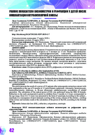
in LibraryThe article presents the result of a survey of 35 patients after extracapsular extraction cataract with IOL implantation at the age of 8 months to 13 years.Patients underwent: viziometry, biomicroscopy, keratorefractometry, skiascopy, A, B-scan ultrasound, ophthalmoscopy, consultation of related specialists. The calculation of the optical power of IOL was performed using the SRK II formula. Refraction in the early postoperative period in children from 8 months to 6 years corresponded to the age range, from 6 to 13 years was presented in the form of ametropia.According to the authors , ametropia is a consequence of post-traumatic scarring of the cornea, tension of the stitches , swelling of the cornea and mistakes made at the calculation of the power of the IOL according to the formula SRK II.
-
 Comparative short term study of posterior chamber phakic intraocular lenses for the correction of high myopia. (ICL VS IPCL)
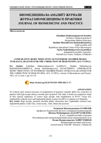
Journal of Biomedicine and PracticeTo evaluate early clinical outcomes of implantation of posterior chamber pIOLs for correction of patients with high myopia during 6 months post-op period. This study is based on comparative study of first clinical experience to correct high myopia with implantation of EVO+ Visian ICL (implantable collamer lens) and Implantable phakic contact lens IPCL to patients in Uzbekistan.
-
 RESULTS OF LENSECTOMY WITH IMPLANTATION OF TWO BACK-TO-BACK IOLS IN HIGH-GRADE MYOPIAThe aim of the study was to evaluate the effectiveness of lensectomy (LE) with the implantation of two intraocular lenses (IOL) by the “back-to-back” method for high myopia using the operating navigation system “VERION. Material and methods. The study included 68 patients (114 eyes) diagnosed with high myopia, which were divided into two groups: the main group included 40 patients (58 eyes) who underwent LE with implantation of two IOLs, the control group consisted of 28 patients (56 eyes), with which the LE was performed by the traditional method. Results and discussion.During implantation of 2 IOLs, an increase in the volume of the lens bag to 1.15 ± 0.05 mm and a lesser displacement of the vitreous body occur, in contrast to a lensectomy with one IOL (LT 0.52 ± 0.02). Conclusion. Analysis of the research results allows us to recommend the removal of a clear crystalline lens with the implantation of two lOLs using the back-to-back method with high myopia, with the aim of creating stability of the anatomical and topographic relative position of intraocular structures and the possibility of reducing the risk of retinal detachment, in contrast to the traditional method of surgical treatment.
Medicine and innovations -
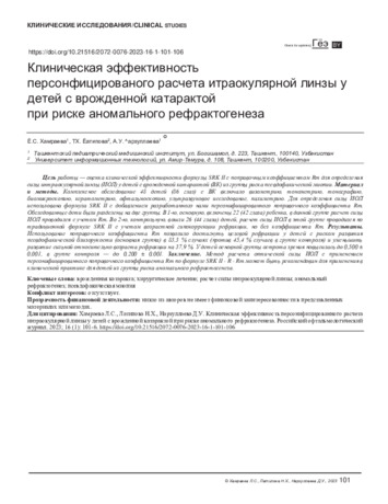 Clinical efficacy of individual intraocular lens calculation in children with congenital cataract at risk of abnormal refraction
in LibraryTo assess the clinical efficacy of the SRK II formula with a correction factor Rm in children with congenital cataracts who are at risk of pseudophakic myopia. Material and methods. A complex examination of 48 children (86 eyes) with congenital cataracts involved visometrics, tonometry, tonography, biomicroscopy, keratorefractometry, ophthalmoscopy, ultrasonography, and pachymetry. To determine the IOL power, we used the SRK II formula supplemented with the individual correction factor Rm, proposed by the authors. The examined children were divided into 2 groups. The main group 1 included 22 patients (42 eyes), for which the IOL power was calculated with the Rm factor. The control group 2 consisted of 26 patients (44 eyes) for which the IOL power was calculated according to the traditional SRK II formula using age-related hypocorrection of refraction but without the Rm coefficient. Results. The correction factor Rm, allowed us to achieve the targeted refraction in children who were at risk of developing pseudophakic myopia in 83.3 % of cases of the main group (versus 45.4 % of the control group cases) and reduce the development of high age-related refraction) by 37.9 %. In children of the main group, visual acuity reached, on average, 0.5 ± 0.001, while in the control group it was also higher but only reached 0.200 ± 0.001. Conclusion.The method of calculating the IOL optical power involving an individual correction factor Rm, according to the formula: SRK II – R – Rm can be recommended for clinical practice focused on children at risk of abnormal refractogenesis.
-
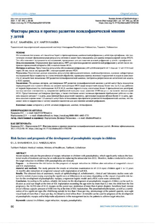
Recent studies indicate the prevalence of myopic refraction in children with pseudophakia, which significantly reduces the functional results of treatment and may be an indication for replacing the intraocular lens (IOL). Therefore, studies conducted to achieve the target refraction in children with pseudophakia are relevant.
Purpose — to determine the risk factors for the prognosis of myopic refraction in children after extraction of congenital cataract and IOL implantation.
Material and methods. The study presents the results of refraction examination in 69 (110 eyes) children aged 1 to 12 years 36 months after extraction of congenital cataract with implantation of soft IOL.
Results. The obtained data of anamnesis, results of ophthalmological, echobiometric, clinical and laboratory studies were subjected to statistical processing assessing the significance of differences in outcomes depending on the impact of possible risk factors for the development of pseudophakic myopia; a regression logistic model and a ROC-curve were constructed.
Conclusion. According to the authors, reliable risk factors for the development of pseudophakic myopia in children can be such indicators as axial eye length at the time of IOL implantation exceeding the age norm by more than 0.2 mm; the child from the first pregnancy; the AL/CR ratio of 23.0; myopia on the paired eye; strabismus of more than 4 prism diopters; hereditary load; tension of eye's fibrous capsule: pressure of SI 80 mm Hg at the time of IOL implantation. The presented reliable factors, as well as a combination of less significant signs (the child being outside for less than 1 hour per day, intermarriage of the patients parents, near-sight visual loads ot more than 3 hours per day, the blood Ca level of less than 1.8 mmol/L) can be used for prognosis of the development of pseudophakic myopia and to help make adjustments in the management tactics for patients to achieve target refraction.
-
 Functional results low-invasive by energy industry with implantation of IOL at 40 patients with senile cataract are studied. Visual acuity at all patients at receipt fluctuated from the correct projection of light to 0,05. Before operative measure of 30 patients (75%) distinguished only light with the correct svetoproyektion, at 10 - visual acuity has made 0,02-0,05 (25%). Within the first week after operation visual acuity has increased to 0,3 at 21 patients (52,5%), 0,4 - at 12 (30%). The maximum visual acuity without correction which managed to achieve as a result of operation, has made 0,8-0,9 at 7 patients - 17,5%.
 -
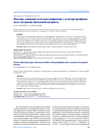 Factors influencing target refraction in children with pseudophakia after extraction of congenital cataract
in LibraryThe article describes the factors affecting the target refraction of pseudophakic eyes of children after extraction of congenital cat-
aracts. The factors include features of the echobiometric parameters of the eye, refraction, comorbidity of congenital cataracts and
ocular pathologies, margins of error in calculating strength of the intraocular lens, localization and structure of the artificial lens,
as well as correction of obscure or refractive amblyopia in pseudophakic eyes. Development of the algorithm for correction of re-
sidual refraction of pseudophakic eyes in children both before and after IOL implantation with consideration of each of those fac-
tors currently remains a relevant problem. -
 Features of the course of the post-operative period in frequently affect children after extraction of congenital cataract
in LibraryThe main link in the treatment of congenital cataracts (VK) is the extraction of the lens with the
subsequent correction of ametropia by various methods, one of which is the implantation of an
intraocular lens (IOL). The aim of the study was to study the features of the course of the postoperative period in frequently ill children (FBI) after VC extraction. We examined 35 children
(59 eyes) with VK who are undergoing surgical treatment in the eye department of the clinic of the Tashkent Pediatric Medical Institute. A comparative analysis showed significant changes in the hydrodynamic parameters of the eyes and a high percentage of intra- (hyphema in 71%, fibrin
reaction in 59%, prolapse of the vitreous in 47% of cases) and postoperative complications (corneal
edema in 36 %, inflammatory exudative reaction in the anterior Chambers: 1+, 2+ in 64% of cases) in FWB compared to children who are not in the category of frequently ill. The revealed feature
indicates the need for adequate management of the preoperative and postoperative periods in
Frequently ill children -
The paper presents the results of extraction of congenital cataract with IOL implantation in 50 (86 eyes) children aged 7 months to 12 years. In the examined patients, intraoperative complications were noted, such as posterior capsule rupture (8%), hyphema (6%), postoperative: fibrin effusion on the IOL (7%) and corneal edema (4.6%). With congenital bilateral cataracts after treatment, visual acuity increased on average from pr.l.certae to 0.1; with unilateral - from pr.l.certae to 0.01.
-
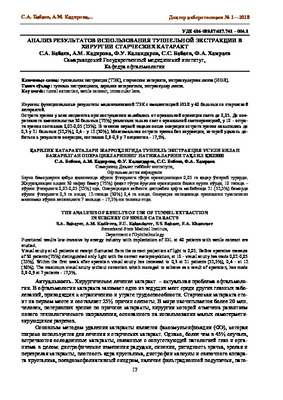
 Преимущества методики удаления катаракты через склеро роговичный тоннель с имплантацией иол
in LibraryВ настоящее время в хирургии катаракт отмечается тенденция к уменьшению разреза и проведение операции бесшовным методом с целью доведения до минимума травмы, инфекции, послеоперационного астигматизма, времени операции, стоимости затраченных средств на лечение и 100% возможности имплантации ИОЛ. Известно множество методик удаления ядра через малые разрезы. Наиболее распространенная методика - удаления ядра хрусталика через малый разрез в верхненазальном квадранте, предложенная докторами К. Kowano, A. Momose (Япония). В 1999 году С.Н. Федоровым и Б.Э. Малюгиным предложена методика удаление ядра через склеророговичный туннель в верхнем квадранте и внедрена в офтальмологическую клинику I-ТашГосМИ А.А. Ахроровым с 2000 года.
-
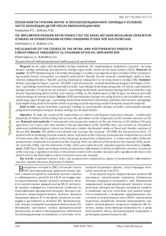 Peculiarities of the course of the intra- and postoperative period in conditionally frequently ill children after iol implantation
in LibraryTo study the results of IOL implantation in children with frequent respiratory diseases - conditionally frequently ill children (FCCI) taking into account the parameters of the composition of the chamber moisture of the eye. Material and methods: A retrospective and prospective analysis of the case histories of 50 children (50 eyes) aged from 1 to 5 years, who were treated in the eye department of the TashPMI clinic, was carried out. All patients underwent ophthalmological, clinical and laboratory studies: biochemical studies of blood and chamber moisture of the eye (EC). Results: The children were divided into 2 groups: the 1st group - 28 UCBD, the 2nd group (control) - 22 patients with no pathology from the somatic status. In patients of the 1st group, intraoperative complications occurred 1.8 times more often than in patients of the 2nd group, postoperative complications - 2.5 times more often. Of the late postoperative complications in patients of the 1st group, there was fibrosis of the posterior lens capsule (61%), poste-rior synechia (18%), and IOL dislocation (14%), which were indications for repeated surgical interventions. Conclu-sions: UCBD has a higher percentage of early postoperative inflammatory and late proliferative reactions. In patients of the 1st group, a significant increase in the protein content in the chamber moisture and a significant decrease in the protein level in the blood before cataract extraction were also revealed.
-
 The effectiveness of the operation of phacoemulsification with intra ocular lenses implantation with mature senile cataractFunctional outcomes were studied surgery phacoemulsification with IOL implantation in patients with senile cataract. We performed an analysis of surgical treatment of 22 patients (22 eyes) with this eye disease. Visual acuity at delivery ranged from the correct projection of light to 0.01. By the degree of core density of 54.5% (12 eyes) - had grade 2, 36.4% (8 eyes) grade 3 and 9.1 % (2 eyes) - 4 degree very dense cataract are drilling core. Discharge of patients from hospital acuity of 0.6-1.0 was observed in 45.4% cases (10 eyes), 0.4-0.5- in 31.8% cases (7 eyes). 0.2-0.3- in 22.7% cases (5 eyes). Poor eyesight in the postoperative comorbidity explained by the retina.
Doctor's Herald -
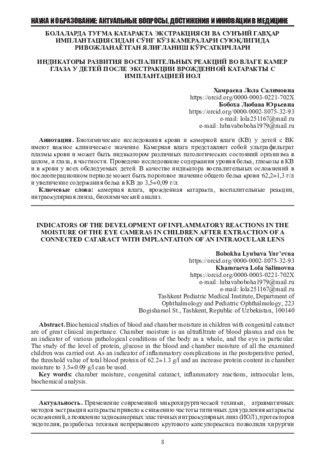 Indicators of the development of inflammatory reactions in the moisture of the eye cameras in children after extraction of a connected cataract with implantation of an intraocular lens
in LibraryBiochemical studies of blood and chamber moisture in children with congenital cataract
are of great clinical importance. Chamber moisture is an ultrafiltrate of blood plasma and can be
an indicator of various pathological conditions of the body as a whole, and the eye in particular.
The study of the level of protein, glucose in the blood and chamber moisture of all the examined
children was carried out. As an indicator of inflammatory complications in the postoperative period,
the threshold value of total blood protein of 62.2±1.3 g/l and an increase protein content in chamber
moisture to 3.5±0.09 g/l can be used. -
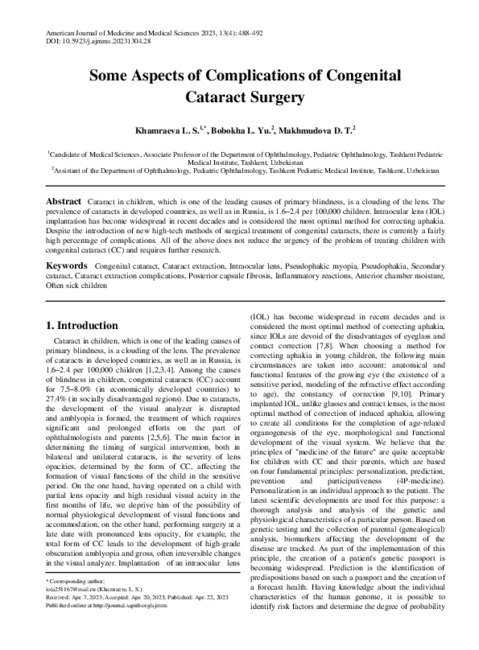
Cataract in children, which is one of the leading causes of primary blindness, is a clouding of the lens. The prevalence of cataracts in developed countries, as well as in Russia, is 1.6–2.4 per 100,000 children. Intraocular lens (IOL) implantation has become widespread in recent decades and is considered the most optimal method for correcting aphakia. Despite the introduction of new high-tech methods of surgical treatment of congenital cataracts, there is currently a fairly high percentage of complications. All of the above does not reduce the urgency of the problem of treating children with congenital cataract (CC) and requires further research.
-
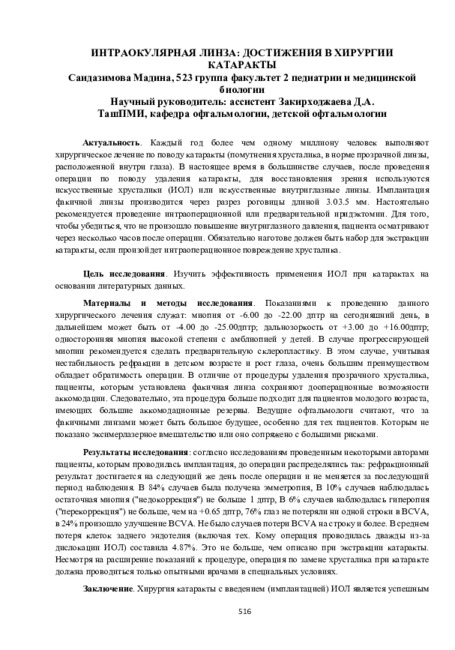
Каждый год более чем одному миллиону человек выполняют хирургическое лечение по поводу катаракты (помутнения хрусталика, в норме прозрачной линзы, расположенной внутри глаза). В настоящее время в большинстве случаев, после проведения операции по поводу удаления катаракты, для восстановления зрения используются искусственные хрусталики (ИОЛ) или искусственные внутриглазные линзы. Имплантация факичной линзы производится через разрез роговицы длиной 3.03.5 мм. Настоятельно рекомендуется проведение интраоперационной или предварительной иридэктомии. Для того, чтобы убедиться, что не произошло повышение внутриглазного давления, пациента осматривают через несколько часов после операции. Обязательно наготове должен быть набор для экстракции катаракты, если произойдет интраоперационное повреждение хрусталика.
-
 INTRАOCULAR AMMETROPIA CORRECTION WITH PHAKIC LENSESThe analysis of literature sources (database PubMed and Google scholar) concerning modern views on the use of Phakic intraocular lenses in order to correct the refractive error of the eye. The current models of these lenses are quite sophisticated and many times increase the possibilities of their use than before. With the help of PIOL it is possible to correct any degree of myopia and hypermetropia, as well as astigmatism. There are examples of using such lenses to correct presbyopia. Currently, lenses are predominantly used in the posterior chamber of the eye. In the long term (on average after 8-10 years) complications are possible in some (up to 10%) patients. When the PIOL is inserted into the anterior chamber, the development of endothelial dystrophy of the cornea or the appearance of glaucoma is possible; when the PIOL is inserted into the posterior chamber, cataracts may develop.
Journal of oral medicine and craniofacial research -
Миопия является одним из самых распространенных глазных заболеваний в мире и наиболее частой причиной снижения остроты зрения, встречаясь в 12-30% случаев среди офтальмопатологии. Высокий интерес к данной проблеме в последние годы связан с увеличением распространения близорукости среди населения всего мира, а именно, ее высоких степеней от -6,0 дптр и выше.
За последние десятилетия отмечен рост близорукой рефракции, особенно среди учащихся и работающих с компьютером.
Частота высокой близорукости, одной из наиболее тяжёлой рефракционной патологии, по данным разных авторов, колеблется от 6-18 до 27-33,2% [1, 41, 45, 89, 97]. В последние годы привлекает все большее внимание исследователей коррекция ослабленного зрения при близорукости высокой степени, в частности врожденной. Врожденная близорукость в 55-60 % случаев бывает высокой и является одной из главных причин инвалидности по зрению [52]. За последние десятилетия число лиц с близорукостью возросло, всего в мире очки носят около 1 млрд, человек. Близорукость чаще обнаруживается у детей 7-12 лет, усиливается в подростковом возрасте, а в 18-40 лет стабилизируется. Это очень важный аспект, т.к. прогрессирование близорукости в среднем возрасте чаще всего связано с увеличением преломляющей силы оптического аппарата глаза. В последнее десятилетие число людей с миопией в странах Европы увеличилась и составляет 30% -40% всего населения.В странах Юго-Восточной Азии достигает 70% и более. Миопия высокой степени развивается у 2% населения и является одной из необратимых причин слепоты у лиц молодого и среднего возраста. В Европе 8,8% стойкой слепоты связаны с миопией высокой степени, в США она занимает второе место среди причин инвалидности.
Низкая острота зрения у близоруких нарушает их трудоспособность, ограничивает возможность выбора профессии и полноценную жизненную активность. Коррекция миопии высокой степени с целью сохранения полноценной остроты зрения актуальна, как в социальном, так и в научном плане. Лица с близорукой рефракцией нуждаются в очковой коррекции. Наблюдения показали, что, если определенный контингент постоянное ношение очков воспринимает как огромное благо, то значительная часть близоруких лиц, особенно молодые женщины нашего региона, категорически отказываются от ношения очков, обрекая себя на зрительный дискомфорт.
Коррекция ослабленного зрения при миопии - актуальнейшая проблема офтальмологии. Она обусловлена как распространенностью миопии преимущественно у молодых лиц, то есть социальноактивного контингента, так и тенденцией к повышению степени ее в связи с возрастающей зрительной и психологической нагрузкой.
Существующие традиционные методы коррекции зрения при высокой миопии не всегда устраивают пациентов в современных сложных социально-экономических условиях.
Традиционным способом коррекции аметропий является очковая. Более 80% людей с нарушением рефракции предпочитают этот вид коррекции из-за ее доступности, отсутствием серьезных осложнений при ее использовании. Однако, очковая коррекция не всегда применима. Очки трудно переносятся при анизометропии и при высоких степенях аметропий. Есть ограничения по профессиональным состояниям больных. Нередко, даже при хорошей переносимости, они не устраивают больных по косметическим соображениям. Очковые линзы до сих пор остаются самым доступным и распространённым средством коррекции миопии. Самым серьёзным и неустранимым недостатком очковых линз является их влияние на размеры ретинального изображения объекта.Второе обстоятельство, часто заставляющее пациентов обращаться к другим видам коррекции, связано с косметическими недостатками очков. Проблема очков доводит до трагедии молодежь, особенно девушек в Узбекистане, где большинство предвзято относятся к «очкарикам». Кроме того, недостатками очковой коррекции являются: сужение полей зрения, зрительный дискомфорт, вызываемый климатическими условиями, частая смена очков (1 раз в 1 -2 года).
В случаях невозможности или непереносимости очковой коррекции альтернативой является использование контактных линз. От 15% до 18% больных с нарушением рефракции используют этот вид коррекции [35, 36]. Контактные линзы свободны от присущих очковым линзам косметических дефектов и, составляя единую оптическую систему с глазом, практически не влияют на величинуретинального изображения. Тем не менее, ряд обстоятельств (как объективного, так и субъективного характера) часто заставляет пациентов отказываться от ношения контактных линз, в частности:
- так называемые манипуляционные сложности;
- необходимость ухода за контактными линзами;
- потенциальная возможность осложнений, связанных с ношением контактных линз.
Контактные линзы - хотя и являются физиологической коррекцией, но не всегда достигается удовлетворительная адаптация к ним в индивидуальных случаях высокой миопии. Негативное влияние на переносимость и возможные осложнения (кератиты, эрозия роговицы, конъюнктивиты) оказывают особенности климатических условий в регионе Средней Азии, отличающиеся повышенной запыленностью и низкой атмосферной влажностью воздуха, из-за чего контактная коррекция не может быть широко рекомендована.
От 1 до 5 % людей с нарушением рефракции не желают или не могут использовать очки и контактные линзы и решаются на хирургические виды коррекции.
Таким образом, комплексный подход к проблеме оптической коррекции миопии высокой степени в настоящее время осуществляется по следующей схеме: очковая коррекция —► коррекция контактными линзами —» хирургическая коррекция. Каждое последующее звено в этой схеме является более сложным и применяется при безуспешности или невозможности использования более простого способа коррекции. Своевременная коррекция высокой миопии необходима для полноценного развития и функционирования зрительного анализатора, что обуславливает целесообразность проведения рефракционных операций у больных с непереносимостью очковой и контактной коррекции.
В настоящее время сложились два подхода к проблеме лечения близорукости высокой миопии - это применение хирургических методик, направленных исключительно на быстрое достижение рефракционного эффекта и более серьёзное отношение к миопии как к болезни, при которой в той или степени поражаются все структуры глазного яблока [24, 73, 106, 107].
Изменить рефракцию оптических сред глаза можно тремя путями.
Первый путь - экстраокулярный, когда дополнительная оптическая система размещается перед глазным яблоком (очки) или непосредственно на нем (контактные линзы). Этот ставший традиционным путь не предусматривает изменения структуры преломляющих сред самого глаза и может рассматриваться как неинвазивный.
Второй путь - это изменение преломляющей способности роговой оболочки дозированным хирургическим воздействием с роговой оболочки дозированным хирургическим воздействием с помощью ножа или лазерного излучения.
Третий путь - интраокулярный, который предусматривает изменение рефракции глаза с помощью операций со вскрытием глазного яблока.
Обилие разработанных к настоящему времени методов воздействия на оптику глаза и конкретных оперативных вмешательств неизбежно ставит как перед пациентом, так и перед врачом проблему выбора наиболее адекватного для каждого конкретного случая метода операции.
В настоящий момент приходится констатировать, что идеального и абсолютно безопасного метода хирургической коррекции аметропий не существует, и каждый потенциальный пациент должен быть информирован об этом. Индивидуальный выбор оптимального метода зависит от существующих в стране или регионе возможностей, как в отношении технического оснащения, так и опытности хирургов в проведении данного вида операций, степени и вида аметропии, а также возраста пациента, общего состояния его здоровья и, наконец, его финансовых возможностей.
В настоящее время арсенал хирургической коррекции аномалий рефракции огромен. Как хирургам, так и пациентам предоставлена уникальная возможность, выбирать методы и способы коррекции рефракционных дефектов [2, 3, 8, 50, 64]. Рефракционные операции - понятие собирательное и включает в себя вмешательства, которые могут изменить оптические свойства и параметры различных частей оптической системы глаз (длины оси, силу роговичной и хрусталиковой линз.)
Выбор метода хирургического лечения - актуальная и жизненно важная проблема. От того, насколько точным будет выбор, зависит исход операции, а значит, и социальная, и профессиональная реабилитация пациента. -
 Comparative age-related assessment of the anterio-posterior axis length in children with normal eyes and with unilateral congenital cataract or congenital glaucoma
in LibraryThe length parameter of anterior-posterior axis is compared for children’s hyperopic eyes (302 eyes, hyperopia of 0.5 to 3.0 D). 109 eyes (109 children) with unilateral congenital cataract (UCC), and 132 eyes (90 children) with congenital glaucoma, aged 1 month to 15 years. Extended observation of anterior-posterior axis growth and refraction in eyes with artifakia showed a partial tendency to myopisation (3— 7%), which requires additional research of the pathogenetic process and should be taken into account when calculating the optical power of 1OL. The analysis of data on age-related growth of the anterio-posterior axis of eyes with congenital glaucoma showed the agreement with average figures for the developed and advanced stages of the condition. In contrast, terminal and advanced glaucoma (in children aged 2 to 3) the obtained data showed a reliable increase as compared to previously published values
-
Features of calculating the optical power of an intraocular lens in children with congenital cataracts at the risk of developing pseudomyopic myopia.
in LibraryThe article presents the results of the clinical efficacy of the calculation formula with the correction factor of the optical force of the intraocular lens in children with congenital cataracts at risk of development of pseudophakic myopia. Personalized Rm corrective coefficient in the formula for calculating the force of IOL in children with risk of development of pseudophakic myopia makes it possible to achieve target refraction in 83.3% cases and reduce the development of reduce myopic refraction.
-
 Некоторые аспекты профилактики постоперационных инфекционных осложнений в хирургии катарактыПослеоперационные инфекционные осложнения, в том числе эндофтальмит, являются крайне опасными осложнениями глазной хирургии, в структуре которой преобладает экстракция катаракты с имплантацией ИОЛ. По данным различных авторов частота возникновения послеоперационного эндофтальмита колеблется в пределах от 0,05 до 1,77%
Doctor's Herald -
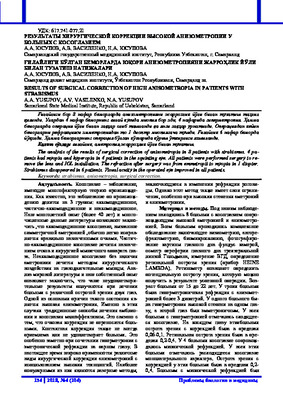 Results of surgical correction of high anisometropia in patients with strabismusThe analysis of the results of surgical correction of anisometropia in 8 patients with strabismus. 4 pa-tients had myopia and hyperopia in 4 patients in the squinting eye. All patients were performed surgery to re-move the lens and IOL installation. The refraction after surgery was from emmetropii to myopia in 1 diopter. Strabismus disappeared in 6 patients. Visual acuity in the operated eye improved in all patients
Journal problems of biology and medicine -
 Clay is the most common soil on earth. It is formed after the disintegration and weathering of rocks into tiny particles, and is the last stage of their degradation. Clay occurs in solid arrays or thin layers (lenses) close to the surface.