Library search
Search Results
-
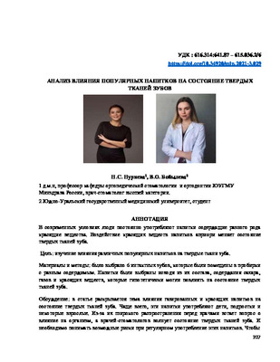 studying the enects ot ditterent beverages on hard tooth tissues is very important tor any person. And you need to understand the possible risks when regularly using them. Since there arc components in the beverage composition that can cause pigmentation and demineralization of hard tissues. Federal State Budgetary Educational Institution of Higher Education "South-Ural State Medical University" of the Ministry of Healthcare of the Russian Federation. Bobyleva Valeria Olegovna.
 -
 CHANGES IN THE MICROELEMENT COMPOSITION OF THE HIPPOCAMPUS AND HARD TISSUES OF RAT TOOTH AS A RESULT OF STRESS IMPACT ON THE ORGANISMToday, many scientists are actively discussing G. Selye’s hypothesis about the effect of stress on the body, including the occurrence of dental caries. The chemical composition of the body is individual, it can change under the influence of various factors, including stress. As a result of the studies, it was found that the ratio of trace elements of variable valency in the hard tissues of the tooth and brain is different. An excess of trace elements in the tooth tissues of experimental groups of rats suggests a shift in redox reactions, which can lead to the development of a pathological process.
Stomatologiya -
 Among several methods of determining the colour of a tooth, technology which determines the colour of the tooth with the help of high-resolution camerais the most effective one. According to the results of a research, works done with the help of dental images look more natural and are maximally close to the natural colour and shades of the teeth. When a standard colour scale was used, patients expressed their dissatisfaction with the л-ague and unnatural colour of the teeth. Therefore, it is recommended to use a standard method of determining the colour of teeth through colour scale in combination with high-resohition cameras.
 -
 Описано собственное наблюдение автора больного с жалобами на боли в области верхней челюсти слева, на прохождении струи воздуха изо рта в нос. Из анамнеза: при удалении 26 зуба врач воспользовался прямым элеватором, после чего произошел отлом дна пазухи с альвеолярным отростком и 26. 27, 28 зубами. Произведен разрез по переходной складке от 23 зуба до передневерхнего угла раны, а также разрез на слизистой оболочке неба по внутреннему краю альвеолярного отростка от 26 зуба до 23 зуба, далее огибая, разрез продолжен параллельно небному шву до уровня 28 зуба и таким образом сформирован языкообразный лоскут. Наложено соустье на нижний носовой ход. Пазуха затампонирована. Лоскуты ушиты между собой после мобилизации. Больной выписан с выздоровлением.
 -
 Medical and social problem of caries affects the population of all countries. Nowadays, many believe that stress plays a role in the occurrence of caries. The purpose of this study was to identify what structural changes occur in the brain and tooth of a rat as a result of exposure to stress factors on the body and the possibility of interconnection. Studying the effects of various types of stress on the rat organism, we found that pathological changes affect such a vital organ as the brain and are dystrophic. According to the severity of the changes occurring in the tissues of the brain and tooth are interrelated. In the teeth, changes initially occur in the dental pulp and are proliferative and dystrophic in nature. In order for pathological changes to occur, affecting the tissues of the tooth, a combmation of a number of factors is necessary, namely, low resistance of the body, sufficient strength of stress and the immune response.
 -
В согласно статистике, 50% от общего количества травм твёрдых тканей челюстно-лицевой области составляют травмы зубо-альвеолярной сферы [1].Из них травмы,наблюдаемые при прорезывании зубов,встречались от 0,9% до 3,9%[2].Полный вывих постоянных зубов составляет около 3% от общего числа травм у детей[3-4].Работа врача-стоматолога нацелена на сохранение зуба как органа. Разновидностью зубосохраняющих операций является реплантация зуба [5]. Дальнейшая разработка технологий реплантации позволяет на более высоком научно-практическом уровне решать многие проблемы восстановления зубных рядов и изготовления полноценных зубных протезов [6-7]. Соответственно, в настоящее время одной из актуальных проблем, стоящих перед стоматологами, является совершенствование метода –реплантации-сохранения зуба как органа.
-
 Assessment of the functional state of the endothelium in patients with viral hepatitis before tooth extraction
in LibraryTooth extraction is the most common operation, after which hemorrhagic complications often occur, especially in patients with chronic viral liver disease. This condition is caused by damage to the endothelial lining of blood vessels. Based on this, the purpose of this study was to study the features of endothelial dysfunction before tooth extraction in patients with viral hepatitis. 58 patients with hepatitis B and C with different prescription periods of the disease were examined. In patients with viral hepatitis, an increase in platelet aggregation activity on the effect of an ADP inducer (Tma) by 45% was noted before tooth extraction. The lengthening of the activated recalcification time (AVR) by 37% observed by us in patients with viral hepatitis reflects a deficiency of plasma factors (XII,XI,XIII) of the blood coagulation system and indicates a state of hypocoagulation. Against this background, high values of alpha-2 macroglobulin in the blood (4 times) and Willebrand factor (15%) and a significant decrease (by 35%) in the content of protein C in the blood of the examined patients were noted. The obtained results of the study indicate that these patients have a narrow band of maintaining hemostatic balance, and the existing balance can easily be transformed into hypo- or hypercoagulation, which requires preventive measures to prevent complications after tooth extraction.
-
 The changes of hard tissue and dental pulp in chronic periodontitis were studied with the Scanning electron microscopy. It was revealed that in parodontitis such changes as looseness of enamel prisms, polymorphism of dentinal tubules, enlargement of their lumen and interlayers between them take place. The chaotic arrangement of fibers is characteristic for cement. These indicate on pronounced trophism disorders of dental hard tissues, which apparently due to lower trophism function of the pulp.
 -
 ASSESSMENT OF THE FUNCTIONAL STATE OF THE ENDOTHELIUM IN PATIENTS WITH VIRAL HEPATITIS BEFORE TOOTH EXTRACTIONAbstract Tooth extraction is the most common operation, after which hemorrhagic complications often occur, especially in patients with chronic viral liver disease. This condition is caused by damage to the endothelial lining of blood vessels. Based on this, the purpose of this study was to study the features of endothelial dysfunction before tooth extraction in patients with viral hepatitis. 58 patients with hepatitis В and C with different prescription periods of the disease were examined. In patients with viral hepatitis, an increase in platelet aggregation activity on the effect of an ADP inducer (Tma) by 45% was noted before tooth extraction. The lengthening of the activated recalcification time (AVR) by 37% observed by us in patients with viral hepatitis reflects a deficiency of plasma factors (XII, XI, XIII ) of the blood coagulation system and indicates a state of hypocoagulation. Against this background, high values of alpha-2 macroglobulin in the blood (4 times) and Willebrand factor (15%) and a significant decrease (by 35%) in the content of protein C in the blood of the examined patients were noted. The obtained results of the study indicate that these patients have a narrow band of maintaining hemostatic balance, and the existing balance can easily be transformed into hypo- or hypercoagulation, which requires preventive measures to prevent complications after tooth extraction.
Medicine and innovations -
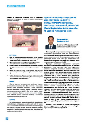 Dental Implantation In the place of a 16-year-old re-implanted tooth Tooth replacement Is о promising tooth-preserving operation, with Inconclusive conservative, endodontic treatment. Although Infrequent resort to re-Implantatlon, this possibility must be considered among other treatment options. Most cases of resorption of the Implanted tooth are diagnosed In the first 2-3 years after the re-Implantatlon, however, resorption can occur even after 5 or 10 years or more. According to our results and reviews, the literature shows that re-Implantatlon Is a reliable and predictable procedure used to preserve the natural dentition. This operation can be carried out much more often than at the present time. The tooth replacement not only preserves the tooth, but also the bone tissue of the alveolar process, preventing atrophy and deformation. Also, the reconstructed root part can serve as a bone support for dental Implants. Considering the possibility of dental Implantation on the site of Implanted teeth, this tactic Is recommended for wide Introduction Into practical dental Implantology.
 -
 The use of "bioactive glass" for osteoplasty of the hole of the removed tooth in patients with diabetes mellitusAfter filling socket extraction of a tooth with osteoplastic material appear risk of secondary infection and progress suppurative inflammation witch one depend on properties of a substance osteoplastic material. Operation extraction of a tooth with filling socket with osteoplastic material prevent inflammation and atrophy of the bone on the strength optimization reparation process in bone Reasonability using bioglass of domestic growth ‘Bioactive glass”. Bioactive glass rating to the grope surfaceactive biomaterials , on surface of witch one happen there row specific reaction . reduce to formation amorphous calcium phosphate and crystalic cacium hydroxyapatite , what is good for bone formation. Recover bone tissue one can base on unique granulated bioactive glass. In osteoplastic socket after extraction of a tooth by patient take clinical laboratory research and foundation indications application of this material researcher . witch one very important importance for prevent deformation aleveolar bone . define target and tasks research.
Stomatologiya -
 Construction of mathematical models of the degree of evaluation of efficiency and prognosis in determining the electrical excitability of the tooth by the express methodThe construction of mathematical integral characteristics allows to adequately assess the efficiency of determining the electrical excitability of the tooth in the express mode, taking into account the sex of the patient, depending on the degree of clinical changes in the enamel, the presence and depth of carious cavities, various changes in the periodontal gap, the degree of bone resorption and tooth trauma. As a basis of calculations of the software product, the parameters of dental X-ray images are taken.
Stomatologiya -
 REPLACEMENT OF THE WELL OF THE REMOVED TOOTH WITH AN AUTOGENOUS BONE GRAFT IN ORDER TO PREVENT THE OCCURRENCE OF DEFECTSIn many (63%) cases, after tooth extraction, there are bone defects and deformities in the alveolar processes of the jaws. Preservation and restoration of the volume of bone tissue of the alveolar processes of the jaws after tooth extraction, periodontal and tooth-preserving operations is an important problem of surgical dentistry and maxillofacial surgery. The difference between the bone tissue of the jaws and any other segment of the skeleton is that when the functional load is distributed or lost, the processes of resorption begin. In this case, bone loss occurs not only in the area of the removed tooth, but affects about 20% of the volume of the hole around it. 2-3 years after removal, there is usually a decrease in the anatomical size by 40-60% of the alveolar ridge, and this is typical for all population groups (A. Ashman). In the available literature, we have not found any works devoted to a detailed study of changes in bone density in the osteoplasty zone using modern methods of replacing jaw defects.
Journal of oral medicine and craniofacial research -
 APPLICATION OF HYALURONIC ACID TO OPTIMIZE REPARATIVE REGENERATION IN THE HOLE EXTRACTED TOOTH IN PREPARATION FOR DENTAL IMPLANTATIONSIn clinical situations where you plan to install the delayed dental implants after tooth extraction, to ensure optimization and positive results of operations predictability, you must prepare the hole of removed tooth to dental implantation. To achieve the full remodeling of the bone tissue in optimal timing and successful osteointegration dental implant, it is considered more appropriate to use osteoplasticheskih materials. One of the promising achievements in medicine in General, and particularly in dentistry, was the use of hyaluronic acid to create optimal conditions for the regenerative processes in the wound. In this review, article the comparative analysis of some of the approaches of preparing wells of a remote tooth the bone with dental implant otstrochennoj.
Stomatologiya -
 The role of magnetic resonance imaging in the comprehensive radial diagnosis of volumetric masses of the eye organ
Catalog of abstractsRelevance of the problem. The difficulties of diagnostics of orbital diseases are well known. Especially difficult is intraspecies differentiation among the multitude of tumour, pseudotumour, inflammatory, vascular, endocrine and other diseases occurring here, manifested by the symptom complex of unilateral exophthalmos [Beradze I.N., 1978; Brovkina A.F., 1993].
Malignant intraocular neoplasms are the main cause of death of patients with diseases of the organ of vision, with 45-48% of patients dying from metastases in the first 5 years after enucleation [Alekseeva I.B., 1990, Barkhash S.A.1978, Brovkina A.F..1991, 1997; Keizer R.W.. Viclvoyc G.L.,1986],
Retinoblastoma is the most frequent malignant neoplasm in children. According to different authors, the frequency of its occurrence is 1 case per 14000 - 35000 newborns. [Bobrova N.F. and Vit V.V., 1993; Brovkina A.F., 1997; Provenzale J.M., et al., 1995; Skulski M., et al., 1997; Weber A.L., Mafee M.F, 1992; Wilms G., et al., 1989]. The frequency of patients with the most malignant intraocular tumour in adults - uveal melanoma has recently reached 7-9 people per 1 million population [Brovkina A.F., 1997; Kotslyansky E.O., 1989; Yushko N.A., Peskova L.I., Kalenich L.A., 1989; Peyster R.G., Augsburger J..I., Shields J.A., 1988; Romani A.. Baldeschi L., ct al 1998; Scott I.U., 1998].
The fundamental difference in treatment tactics, depending on the stage of development, size and topography of the tumour, as well as the seriousness of the prognosis in retinoblastomas and melanomas sharply increase the requirements for the accuracy of their differential diagnosis. At the same time, the number of diagnostic errors in ocular tumours continues to be 10-30% even when complex clinical and instrumental examination is applied in specialised ophthalmological centres [Ternovoy S.K., Panfilova G.V., Rogozhin V.A., 1979; Friedman F.E., Malyuta G.D., Kodzov M.V., 1995; Song G.X., 1991].
Widely used in ophthalmological practice traditional diagnostic methods (ophthalmoscopy, gonioscopy, diaphanoscopy, fluorescence angiography, laboratory tests) are insufficient to obtain comprehensive information about the localisation, nature of growth and prevalence of volumetric pathological formations of the eye and orbit. This circumstance and not quite satisfactory results of surgical treatment are the causes of high mortality of patients [Muratova T.T., Nigmanova N.H., Kozlovskaya G.M.. 1989, Naches A.I., 1980; Cheremisin V.M., Trufanov G.E., Kholin A.V., 1991]. Untimely or erroneous recognition of pathological processes of the orbit leads to a sharp deterioration of visual functions, up to blindness, and in some cases to the death of the patient [Yuzhakov A.M., Travkin A.G., Kiseleva O.A., 1991]. All this determines the importance of timely and accurate diagnosis of diseases of the orbit, on the one hand, and the difficulty of such diagnosis - on the other [Gabunia R.I., Kolesnikova E.K., Tumanov L.B., 1982].
The fact that the orbit is closed from direct inspection and palpation by bone walls and the eyeball, indicates the advantage of radial diagnostics in comparison with other methods of examination. In the arsenal of clinicians there is a great variety of methods of clinical-radial diagnostics of orbital pathology, however, at present the information in the literature about their resolving capabilities and significance in comparative aspect is incomplete and not fully studied. The priority of using one or another instrumental investigation, their sequence and expedient combination have not been determined yet. This makes it difficult to choose the optimal standardised approach for diagnosis and adequate treatment [Cheremisin V.M., Trufanov G.E., 1993, Weber A.L., Sabates N.R., 1996; Wenig V.M., Mafee M.F., 1998].
Thus, the study of these and other questions, contributing to the improvement of diagnostics and treatment of patients with neoplasms of the eye and ocular cavity, should be recognised as urgent urgent.
Purpose of the study. Comparative evaluation of magnetic resonance tomography capabilities and development of algorithms for complex radial diagnostics of volumetric formations of the visual organ. To solve this goal we set the following tasks.
1. To study the normal picture of the magnetic resonance image of the visual organ in comparison with other methods of visualisation.
2. To find out the possibilities of magnetic resonance tomography, ultrasound and computed tomography in detection and evaluation of intraocular neoplasms.
3. To determine the role and place of magnetic resonance tomography in differential diagnostics of volumetric pathological formations of the eye cavity in comparison with other radial methods of research.
4. To determine the indications and to develop an algorithm for the complex application of radiography, ultrasound, computer and magnetic resonance tomography for diagnostics of volumetric formations of the eye organ.
Scientific novelty.
The present work is the first to give a detailed and detailed description of the complex clinical and radiation examination, with generalisation and standardisation of magnetic resonance, computer and ultrasound semiotics of volumetric pathological formations of the eye and eye cavity. The conducted clinical and instrumental investigations allowed to determine the diagnostic value and resolving capabilities of each of the applied methods. The ultrasound, CT and MRI signs of volumetric formations of the eye organ were studied, clarified and supplemented taking into account the use of low-field magnetic field and general-purpose ultrasound apparatus. The developed standardised diagnostic algorithm of examination of patients with this pathology is new, thanks to which the pre-oppositional diagnosis of tumour and other diseases of the visual organ is improved and the total radiation load on the patient is reduced.
Conclusions
1. MPT will provide an opportunity to study the weight of the soft tissue and anatomical components of the ocular cavity, up to the optic nerve sheath and perineural liquor space, the orbital apex and chiasmal-sellar region, as well as to assess the condition of adjacent structures of the brain and facial skull. The method is limited in the evaluation of changes in the bony walls of the orbital cavity.
2. MRI is inferior in detecting characteristic signs of retinoblastoma (presence of calcification). The sensitivity of MRI was 66.6%, while for ultrasound and CT these values were 96.1 and 100%, respectively. But when the tumour spreads rstrobulbarly outside the eyeball (at 3-4 stages) the informativeness of MRI increases significantly. In uveal melanoma the sensitivity and specificity of MRI reaches 100%.
3. Both MRI and CT have a high detection rate (98.1% and 95.8% respectively) of benign orbital tumours of both primary and secondary origin. However, MRI is the preferred method of investigation. MRI is especially informative when a cranioorbital tumour and pseudotumour are suspected. The sensitivity of the method is 90.9% and 91.6%, respectively
4. In some cases ultrasound can be used to differentiate between encapsulated and diffuse neoplasms, which facilitates the diagnosis. However, when the pathological process is localised near the orbital apex, the diagnostic value of ultrasound decreases. In such cases it is advisable to use MRI.
5. In detection of primary and secondary malignant tumours of the orbital cavity both MRI and CT are quite informative (sensitivity 97,2% and 95,4% respectively), but the most comprehensive information about the state of bone walls will be provided by CT. When the process spreads intracranially, the value of MRI increases significantly, especially with the use of contrast enhancement.
6. The developed algorithm of complex clinical and radiation examination of patients with the use of ultrasound, CT and MRI is the most effective in the diagnosis of volumetric pathological formations of the eye and eye cavity, allowing to reduce to an adequate minimum the total radiation load on the patient and diagnostic period, excluding duplication of research techniques and choosing the most informative in each case, which in turn allows to develop appropriate treatment tactics and reduce the level of disability of the patient. -
 DEFINITION OF DISCRIMINATORY SENSITIVITY OF LANGUAGE WITH GLOSSALGIA IN PATIENTS WHO UNDERWENT COVID-19 AT THE STAGE REHABILITATIONAn article is presented on the topical problem of therapeutic dentistry to determine the discriminatory sensitivity of the tongue in glossalgia in patients who have undergone COVID-19 at the stage of rehabilitation Purpose: to determine the discriminatory sensitivity of the tongue in glossalgia in patients undergoing COVID-19 in the rehabilitation stage. Materials and methods: the study included 88 patients aged 35 - 70 years, of which 51 patients with glossalgia who underwent COVID 19 and had inflammatory diseases of the oral mucosa were included in the main group, of whom 36 were women, 15 were men; 37 patients with glossalgia who did not suffer from COVID 19, of which 29 were women, 8 were men in the comparison group; 20 healthy individuals served as controls. Examination of the oral cavity of patients with glossalgia was carried out when patients applied to the therapeutic dentistry polyclinic of the TGSI, while filling out a dental questionnaire, carried out photo and video documentation. The average age of the patients was 52.57 ± 1.20 years. Determination of the deep discriminator}' sensitivity of the tongue was carried out, based on the measurement in units of length (mm) by Weber’s compass of the zone of the mucous membrane of the tongue. The studies were carried out at a room temperature of 18-20 degrees using a Weber compass. The branches of the Weber compass simultaneously touch the examined area of the mucous membrane of the tip of the tongue. At the same time, the minimum distance between the sections of the mucous membrane of the tongue was determined, when the patient under study clearly distinguished the touch of both branches of the compass. Research results and discussion.lt should be noted that when measuring with Weber's compass, the discriminatory sensitivity of the tip of the tongue was 2.92 ± 0.01 mm in the main group, 2.45 ± 0.01 mm in the comparison group, in the control group in healthy individuals the discriminatory sensitivity was 1.1 ± 0.01 mm. From the obtained measurement results, carried out in patients of both clinical groups, it follows that deep tactile sensitivity was significantly (p <0.01) reduced in patients with glossalgia, especially in persons who had undergone COVID 19 (main group) and patients with glossalgia who did not COVID 19 versus control group Conclusions. The presented study results obtained in patients with glossalgia who underwent COVID-19 showed a significant (p <0.01) decrease in the discriminatory sensitivity of the language, which once again proves the psychoemotional nature of glossalgia who underwent COVID-19 during the rehabilita.
Medicine and innovations -
 Реставрация культевой части зуба в клинике ортопедической стоматологии
Actual problems of dentistry and maxillofacial surgery 4Проблема ортопедического лечения обширных и полных дефектов коронковой части зуба является актуальной вследствие ее распространенности, существующих недостатков известных методов протезирования и их недостаточной эффективности. Сохранение зубов с дефектами коронковой части и их ортопедическое лечение позволяет предупредить возникновение дефектов и деформаций зубных рядов, сохранить естественный механизм передачи жевательного давления, восстановить анатомическую форму и функцию разрушенного зуба.
-
В работе показаны достоинства и недостатки методов реплантации и аутотрансплантации зубов. Представлены результаты экспериментов на беспородных собаках, которым выполнены операции реплантации и аутотрансплантации с использованием внутрикостной фиксации зубов по разработанной автором технологии. Существующие методы искусственного замещения дефектов зубного ряда мало устраивает больных как в эстетическом, так и в функциональном отношении. А также ортопедические способы восстановления зубного ряда требуют больших материальных затрат. По данным Э.Я.Вареса (1991) и А.Д.Шварца (1994) средняя продолжительность функционирования консольных и мостовидных протезов не превышает 6-7 лет, в итоге возникает необходимость повторного протезирования. При этом зубы, ранее находившиеся под коронками, зачастую оказываются разрушенными, что требует их удаления и препаровки под коронки следующих, рядом расположенных интактных зубов. Это в конечном итоге приводит к потере зубов. Альтернативой дентальной имплантации и традиционному протезированию являются реплантация и аутотрансплантация зубов, разработка технологий которых позволит на совершенно ином научно-практическом уровне решить многие задачи ортопедической реставрации, и, в конечном счете, улучшить качество жизни пациента. Известно, что реплантация зубов – это органосохраняющая операция, предотвращающая атрофию костной ткани, выдвижение зубов-антагонистов и смещение соседних зубов, устраняющая косметические дефекты зубного ряда. Основным достоинством реплантации и аутотрансплантации зубов является то, что это органосохраняющие операции, которые предотвращают атрофию костной ткани, смещение соседних зубов, устраняют косметические дефекты зубных рядов, а также стабилизируют функцию зубочелюстной системы. Одним из наиболее сложных этапов проведения реплантации и аутотрансплантации зубов является шинирование их к соседним интактным зубам с целью их полной иммобилизации. Ряд авторов предлагает проводить реплантацию и аутотрансплантацию зубов без последующего их шинирования, но полностью отказаться от него нельзя. Шинирование позволяет снизить интенсивность и длительность клинических проявлений в
области реплантируемого зуба, ведущих к его отторжению. Съемные шины различных видов, предлагаемые в настоящее время, в большинстве случаев вызывают дискомфорт при длительном ношении, приводят к нарушению функции фонетики и жевания, а также к возникновению воспалительного процесса в местах контакта с поверхностью зуба из-за скопления зубного налета. Ещё один недостаток съёмных шинирующих конструкций, удлиняющих период реабилитации – их неустойчивое крепление к зубам. Следствием неустойчивого крепления является нарушение первичной стабилизации реплантируемых и аутотрансплантируемых зубов и в результате – замедление процесса остеоинтеграции. Таким образом, реплантация зубов позволяет сохранить зубы с очагом инфекции в периапекальной области и является достойной альтернативой съемному/ несъемному протезированию и имплантации.
-
 ESTIMATE OF THE EFFECTIVENESS OF OSTEOPLASTIC MATERIAL OSTEON COLLAGEN 3 AFTER THE SOCKET PRESERVATION TECHNIQUE USING A DPTFE MEMBRANEMechanism of irreversible changes is started in the alveolar ridge, when the tooth is removed from its socket, in particular the alveolar ridge itself in the region of this extraction well begins to decrease in volume and morphologically transform. [5] These changes subsequently create unfavorable conditions for prosthetics in the extraction site and sometimes the impossibility of installing a dental implant. [6,7] Preventive procedures of the dental surgeon by mean specific manipulations with the tooth socket will be preservation of its volume and help not only to carry out the implantation procedure in this place, but also to achieve an excellent aesthetic and functional result during prosthetics on the implant. This article open the possibility of using a non-resorbable membrane dPTFE in well socket preservation techniques for multiple tooth removal using sole a single xenogenic bone-plastic material Osteon collagen 3
Medicine and innovations -
 Подготовка к отсороченной имплантации путём концеврации лунки удаленного зуба применением исскуственного костнозамещающего материала
Topical issues of surgical dentistry and dental implantologyВ последнее время развитием возможностей и расширением показаний имплантологического лечения операция удаления зуба приобрела особое значение в практике хирурга стоматолога и имплантолога. Удаление зуба — наиболее часто выполняемая в хирургической стоматологии процедура, вследствие которой происходят естественные процессы атрофии альвеолярной кости и окружающих мягких тканей. Среднее значение атрофии альвеолы через 1 год после удаления составляет 4 мм по горизонтали и 1,8 мм по вертикали. При этом максимальная потеря 3 мм по горизонтали происходит в первые 3 месяца после удаления, что составляет 60% всей горизонтальной резорбции.
-
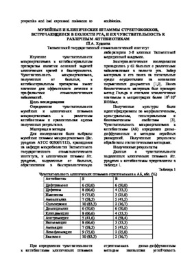 Museum and clinical strains of streptococci found in the oral cavity and their sensitivity to various antibioticsWhen determining antibiotic sensitivity museum and clinical strains of microorganisms found that the sensitivity to antibiotics of clinical strains of 1,3 time lover than the museum strains. Recommendations for antibiotics for use in the practice of medicine, as well as the assessment of their effectiveness to use not only the museum but clinical strains of microorganisms.
Stomatologiya -
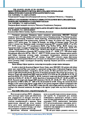 Kidney cancer regions limfatugunlarini-invasive visualization methodsIn order to check the Samarkand Regional Cancer Center, years 2002-2007, 101 patients with kidney cancer were controlled.They were treated in a specialized urology center of Samarkand Regional Cancer Center, Cancer Research Center. Among them, men 57, women -44. Male and female ratio of 1.27: 1. The cancer of right kidney had 56 patients, the cancer of left kidney-45 patients. The average age was 44.6 ± 1.4. Parakaval lymph node authenticated MRT sensitivity 90.3%, CT at 91%, the US accounted for 57.7%. CT specificity 68.6%, CT at 78.1% and MRT at -89.2%. Aortokoval lymph node identified changes in the MRT sensitivity of 92.0%, CT - 82.3%, CT - 68.0%. Specificity of ultrasound at 67.3%, CT - 84.4% and MRT was 93.4%. Paraaortic lymph node changed MRT sensitivity of 94.1%, CT - 82.3%, CT - 75%. An indication of the specificity of MRI at 95%, CT - 80% of the US accounted for 67.6%. Taking into account the above changes in renal cancer to the regional lymph nodes of non-invasive diagnostic method is recognized as the most effective method of MRT. As the MRT is better than SE, with its high sensitivity and specificity. Ac-cording to the ultrasound assessment, the changes in the regional lymph nodes may lead to the diagnostic error.
Journal problems of biology and medicine -
 THE NEW IN EFFICIENCY OF THE SECONDARY PREVENTION OF THE TEETH HYPERESTHESIA AMONG THE YOUNG PEOPLEABSTRACT Hyperesthesia of teeth among the adults is common, poorly treatable and often relapses. In a clinical study, a comparative assessment of the effectiveness of secondary prevention of dental hyperesthesia among the adults using a new domestic gel “Asepta remineralizing” was made in comparison with conventional dental and oral care products. The work was carried out on 65 young patients wrho were divided into 3 study groups taking into account the applied oral care tools. In group 1, patients used “Ascpta PLUS rcmincralization” special toothpaste. In groups 2 and 3 of the study, patients combined this toothpaste with the “Asepta parodontal FRESH” mouthwash or gel “Asepta remineralizing” (Vertex CJSC, St. Petersburg, Russia). To evaluate the results of the study, a relatively new method was used to determine the effectiveness of secondary prevention of dental hyperesthesia, which allows simultaneously measuring the severity of the course of the pathology. The experience with gel “Ascpta remineralizing” has proven to be very effective in the secondary prevention of dental hyperesthesia among the adults. It was established that among the patients, who used “Asepta PLUS remineralization toothpaste” and gel “Asepta remineralizing”, after a day the effectiveness of secondary prevention of dental hyperesthesia was 65.4%, on day 3 - 90.4%, and upon completion of clinical observation of this group - 99.04%, which allows recommending this oral care product to patients with increased sensitivity of solid teeth tissues.
Medicine and innovations -
 РАЗВИТИЕ ТРАНСФОРМАЦИИ КОММЕРЧЕСКИХ БАНКОВ В УЗБЕКИСТАНЕСовременный этап экономического развития характеризуется не только внутренней технологической модернизацией, но и необходимостью эффективных трансформации и адаптации к глобальным вызовам мира. Направление развития коммерческих банков определяет совокупность факторов внешней среды, характеризующейся на данном этапе нестабильностью и повышенными рисками. В условиях глобализации политические события и их экономические последствия, переход к новому технологическому укладу, жесткая регуляторная нагрузка и повышенная конкуренция бросают вызов всем финансово-кредитным организациям.
Priority directions, modern trends and prospects of the development of the financial market -
 CLINICAL MANIFESTATIONS OF СOVID-19 IN PATIENTS WITH CARDIOVASCULAR DISEASES AND MODERATE SEVERITY OF CORONAVIRUS INFECTION 1 AND 3 MONTH AFTER DISCHARGE FROM THE HOSPITALObjective: to investigate in patients with cardiovascular diseases (CVD) hospitalized due to moderate coronavirus infection dynamic changes in clinical symptoms manifestations of COVID-19 1 month after the discharge. Material and methods: the study included 88 patients with diseases of the cardiovascular system hospitalized for coronavirus infection. After 1 month 72 respondents continued the study. Anamnesis collection, detailed survey on clinical manifestations of the disease, Mini-mental State Examination (MMSE) mental status assessment scale was completed. Results: 1 month after discharge, there is a decrease in the number of patients with signs of respiratory system damage, such as cough, shortness of breath, chest congestion, after 1 and 3 months there is a decrease in exercise tolerance – in 80.5% vs 69.5% (out of 95.5% at the hospital stage), general weakness and increased sweating – in 69.5% after 1 month and 38.9% and 50.0%, respectively, after 3 months. 38.9% of patients noted noticeable, previously undetectable, hair loss. Prevalence of neurological symptoms were noted at the hospital stage including dizziness, severe headaches that cannot be relieved by analgesics and nonsteroidal anti-inflammatory drugs (NSAIDs), lethargy, disorientation in place and time, and in some cases even hallucinations. Some of the symptoms persist after 1 and 3 months: 55.5% of patients after 1 month and 36.0% after three months note a decrease in memory, 36% and 8.3% of respondents say they retain a sense of fear and anxiety, 63.9% and 38.9% of patients who noted problems with sleep during the COVID-19 disease, violations persist after 1 month and 3 months, respectively. Some of the respondents over the past month revealed adverse events: destabilization of blood pressure (BP) in the form of episodes of increased and decreased blood pressure during the day (36.0% vs 50,0), hypertensive crisis (14.0% vs 2,8%). Conclusion: 1 month after discharge, respiratory clinical sing and symptoms naturally decrease, but new symptoms appear, such as shortness of breath during exercise, in patients who have not previously noted breathing difficulties, fatigue, shakiness of gait, hair loss, increased sweating appeared. Within three months after discharge, symptoms of central nervous system damage persist in the form of sleep disorders, memory loss 38,9% and 69,5%, respectively, note weakness and a decrease in exercise tolerance, 50,0% continue to have increased sweating.
Journal of Cardiorespiratory Research