Library search
Search Results
-
This clinical example shows the progressive nature of the course of a combined eye injury: contusion and thermochemical
burn as a result of the explosion of the “New Year’s firecracker”, which led to the development of cicatricial, degenerative
processes in the tissues of the eyeball and eyelids. After the injury, the patient underwent three stages of emergency (according
to vital indications for the eye) surgical treatment, as a result of which it was possible to preserve the structures of the eyeball
and visual functions. At the same time, the rehabilitation period has not yet been completed. These injuries, unfortunately, are
one of the causes of low vision and blindness in children and require constant monitoring and treatment. -
Одним из факторов, способствующих развитию глаукоматозного процесса, по мнению ряда авторов, является нарушение баланса между процессами перекисного окисления липидов (ПОЛ) и состоянием антиоксидантной системы в камерной влаге глаза [1,4]. Усиление процессов ПОЛ ведет к угнетению адаптационных механизмов в структуре глаза [1,3], ухудшению микроциркуляции и кровоснабжения трабекулярной ткани глаза, способствующих снижению уровня секретируемого белка в слезной жидкости, зависящего от степени ПОУГ [2,3]. С интенсификацией ПОЛ связывают структурные и функциональные изменения глаза. Все это обусловливает необходимость коррекции нарушенного антиоксидантного статуса в глазном яблоке с использованием.
-
 The relationship between functional and anatomical-optical parameters of the eye in congenital glaucoma in children
in LibraryAim of research. To study the dynamics of the FBS of the eyes in children with congenital glaucoma by age. The studies were carried out in the clinic of TashPMI, on 132 eyes with congenital glaucoma. By age, patients were distributed according to the classification of E.S. Avetisova 2003. At the advanced stage of FBS, the eye exceeded the average statistical norm by 2.9 mm, 2.3 mm, 2.3 mm, at the advanced stage by 4.7 mm, 4.8 mm, 6.3 mm, and at the terminal stage by 7.5 mm, respectively, in each age group of eyes. An increase in the size of the anterior-posterior axis of the eye in congenital glaucoma depends not only on the violation of the hemo hydrodynamic processes of the eye with the accumulation of intraocular fluid, but also depends on the age-related dynamics of eye growth.
-
 The role of eye biomechanical parameters in the development of congenital glaucoma in children
in LibraryTug'ma glaukoma bilan og'rigan bolalarda ko'zning biomexanik ko'rsatkichlari uning turli bosqichlari bilan o'rganildi. Oddiy birlamchi tug'ma operatsiyasiz glaukoma bilan og'rigan 1 oydan 3 yoshgacha bo'lgan 20 bemor (40 ko'z) tekshirildi. Ulardan 1-guruhga (ilg'or bosqich) mos ravishda 7 (14 ko'z), 2-chi (ilg'or bosqich) - 8 (16 ko'z), 3-chi (terminal bosqich) 5 bemor (10 ko'z) kiradi. Tadqiqot usullari orasida visometriya, refraktometriya, oftalmoskopiya, optik diskni qazib olishni aniqlash, ko'zning old-orqa o'lchamini ro'yxatga olish bilan A-skanerlash, 5 g og'irlikdagi elastotonometriya; janubiy; 15d, shuningdek, Filatov-Kalf usuli bilan. Olingan ma'lumotlarning tahlili shuni ko'rsatdiki, elastokrivning ko'tarilish gradienti barcha guruhlarda qayd etilgan, ammo eng yuqori ko'rsatkich terminal bosqichi bo'lgan bolalar guruhida bo'lgan, ko'zning anteroposterior hajmi esa mos ravishda kattalashgan. kasallikning og'irligi bilan. Ko'z ichi bosimining oshishi kasallikning og'irligiga mutanosib ravishda qayd etilgan. Konjenital glaukomaning oddiy shakli rivojlanishi bilan ko'zning biomexanik ko'rsatkichlari bosqichlarga ko'ra ortadi. Shu bilan birga, tolali membrananing qattiqligining pasayishi ko'zning to'qimalarida morfologik o'zgarishlarni va glaukoma jarayonining zo'ravonligini ko'rsatadi.
-
 Optical and biometric indicators of the eye in children with juvenile glaucoma combined with myopiaPurpose. To analyze optical and biometric indicators of children's eyes with various stages of congenital juvenile glaucoma (CJG) combined with myopia. Material and methods. We examined 17 patients (31 eyes) aged 11 to 17 (averagely 14.0 ± 0.2 years) diagnosed with CYG who underwent, prior to surgical or conservative treatment, a regular ophthalmological examination supplemented with the measurement of the central corneal thickness (on an automatic contactless tonometer-pachymeter by NIDEK, USA), and the index of corneal deformation (ICD) by the Shkrebets technique. Results. The analysis showed a possible correlation between 1) tonometric intraocular pressure (Pt) and the axial length of the eye, 2) Pt and the ratio of excavation to optic disk diameter (E/ON), 3) axial length of the eye and the central corneal
in Library -
 The role of eye biomechanical parameters in the development of congenital glaucoma in children
in LibraryThe objective of this review was to study biomechanical parameters of the eye in children with different stages of congenital glaucoma. 20 patients (40 eyes) aged from 1 month to 3 years with simple primary congenital not operated glaucoma were examined. 7 patients (14 eyes) entered in the 1st group (developed stage), 8 patients (16 eyes) — the 2nd group (passed stage), 5 patients (10 eyes) — the 3rd group (end stage) respectively. Research methods included visiometry, refractometry, ophthalmoscopy, definition of the optic disc excavation, ultrasound А-scan of the anterior posterior size of the eye, Filatov’s — Kalfa elastotonometry with weights 5g, 10g, 15g.
The analysis of the data shows that gradient of the elastotonometry graphs elevation registered in all groups, but the highest was in group of children with terminal stage. Thus the anteroposterior size of the eye and intraocular pressure increase corresponding to disease severity. Biomechanical parameters of the eye increase according to the stages of a congenital glaucoma first form. The decrease of sclera rigidity shows morphological changes of the eye tissue and severity of glaucoma process. -
 Purpose: to study the interrelation between the biomechanical and the biometric parameters of the eye in different stages of infantile glaucoma. Material and methods. A total of 19 patients (37 eyes) aged from 3 to 10 years with a non-operated primary infantile glaucoma were examined. Of these 6 patients (9 eyes) had initial stage, 7 patients (9 eyes) had advanced stage), 6patients (9 eyes) hadfar advanced stage, and 7patients (10 eyes) had terminal stage, respectively. A combination of different stages of the disease was found in 11 children, while 8 children had the same stage of the disease in both eyes. Examination methods included visometry, ophthalmoscopy, determining the excavation of the optic disc, A -scan recording the anterior-posterior axis (APA) of the eye, Filatov — Kalfa elastotonometry with weights of 5g, 10g, 15g. Results. The gradient of elastic curve rise was noted in all stages, but it was the highest in the terminal stage, where APA was greater according to the severity of the disease. APA and elastic curve raise was found to correlate in the far-advanced and the terminal stages of glaucoma. Conclusion. The changes in the biomechanical properties of the fibrous membrane of the eye in children with infantile glaucoma depend on the severity of the disease, which is manifested in changes in the biometric parameters. Sclera rigidity is reduced in the far-advanced and terminal stages of infantile glaucoma, which can lead to an underestimation of the true level oflOPand the risk of glaucoma development in children.
 -
 Clinical evaluation of biomechanical features of the fibrous membrane in children with congenital glaucoma
in LibraryThe clinical, functional and biomechanical properties of the fibrous membrane of the eye in children with
primary congenital glaucoma were studied using the method of elastotonometry. The results of a study of the
clinical, functional and biomechanical properties of the eyes in 57 children with primary congenital glaucoma and
in 11 healthy children aged 8 days to 7 years in the eye department of the clinic of the Tashkent Pediatric Medical
Institute are analyzed. Research methods included clinical and functional methods and special (to determine the
rigidity of the membranes of the eye). The results of the study showed that with an increase in elastopod values,
the indicators of corneal deformation decrease as a result of pronounced corneal edema; the level of IOP in the
advanced and terminal stages indicates the weak rigid properties of the fibrous membrane of the eye. Thus, the
elastotonometry method is an objective quantitative diagnostic criterion for assessing the biomechanical proper-
ties of the fibrous membrane of the eye in children with congenital glaucoma. -
 Optical and biometric indicators of the eye in children with juvenile glaucoma combined with myopia
in LibraryPurpose. To analyze optical and biometric indicators of children's eyes with various stages of congenital juvenile glaucoma (CJG) combined with myopia. Material and methods. We examined 17 patients (31 eyes) aged 11 to 17 (averagely 14.0 ± 0.2 years) diagnosed with CYG who underwent, prior to surgical or conservative treatment, a regular ophthalmological examination supplemented with the measurement of the central corneal thickness (on an automatic contactless tonometer-pachymeter by NIDEK, USA), and the index of corneal deformation (ICD) by the Shkrebets technique. Results. The analysis showed a possible correlation between 1) tonometric intraocular pressure (P) and the axial length of the eye, 2) P t and the ratio of excavation to optic disk diameter (E/ON), 3) axial length of the eye and the central corneal thickness at the terminal CYG stage, and 4) the axial length and the refraction at the initial stage of CYG. Conclusion. As the glaucomatous process progresses, children with CYG combined with myopia show an increase of myopic refraction, a decrease in fibrous membrane rigidity, pretrabecular and trabecular changes, axial elongation, increased IOP due to an imbalance between the production of intraocularfluid and its outflow, an expansion of the excavation of the optic disk and a decrease in the central corneal thickness.
-
Миопия является одним из самых распространенных глазных заболеваний в мире и наиболее частой причиной снижения остроты зрения, встречаясь в 12-30% случаев среди офтальмопатологии. Высокий интерес к данной проблеме в последние годы связан с увеличением распространения близорукости среди населения всего мира, а именно, ее высоких степеней от -6,0 дптр и выше.
За последние десятилетия отмечен рост близорукой рефракции, особенно среди учащихся и работающих с компьютером.
Частота высокой близорукости, одной из наиболее тяжёлой рефракционной патологии, по данным разных авторов, колеблется от 6-18 до 27-33,2% [1, 41, 45, 89, 97]. В последние годы привлекает все большее внимание исследователей коррекция ослабленного зрения при близорукости высокой степени, в частности врожденной. Врожденная близорукость в 55-60 % случаев бывает высокой и является одной из главных причин инвалидности по зрению [52]. За последние десятилетия число лиц с близорукостью возросло, всего в мире очки носят около 1 млрд, человек. Близорукость чаще обнаруживается у детей 7-12 лет, усиливается в подростковом возрасте, а в 18-40 лет стабилизируется. Это очень важный аспект, т.к. прогрессирование близорукости в среднем возрасте чаще всего связано с увеличением преломляющей силы оптического аппарата глаза. В последнее десятилетие число людей с миопией в странах Европы увеличилась и составляет 30% -40% всего населения.В странах Юго-Восточной Азии достигает 70% и более. Миопия высокой степени развивается у 2% населения и является одной из необратимых причин слепоты у лиц молодого и среднего возраста. В Европе 8,8% стойкой слепоты связаны с миопией высокой степени, в США она занимает второе место среди причин инвалидности.
Низкая острота зрения у близоруких нарушает их трудоспособность, ограничивает возможность выбора профессии и полноценную жизненную активность. Коррекция миопии высокой степени с целью сохранения полноценной остроты зрения актуальна, как в социальном, так и в научном плане. Лица с близорукой рефракцией нуждаются в очковой коррекции. Наблюдения показали, что, если определенный контингент постоянное ношение очков воспринимает как огромное благо, то значительная часть близоруких лиц, особенно молодые женщины нашего региона, категорически отказываются от ношения очков, обрекая себя на зрительный дискомфорт.
Коррекция ослабленного зрения при миопии - актуальнейшая проблема офтальмологии. Она обусловлена как распространенностью миопии преимущественно у молодых лиц, то есть социальноактивного контингента, так и тенденцией к повышению степени ее в связи с возрастающей зрительной и психологической нагрузкой.
Существующие традиционные методы коррекции зрения при высокой миопии не всегда устраивают пациентов в современных сложных социально-экономических условиях.
Традиционным способом коррекции аметропий является очковая. Более 80% людей с нарушением рефракции предпочитают этот вид коррекции из-за ее доступности, отсутствием серьезных осложнений при ее использовании. Однако, очковая коррекция не всегда применима. Очки трудно переносятся при анизометропии и при высоких степенях аметропий. Есть ограничения по профессиональным состояниям больных. Нередко, даже при хорошей переносимости, они не устраивают больных по косметическим соображениям. Очковые линзы до сих пор остаются самым доступным и распространённым средством коррекции миопии. Самым серьёзным и неустранимым недостатком очковых линз является их влияние на размеры ретинального изображения объекта.Второе обстоятельство, часто заставляющее пациентов обращаться к другим видам коррекции, связано с косметическими недостатками очков. Проблема очков доводит до трагедии молодежь, особенно девушек в Узбекистане, где большинство предвзято относятся к «очкарикам». Кроме того, недостатками очковой коррекции являются: сужение полей зрения, зрительный дискомфорт, вызываемый климатическими условиями, частая смена очков (1 раз в 1 -2 года).
В случаях невозможности или непереносимости очковой коррекции альтернативой является использование контактных линз. От 15% до 18% больных с нарушением рефракции используют этот вид коррекции [35, 36]. Контактные линзы свободны от присущих очковым линзам косметических дефектов и, составляя единую оптическую систему с глазом, практически не влияют на величинуретинального изображения. Тем не менее, ряд обстоятельств (как объективного, так и субъективного характера) часто заставляет пациентов отказываться от ношения контактных линз, в частности:
- так называемые манипуляционные сложности;
- необходимость ухода за контактными линзами;
- потенциальная возможность осложнений, связанных с ношением контактных линз.
Контактные линзы - хотя и являются физиологической коррекцией, но не всегда достигается удовлетворительная адаптация к ним в индивидуальных случаях высокой миопии. Негативное влияние на переносимость и возможные осложнения (кератиты, эрозия роговицы, конъюнктивиты) оказывают особенности климатических условий в регионе Средней Азии, отличающиеся повышенной запыленностью и низкой атмосферной влажностью воздуха, из-за чего контактная коррекция не может быть широко рекомендована.
От 1 до 5 % людей с нарушением рефракции не желают или не могут использовать очки и контактные линзы и решаются на хирургические виды коррекции.
Таким образом, комплексный подход к проблеме оптической коррекции миопии высокой степени в настоящее время осуществляется по следующей схеме: очковая коррекция —► коррекция контактными линзами —» хирургическая коррекция. Каждое последующее звено в этой схеме является более сложным и применяется при безуспешности или невозможности использования более простого способа коррекции. Своевременная коррекция высокой миопии необходима для полноценного развития и функционирования зрительного анализатора, что обуславливает целесообразность проведения рефракционных операций у больных с непереносимостью очковой и контактной коррекции.
В настоящее время сложились два подхода к проблеме лечения близорукости высокой миопии - это применение хирургических методик, направленных исключительно на быстрое достижение рефракционного эффекта и более серьёзное отношение к миопии как к болезни, при которой в той или степени поражаются все структуры глазного яблока [24, 73, 106, 107].
Изменить рефракцию оптических сред глаза можно тремя путями.
Первый путь - экстраокулярный, когда дополнительная оптическая система размещается перед глазным яблоком (очки) или непосредственно на нем (контактные линзы). Этот ставший традиционным путь не предусматривает изменения структуры преломляющих сред самого глаза и может рассматриваться как неинвазивный.
Второй путь - это изменение преломляющей способности роговой оболочки дозированным хирургическим воздействием с роговой оболочки дозированным хирургическим воздействием с помощью ножа или лазерного излучения.
Третий путь - интраокулярный, который предусматривает изменение рефракции глаза с помощью операций со вскрытием глазного яблока.
Обилие разработанных к настоящему времени методов воздействия на оптику глаза и конкретных оперативных вмешательств неизбежно ставит как перед пациентом, так и перед врачом проблему выбора наиболее адекватного для каждого конкретного случая метода операции.
В настоящий момент приходится констатировать, что идеального и абсолютно безопасного метода хирургической коррекции аметропий не существует, и каждый потенциальный пациент должен быть информирован об этом. Индивидуальный выбор оптимального метода зависит от существующих в стране или регионе возможностей, как в отношении технического оснащения, так и опытности хирургов в проведении данного вида операций, степени и вида аметропии, а также возраста пациента, общего состояния его здоровья и, наконец, его финансовых возможностей.
В настоящее время арсенал хирургической коррекции аномалий рефракции огромен. Как хирургам, так и пациентам предоставлена уникальная возможность, выбирать методы и способы коррекции рефракционных дефектов [2, 3, 8, 50, 64]. Рефракционные операции - понятие собирательное и включает в себя вмешательства, которые могут изменить оптические свойства и параметры различных частей оптической системы глаз (длины оси, силу роговичной и хрусталиковой линз.)
Выбор метода хирургического лечения - актуальная и жизненно важная проблема. От того, насколько точным будет выбор, зависит исход операции, а значит, и социальная, и профессиональная реабилитация пациента. -
 Dynamics of microcirculatory changes in the retina in patients after renal transplantationVisual disorders are the main factor determining the quality of life of patients with various somatic pathologies, a large part of which is represented by kidney diseases. The most famous ophthalmic manifestations of uremia are albuminuric retinopathy with eye hemorrhage, bilateral retinal detachment, typical cotton-like foci, and a star shape in the macula. The goal is to assess the state of the organ of vision in patients with end-stage chronic renal failure after kidney transplantation. As a result of studies in patients after 6-8 months, against the background of a decrease in normalization of blood pressure, regression of angiopathy and retinopathy, restoration of nerve conduction, and improvement of visual functions were noted. In patients with end-stage CRF, kidney transplantation is the most effective and only radical method of treating the body, which leads to a pronounced improvement in microcirculation, hemo-and hydrodynamics of the eye, improvement of the functions of the organ of vision, and the condition of the fundus.
Medicine and innovations -
 The role of magnetic resonance imaging in the comprehensive radial diagnosis of volumetric masses of the eye organ
Catalog of abstractsRelevance of the problem. The difficulties of diagnostics of orbital diseases are well known. Especially difficult is intraspecies differentiation among the multitude of tumour, pseudotumour, inflammatory, vascular, endocrine and other diseases occurring here, manifested by the symptom complex of unilateral exophthalmos [Beradze I.N., 1978; Brovkina A.F., 1993].
Malignant intraocular neoplasms are the main cause of death of patients with diseases of the organ of vision, with 45-48% of patients dying from metastases in the first 5 years after enucleation [Alekseeva I.B., 1990, Barkhash S.A.1978, Brovkina A.F..1991, 1997; Keizer R.W.. Viclvoyc G.L.,1986],
Retinoblastoma is the most frequent malignant neoplasm in children. According to different authors, the frequency of its occurrence is 1 case per 14000 - 35000 newborns. [Bobrova N.F. and Vit V.V., 1993; Brovkina A.F., 1997; Provenzale J.M., et al., 1995; Skulski M., et al., 1997; Weber A.L., Mafee M.F, 1992; Wilms G., et al., 1989]. The frequency of patients with the most malignant intraocular tumour in adults - uveal melanoma has recently reached 7-9 people per 1 million population [Brovkina A.F., 1997; Kotslyansky E.O., 1989; Yushko N.A., Peskova L.I., Kalenich L.A., 1989; Peyster R.G., Augsburger J..I., Shields J.A., 1988; Romani A.. Baldeschi L., ct al 1998; Scott I.U., 1998].
The fundamental difference in treatment tactics, depending on the stage of development, size and topography of the tumour, as well as the seriousness of the prognosis in retinoblastomas and melanomas sharply increase the requirements for the accuracy of their differential diagnosis. At the same time, the number of diagnostic errors in ocular tumours continues to be 10-30% even when complex clinical and instrumental examination is applied in specialised ophthalmological centres [Ternovoy S.K., Panfilova G.V., Rogozhin V.A., 1979; Friedman F.E., Malyuta G.D., Kodzov M.V., 1995; Song G.X., 1991].
Widely used in ophthalmological practice traditional diagnostic methods (ophthalmoscopy, gonioscopy, diaphanoscopy, fluorescence angiography, laboratory tests) are insufficient to obtain comprehensive information about the localisation, nature of growth and prevalence of volumetric pathological formations of the eye and orbit. This circumstance and not quite satisfactory results of surgical treatment are the causes of high mortality of patients [Muratova T.T., Nigmanova N.H., Kozlovskaya G.M.. 1989, Naches A.I., 1980; Cheremisin V.M., Trufanov G.E., Kholin A.V., 1991]. Untimely or erroneous recognition of pathological processes of the orbit leads to a sharp deterioration of visual functions, up to blindness, and in some cases to the death of the patient [Yuzhakov A.M., Travkin A.G., Kiseleva O.A., 1991]. All this determines the importance of timely and accurate diagnosis of diseases of the orbit, on the one hand, and the difficulty of such diagnosis - on the other [Gabunia R.I., Kolesnikova E.K., Tumanov L.B., 1982].
The fact that the orbit is closed from direct inspection and palpation by bone walls and the eyeball, indicates the advantage of radial diagnostics in comparison with other methods of examination. In the arsenal of clinicians there is a great variety of methods of clinical-radial diagnostics of orbital pathology, however, at present the information in the literature about their resolving capabilities and significance in comparative aspect is incomplete and not fully studied. The priority of using one or another instrumental investigation, their sequence and expedient combination have not been determined yet. This makes it difficult to choose the optimal standardised approach for diagnosis and adequate treatment [Cheremisin V.M., Trufanov G.E., 1993, Weber A.L., Sabates N.R., 1996; Wenig V.M., Mafee M.F., 1998].
Thus, the study of these and other questions, contributing to the improvement of diagnostics and treatment of patients with neoplasms of the eye and ocular cavity, should be recognised as urgent urgent.
Purpose of the study. Comparative evaluation of magnetic resonance tomography capabilities and development of algorithms for complex radial diagnostics of volumetric formations of the visual organ. To solve this goal we set the following tasks.
1. To study the normal picture of the magnetic resonance image of the visual organ in comparison with other methods of visualisation.
2. To find out the possibilities of magnetic resonance tomography, ultrasound and computed tomography in detection and evaluation of intraocular neoplasms.
3. To determine the role and place of magnetic resonance tomography in differential diagnostics of volumetric pathological formations of the eye cavity in comparison with other radial methods of research.
4. To determine the indications and to develop an algorithm for the complex application of radiography, ultrasound, computer and magnetic resonance tomography for diagnostics of volumetric formations of the eye organ.
Scientific novelty.
The present work is the first to give a detailed and detailed description of the complex clinical and radiation examination, with generalisation and standardisation of magnetic resonance, computer and ultrasound semiotics of volumetric pathological formations of the eye and eye cavity. The conducted clinical and instrumental investigations allowed to determine the diagnostic value and resolving capabilities of each of the applied methods. The ultrasound, CT and MRI signs of volumetric formations of the eye organ were studied, clarified and supplemented taking into account the use of low-field magnetic field and general-purpose ultrasound apparatus. The developed standardised diagnostic algorithm of examination of patients with this pathology is new, thanks to which the pre-oppositional diagnosis of tumour and other diseases of the visual organ is improved and the total radiation load on the patient is reduced.
Conclusions
1. MPT will provide an opportunity to study the weight of the soft tissue and anatomical components of the ocular cavity, up to the optic nerve sheath and perineural liquor space, the orbital apex and chiasmal-sellar region, as well as to assess the condition of adjacent structures of the brain and facial skull. The method is limited in the evaluation of changes in the bony walls of the orbital cavity.
2. MRI is inferior in detecting characteristic signs of retinoblastoma (presence of calcification). The sensitivity of MRI was 66.6%, while for ultrasound and CT these values were 96.1 and 100%, respectively. But when the tumour spreads rstrobulbarly outside the eyeball (at 3-4 stages) the informativeness of MRI increases significantly. In uveal melanoma the sensitivity and specificity of MRI reaches 100%.
3. Both MRI and CT have a high detection rate (98.1% and 95.8% respectively) of benign orbital tumours of both primary and secondary origin. However, MRI is the preferred method of investigation. MRI is especially informative when a cranioorbital tumour and pseudotumour are suspected. The sensitivity of the method is 90.9% and 91.6%, respectively
4. In some cases ultrasound can be used to differentiate between encapsulated and diffuse neoplasms, which facilitates the diagnosis. However, when the pathological process is localised near the orbital apex, the diagnostic value of ultrasound decreases. In such cases it is advisable to use MRI.
5. In detection of primary and secondary malignant tumours of the orbital cavity both MRI and CT are quite informative (sensitivity 97,2% and 95,4% respectively), but the most comprehensive information about the state of bone walls will be provided by CT. When the process spreads intracranially, the value of MRI increases significantly, especially with the use of contrast enhancement.
6. The developed algorithm of complex clinical and radiation examination of patients with the use of ultrasound, CT and MRI is the most effective in the diagnosis of volumetric pathological formations of the eye and eye cavity, allowing to reduce to an adequate minimum the total radiation load on the patient and diagnostic period, excluding duplication of research techniques and choosing the most informative in each case, which in turn allows to develop appropriate treatment tactics and reduce the level of disability of the patient. -
To determine the hydrodynamic parameters of the uninjured fellow eye of children with combined injuries of the organ of vision. A prospective analysis of the hydrodynamic parameters of the fellow eye according to Friedenwald was carried out in 18 patients (18 eyes) aged 3 to 10 years 2–3 and 45–50 days after primary surgical treatment (PSD) of a penetrating wound of the cornea, who were hospitalized in the ophthalmological department of the clinic Tashkent Pediatric Medical Institute. Group I included 8 (44%) children with the following diagnosis: “Combined injury of the organ
of vision. Contusion of the eyeball severe. Complex penetrating wound of the cornea. Group II included 10 (56%) patients with complex penetrating wounds of the cornea. 2–3 days after PST of the wound, group I showed a statistically significant increase in Pt by 2.04±0.03 mm Hg. compared with the control group, while 1–2 days after the first measurement and 45–50 days after PST, the indicators decreased, on average, by 4.4±0.02 mm Hg. without the use of antihypertensive drugs. Changes in the hydrodynamics of the eye in children of group II were not statistically significant. The results of the examination of children revealed a transient increase in tonometric intraocular pressure in the paired uninjured eye 2–3 days after PST of a penetrating wound of the cornea with combined injuries of the organ of vision. -
For parents, a child of any age seems vulnerable, so adults take care of him, they want protection from all difficulties. Unfortunately, a person is not strong and some diseases are very dangerous for the life of children. Some pathologies pass quickly and affect the future life, otherwise others will significantly affect the future life of the child. In order to reduce the impact of pathologies on the child's body, it is possible to diagnose the disease in the early stages, it is necessary to identify and immediately begin treatment. Among these diseases are ophthalmic we can not do without the introduction of diseases. If a child has vision problems from an early age, it can lead to a delay in the development of the child in the future. Ophthalmic diseases the main part: eye injuries, glaucoma, cataracts, glaucoma, retinopathy, myopia, cataract diseases, etc. More than 20% of diseases in ophthalmological practice, depending on the injury, damage the orbit and the eyeball. From an eye injury, then in 13% of cases, subatrophy of the eye develops, in 25% anophthalmos may occur. As for the characteristics of injuries, 10% of children suffer from damage to the organ of vision. This leads to various pathologies of the eye, in 30-60% of cases it can lead to one- or two-sided blindness. The most important traumatic factors in children are: knives, bullets, stones and clubs, hockey sticks, spears, nails, wire, etc. Glaucoma is one of these diseases. The disease also requires special attention. Reason: Prevalence of glaucoma in children Occurs in 1:10,000-1:12,000 cases. Its share in eye pathology is 0.1%. enough. More than 75% of glaucoma cases are bilateral. In parallel, there was glaucoma in 5 to 15% among children, blind and non-blind schoolchildren. Blindness in children, the proportion of this pathology ranges from 2 to 15%. in the Russian Federation Congenital glaucoma accounts for 10.1% of childhood blindness.
-
 Differential approach to the application of biomaterials in reconstructive surgery for thermochemical burns of the eyes in children
Journal of Biomedicine and PracticeSimblefaron is a scar fusion of the posterior conjunctival surface of the eyelid with the eyeball. It often develops as a complication of thermal burns of the eyes. Eye burns are severe damage to the organ of vision and occupy a significant share (6.9-30.5%) in the structure of eye injuries. The results of the study showed that the stage-by-stage surgical plastic surgery of the anterior segment of the eye performed by us, which consists in a combination of plastics of the fornix with flaps from the automucosal cavity of the mouth (lips) and transplantation of the amniotic membrane onto the surface of the cornea and conjunctiva with the capture of the limbal zone, allows you to eliminate the defect of the cornea and maintain its transparency and also contributes to the restoration of the conjunctival arches, which can significantly reduce the likelihood of developing complications of burn injury and shorten the rehabilitation period, improve the functional results of treatment.
-
 Clinical and functional evaluation of the effectiveness of endonasal electrophoresis in combination with electrical stimulation in the complex therapy of glaucomatous optic neuropathy
in LibraryClinical and functional assessment of the results of complex treatment of optic glaucoma neuropathy using tanakan in the form of endonasal electrophoresis in combination with electrical stimulation according to optical coherence tomography of the eye and ultrasound Doppler mapping. Material and methods: 43 patients (74 eyes) with glaucomatous optic neuropathy aged from 58 to 76 years were examined. Results: The used method effectively delays the development of optic nerve atrophy and, along with the improvement of visual functions, lengthens the positive effect of the main treatment, which was confirmed by a significant improvement in hemodynamic parameters ac-cording to the ultrasound Doppler study. Conclusions: The proposed method will improve the efficiency of treatment of patients with compensated open-angle glaucoma and improve the vision prognosis and the quality of rehabilitation measures.
-
 Progressive thinning of nerve fibers and retinal ganglion cells in traumatic optic neuropathy: 2 clinical cases
in LibraryThe aim of this work was an optical coherence tomography study to assess the state of the optic nerve in contusion of the eye. In 12 patients with eye contusion, an OCT study of the retina and optic nerve of the injured and fellow eye was performed. The layer of nerve fibers and ganglion cells of the retina was measured. Thus, broad-spectrum SS-OCT allowed us to obtain a wide-spectrum image of the macula and peripapillary areas, which is useful for monitoring various sequential developmental conditions in patients with TON as well as those with TON.
-
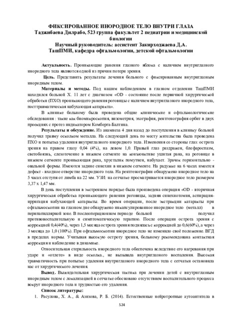
Проникающие ранения глазного яблока с наличием внутриглазного инородного тела являются одной из причин потери зрения.
-
 Возможности применения ретиналамина в терапии возрастных дистрофических заболеваний глаза
in LibraryДистрофические заболевания глаза, такие как глаукома, возрастная макулярная дегенерация, диабетическая ретинопатия, являются.основнымй причинами слабовидения и слепоты у лиц пожилого возраста. Кроме того, наблюдается тенденция к росту частоты данных заболеваний, как пожилом так и в молодом возрасте. Поэтому поиск лекарственных средств, позволяющих осуществить патогенетически обоснованную, эффективную, безопасную терапию и реабилитацию пациентов с различными заболеваниями сетчатки и зрительного нерва, является актуальной задачей современной офтальмологии. Достойное место в клинической практике занимают биогенные пептиды. Ретиналамин, входящий в эту группу, представляет собой комплекс водорастворимых полипептидных фракций.
-
Целью данного исследования явилась сравнительная оценка зрительного восприятия пациентов с артифакией в сравнении со здоровыми лицами. Материалы и методы. Нами было обследованы 48 пациентов (82 глаза) с артифакией, средний возраст которых составлял 58,6 ± 4,2 года. Критериями отбора пациентов в группу исследования были: отсутствие сопутствующей офтальмопатологии, гладкое течение операции и послеоперационного периода, ОЗ каждого глаза при выписке в среднем 0,7 и более без коррекции. Пациенты были обследованы через год после хирургического вмешательства.
-
 Эффективность интравитреального введения фортума и дексаметазона в лечении посттравматического эндофтальмитаАктуальность проблемы. Одним из наиболее опасных и грозных осложнений проникающих ранений глаза с внедрением инородного тела является развитие внутриглазной инфекции, оказывающей существенное неблагоприятное влияние на течение и исход травмы глаза. Проблема диагностики, лечения и профилактики внутриглазной инфекции постоянно находится в центре внимания офтальмологов. Актуальность данной проблемы обуславливается тяжестью развития инфекционных процессов, нередко приводящих не только к утрате зрительных функций, но и к потере самого глаза как анатомического органа
Doctor's Herald -
 Treatment of burn wounds in dogsThermal burns in animals II, III and IV degrees are a serious problem, which occupies a central place in veterinary surgery and combustiology. In farm animals burns often occur in fires in livestock buildings, and domestic animals (dogs, cats) – due to scalding with boiling water or other liquids. In areas where military operations are conducted, thermal burns are possible when using incendiary shells, incendiary compositions, etc. At the present time, despite the advances made in the treatment of burns, burn disease, mortality among the victims remains high. This is facilitated by the emergence of a painful shock and burn toxemia, as well as the development of pathogenic organisms at the surface of burn wounds as wet coagulated biological tissue at a temperature of the animal’s body, provides optimal conditions for intensive growth of microorganisms. Just be aware that the wound process in burn injury has three phases (as И.И. Кузин): an inflammation phase (melts necrotic masses and cleansing wounds from the decay products of damaged tissues and pollution), the regeneration phase (formation and maturation of the granulation tissue) and stage organization and epithelialization of scar. At various stages of the healing of burn wounds are subject to different treatment principles on which depends the choice of local treatments for burns. After providing first aid in the first phase of healing burn wounds should prevent the spread of bacterial infection, which is used antibacterials and antiseptics. Further, when the wound is cleansed of necrotic tissue, should pay attention to the impact on the healing process and to use the funds, promotes rapid tissue regeneration. Therefore, treatment should include pain relief and be directed to fighting infection and intoxication of the organism, as well as take into account the peculiarities of healing burn wounds in different phases of wound healing. Therefore, the aim of the study was to find the optimal combination of medicinal substances and drugs that meet these requirements.
Prospects for the development of veterinary science and its role in ensuring food safety -
 COMORRIDITY OF ISCHEMIC DISEASES OF THE ORGAN OF VISION AND CHRONIC BRAIN ISCHEMIA IN ATHEROSCLEROSISThe purpose of this research was to study the features of the comorbid course of ischemic diseases of the organ of vision and chronic disorders of cerebral circulation in atherosclerosis. The material for this study was the results of complex examinations of 42 patients (84 eyes) with ischemic diseases of the organ of vision in combination with chronic brain ischemia. Important in the development chronic brain ischemia and progression of ischemic diseases of the organ of vision holds the consistency of collateral circulation. So. good consistency of collateral hemodynamics eliminates ischemia of brain and eyes tissue, and suffer less visual function.
Stomatologiya -
 The crystalloscopic picture of native tears of healthy people aged 20 to 70 years is studied. In the facies of the tears of young people (20-35 years old) there are crystals forming the figures of snowflakes and chamomiles. Crystallization in the form of branches of pine or spruce is characteristic for individuals of 40 and 50 years. In individuals older than 50 years, the crystallization of tears in the form of fern stems is observed, from which secondary branches of different length and thickness leave. At the same time, the systemic nature of their branching is preserved. In this age group, in the tear facies, we did not observe crystal patterns in the form of snowflakes and chamomiles.The study showed that the crystalloscopic picture of tear fluid of healthy people of different age groups is different, which should be taken into account when studying the crystallogram of tears, both in normal and in different pathologies.
 -
 INTRАOCULAR AMMETROPIA CORRECTION WITH PHAKIC LENSESThe analysis of literature sources (database PubMed and Google scholar) concerning modern views on the use of Phakic intraocular lenses in order to correct the refractive error of the eye. The current models of these lenses are quite sophisticated and many times increase the possibilities of their use than before. With the help of PIOL it is possible to correct any degree of myopia and hypermetropia, as well as astigmatism. There are examples of using such lenses to correct presbyopia. Currently, lenses are predominantly used in the posterior chamber of the eye. In the long term (on average after 8-10 years) complications are possible in some (up to 10%) patients. When the PIOL is inserted into the anterior chamber, the development of endothelial dystrophy of the cornea or the appearance of glaucoma is possible; when the PIOL is inserted into the posterior chamber, cataracts may develop.
Journal of oral medicine and craniofacial research