Library search
Search Results
-
 STRUCTURAL AND HEMADINAMICAL FEATURES OF THE HEART IN ARTERIAL HYPERTENSION IN WOMEN DURING MENOPAUSE IN RELATIONSHIP WITH PSYCHOEMOTIONAL STATUSCharacteristics of the left ventricle of the heart in women, with arterial hypertension (AH) and climacteric syndrome in 30 patients and 30 women with AH without menopausal syndrome were studied, the control group consisted of 20 women without signs of cardiovascular pathology. In women with arterial hypertension and climacteric syndrome, a high level of personality and a moderate level of reactive anxiety prevailed. The presence of diastolic dysfunction in women with hypertension and climacteric syndrome was revealed.
Journal of Cardiorespiratory Research -
 CLINICAL MANIFESTATIONS OF ACUTE MYOCARDITIS IN CHILDREN ON THE BACKGROUND OF BRONCHO-PULMONARY DISEASESBroncho-pulmonary diseases in children is one of the urgent problems in pediatrics, which is determined by the continuing high incidence and severe prognosis, especially in young children. The aim of the study was to determine the clinical characteristics of acute myocarditis in children with acute broncho-pulmonary diseases. We examined 84 children aged 6 months to 6 years with broncho-pulmonary diseases, which we divided into 2 groups. Group I (control) included 42 children who had only respiratory complaints. Group II (main) included 42 sick children with broncho-pulmonary diseases, who had a violation of the cardiovascular system, the presence of which was confirmed by instrumental methods. The results obtained emphasize that against the background of broncho-pulmonary diseases, all symptoms of acute heart failure are masked, the cause of which in most cases is acute coronary insufficiency; changes in the heart muscle in this pathology in children increases the risk of severe unwanted heart complications.
Modern Science and Research -
 THE EPIDEMIOLOGY PREVALENCE OF RESPIRATORY ALLERGIC DISEASES IN THE BUKHARA REGIONThe beginning of the 21st century was marked by an increase in allergic diseases (AL). The prevalence of allergic diseases (more than 20% of the population) has made the allergy problem a global medical and social problem. Allergiesarecalled "diseases of civilization.". Study and analysis of the prevalence of respiratory allergic diseases in the Bukhara region.. To clarify the situation with allergic diseases in the Bukhara region, we studied the treatment of the population on the basis of outpatient records and the medical history of clinics. The total number of patients (both sexes, different age groups) was 1035 people. A patient questionnaire map was compiled. For Bukhara region, allergic diseases remain as relevant as frequently encountered other diseases. The prevalence of allergic diseases has its own characteristics and differences for almost every region, but for some it is advisable to conduct additional studies.
Medical science of Uzbekistan -
Purpose: transition of anemia chronic disease along with other chronic diseases
Materials and methods: 82 patients with chronic anemia were observed
Results: Anemia occurs in infectious diseases, non-infectious diseases, asthma, and autoimmune diseases. The frequency of anemia in chronic diseases is 100%. In terms of prevalence, anemia ranks second after iron deficiency anemia in the elderly, from 2.9 to 61% in men and 3.3 in women. from 41%. Chronic anemia occurs in half of all patients with systemic diseases of the connective tissue. In chronic diseases of the kidney, the hemoglobin level is below 100 g/l. In the treatment of anemia in this category, the main disease is treated first (in infectious diseases, antibacterial treatment against infection, basic and anti-inflammatory treatment in rheumatic diseases, surgery treatment if there is an indication (abscess in the head of the abdomen, purulent pyelonephritis, etc.
Recommending iron preparations and vitamin 12 to these patients will be ineffective because the underlying disease must be treated:
Conclusion: Anemia aggravates the course of a chronic disease if it is accompanied by other chronic diseases.
-
 Inflammatory diseases of female genital organs take the first place among gynecological diseases. There are three principles for the classification of diseases of inflammation of the female genital organs. Inflammatory diseases of the female genital organs and their treatment are described in detail in this article.
 -
For parents, a child of any age seems vulnerable, so adults take care of him, they want protection from all difficulties. Unfortunately, a person is not strong and some diseases are very dangerous for the life of children. Some pathologies pass quickly and affect the future life, otherwise others will significantly affect the future life of the child. In order to reduce the impact of pathologies on the child's body, it is possible to diagnose the disease in the early stages, it is necessary to identify and immediately begin treatment. Among these diseases are ophthalmic we can not do without the introduction of diseases. If a child has vision problems from an early age, it can lead to a delay in the development of the child in the future. Ophthalmic diseases the main part: eye injuries, glaucoma, cataracts, glaucoma, retinopathy, myopia, cataract diseases, etc. More than 20% of diseases in ophthalmological practice, depending on the injury, damage the orbit and the eyeball. From an eye injury, then in 13% of cases, subatrophy of the eye develops, in 25% anophthalmos may occur. As for the characteristics of injuries, 10% of children suffer from damage to the organ of vision. This leads to various pathologies of the eye, in 30-60% of cases it can lead to one- or two-sided blindness. The most important traumatic factors in children are: knives, bullets, stones and clubs, hockey sticks, spears, nails, wire, etc. Glaucoma is one of these diseases. The disease also requires special attention. Reason: Prevalence of glaucoma in children Occurs in 1:10,000-1:12,000 cases. Its share in eye pathology is 0.1%. enough. More than 75% of glaucoma cases are bilateral. In parallel, there was glaucoma in 5 to 15% among children, blind and non-blind schoolchildren. Blindness in children, the proportion of this pathology ranges from 2 to 15%. in the Russian Federation Congenital glaucoma accounts for 10.1% of childhood blindness.
-
 The role of magnetic resonance imaging in the comprehensive radial diagnosis of volumetric masses of the eye organ
Catalog of abstractsRelevance of the problem. The difficulties of diagnostics of orbital diseases are well known. Especially difficult is intraspecies differentiation among the multitude of tumour, pseudotumour, inflammatory, vascular, endocrine and other diseases occurring here, manifested by the symptom complex of unilateral exophthalmos [Beradze I.N., 1978; Brovkina A.F., 1993].
Malignant intraocular neoplasms are the main cause of death of patients with diseases of the organ of vision, with 45-48% of patients dying from metastases in the first 5 years after enucleation [Alekseeva I.B., 1990, Barkhash S.A.1978, Brovkina A.F..1991, 1997; Keizer R.W.. Viclvoyc G.L.,1986],
Retinoblastoma is the most frequent malignant neoplasm in children. According to different authors, the frequency of its occurrence is 1 case per 14000 - 35000 newborns. [Bobrova N.F. and Vit V.V., 1993; Brovkina A.F., 1997; Provenzale J.M., et al., 1995; Skulski M., et al., 1997; Weber A.L., Mafee M.F, 1992; Wilms G., et al., 1989]. The frequency of patients with the most malignant intraocular tumour in adults - uveal melanoma has recently reached 7-9 people per 1 million population [Brovkina A.F., 1997; Kotslyansky E.O., 1989; Yushko N.A., Peskova L.I., Kalenich L.A., 1989; Peyster R.G., Augsburger J..I., Shields J.A., 1988; Romani A.. Baldeschi L., ct al 1998; Scott I.U., 1998].
The fundamental difference in treatment tactics, depending on the stage of development, size and topography of the tumour, as well as the seriousness of the prognosis in retinoblastomas and melanomas sharply increase the requirements for the accuracy of their differential diagnosis. At the same time, the number of diagnostic errors in ocular tumours continues to be 10-30% even when complex clinical and instrumental examination is applied in specialised ophthalmological centres [Ternovoy S.K., Panfilova G.V., Rogozhin V.A., 1979; Friedman F.E., Malyuta G.D., Kodzov M.V., 1995; Song G.X., 1991].
Widely used in ophthalmological practice traditional diagnostic methods (ophthalmoscopy, gonioscopy, diaphanoscopy, fluorescence angiography, laboratory tests) are insufficient to obtain comprehensive information about the localisation, nature of growth and prevalence of volumetric pathological formations of the eye and orbit. This circumstance and not quite satisfactory results of surgical treatment are the causes of high mortality of patients [Muratova T.T., Nigmanova N.H., Kozlovskaya G.M.. 1989, Naches A.I., 1980; Cheremisin V.M., Trufanov G.E., Kholin A.V., 1991]. Untimely or erroneous recognition of pathological processes of the orbit leads to a sharp deterioration of visual functions, up to blindness, and in some cases to the death of the patient [Yuzhakov A.M., Travkin A.G., Kiseleva O.A., 1991]. All this determines the importance of timely and accurate diagnosis of diseases of the orbit, on the one hand, and the difficulty of such diagnosis - on the other [Gabunia R.I., Kolesnikova E.K., Tumanov L.B., 1982].
The fact that the orbit is closed from direct inspection and palpation by bone walls and the eyeball, indicates the advantage of radial diagnostics in comparison with other methods of examination. In the arsenal of clinicians there is a great variety of methods of clinical-radial diagnostics of orbital pathology, however, at present the information in the literature about their resolving capabilities and significance in comparative aspect is incomplete and not fully studied. The priority of using one or another instrumental investigation, their sequence and expedient combination have not been determined yet. This makes it difficult to choose the optimal standardised approach for diagnosis and adequate treatment [Cheremisin V.M., Trufanov G.E., 1993, Weber A.L., Sabates N.R., 1996; Wenig V.M., Mafee M.F., 1998].
Thus, the study of these and other questions, contributing to the improvement of diagnostics and treatment of patients with neoplasms of the eye and ocular cavity, should be recognised as urgent urgent.
Purpose of the study. Comparative evaluation of magnetic resonance tomography capabilities and development of algorithms for complex radial diagnostics of volumetric formations of the visual organ. To solve this goal we set the following tasks.
1. To study the normal picture of the magnetic resonance image of the visual organ in comparison with other methods of visualisation.
2. To find out the possibilities of magnetic resonance tomography, ultrasound and computed tomography in detection and evaluation of intraocular neoplasms.
3. To determine the role and place of magnetic resonance tomography in differential diagnostics of volumetric pathological formations of the eye cavity in comparison with other radial methods of research.
4. To determine the indications and to develop an algorithm for the complex application of radiography, ultrasound, computer and magnetic resonance tomography for diagnostics of volumetric formations of the eye organ.
Scientific novelty.
The present work is the first to give a detailed and detailed description of the complex clinical and radiation examination, with generalisation and standardisation of magnetic resonance, computer and ultrasound semiotics of volumetric pathological formations of the eye and eye cavity. The conducted clinical and instrumental investigations allowed to determine the diagnostic value and resolving capabilities of each of the applied methods. The ultrasound, CT and MRI signs of volumetric formations of the eye organ were studied, clarified and supplemented taking into account the use of low-field magnetic field and general-purpose ultrasound apparatus. The developed standardised diagnostic algorithm of examination of patients with this pathology is new, thanks to which the pre-oppositional diagnosis of tumour and other diseases of the visual organ is improved and the total radiation load on the patient is reduced.
Conclusions
1. MPT will provide an opportunity to study the weight of the soft tissue and anatomical components of the ocular cavity, up to the optic nerve sheath and perineural liquor space, the orbital apex and chiasmal-sellar region, as well as to assess the condition of adjacent structures of the brain and facial skull. The method is limited in the evaluation of changes in the bony walls of the orbital cavity.
2. MRI is inferior in detecting characteristic signs of retinoblastoma (presence of calcification). The sensitivity of MRI was 66.6%, while for ultrasound and CT these values were 96.1 and 100%, respectively. But when the tumour spreads rstrobulbarly outside the eyeball (at 3-4 stages) the informativeness of MRI increases significantly. In uveal melanoma the sensitivity and specificity of MRI reaches 100%.
3. Both MRI and CT have a high detection rate (98.1% and 95.8% respectively) of benign orbital tumours of both primary and secondary origin. However, MRI is the preferred method of investigation. MRI is especially informative when a cranioorbital tumour and pseudotumour are suspected. The sensitivity of the method is 90.9% and 91.6%, respectively
4. In some cases ultrasound can be used to differentiate between encapsulated and diffuse neoplasms, which facilitates the diagnosis. However, when the pathological process is localised near the orbital apex, the diagnostic value of ultrasound decreases. In such cases it is advisable to use MRI.
5. In detection of primary and secondary malignant tumours of the orbital cavity both MRI and CT are quite informative (sensitivity 97,2% and 95,4% respectively), but the most comprehensive information about the state of bone walls will be provided by CT. When the process spreads intracranially, the value of MRI increases significantly, especially with the use of contrast enhancement.
6. The developed algorithm of complex clinical and radiation examination of patients with the use of ultrasound, CT and MRI is the most effective in the diagnosis of volumetric pathological formations of the eye and eye cavity, allowing to reduce to an adequate minimum the total radiation load on the patient and diagnostic period, excluding duplication of research techniques and choosing the most informative in each case, which in turn allows to develop appropriate treatment tactics and reduce the level of disability of the patient. -
 Characteristics of fecale microflora for gastrointestinal diseases of newborn lambsDuring the period of colostrum nutrition, digestive disorders in lambs can be caused by dyspepsia (acute digestive disorders), wound infection and infectious diseases. At the same time, a versatile relationship of cause-and-effect factors is observed, which in some cases makes it difficult to determine the causes of these diseases. The preservation of newborn lambs and the cultivation of healthy, well-developed young animals is the basis for increasing the yield of sheep production. The main losses of young animals are due to gastrointestinal diseases, and the close functional connection of all organs and systems of the body forces us to talk not about gastrointestinal diseases, but about diseases with a predominant lesion of the digestive organs, since the entire body of lambs is involved in the pathological process In acute functional indigestion in young animals, opportunistic and toxigenic microflora is activated. Newborn animals are especially sensitive to these factors, because. their own defenses of the body are not sufficiently developed and the microecological system of the intestine is not formed. A decoction of willow bark (Salix alba L.) 2-3-fold application allows you to restore the composition of the intestinal microflora, normalize the functional activity of the intestine in gastrointestinal diseases of newborn lambs. The results of our studies indicate that a decoction of willow bark with 2-3-fold application allows you to restore the qualitative and quantitative composition of the intestinal microflora, normalize the functional activity of the intestine in newborn lambs with dyspepsia.
Prospects for the development of veterinary science and its role in ensuring food safety -
 THE IMPORTANCE OF PRODUCTION DUST IN DISEASES OF THE BRONCHI-PULMONARY SYSTEMThe article considered the relevance of harmful occupational factors - production skis, timely diagnosis of occupational disease, treatment, and Prevention, which are one of the etiologies of upper respiratory diseases. Under the influence of a harmful occupational factor, workers are more noticeable due to the increasing problem of labor intensity, quality, environmental pollution, occupational diseases. In recent years, the analysis of professional morbidity in the Republic of Uzbekistan has shown that they are often noted among those who work in the field of economy, agriculture. These include chronic bronchitis, acute and chronic poisoning from pesticides, toxic hepatitis, silicosis, dusty bronchitis, cochlear neuritis, vibration sickness, brucellosis, allergic dermatitis, etc. Available harmful and negative factors: influencer and poisonous substances in the form of gases, dust in the form of smoke, steam, allergens have a harmful effect on the respiratory system. The dry climate in Uzbekistan often causes the appearance of respiratory diseases. In most cases, eliminating the cause of diseases of the pulmonary and bronchial system may be sufficient to alleviate the patient's condition and restore strength, improve the quality of his life.
Journal of Cardiorespiratory Research -
 GOITRE IS A GROUP OF DISEASES CHARACTERIZED BY ENLARGEMENT OF THE THYROID GLANDAccording to the World Health Organization, more than 665 million people worldwide suffer from endemic goiter and other thyroid diseases. And 1.5 billion people are at risk of developing iodine deficiency diseases. Among these diseases, tuberculosis is becoming an urgent issue for most countries of the world, including Uzbekistan, in recent years.
Modern Science and Research -
 The influence of general somatic diseases on the state of the oral organs (review of the literature)
Journal of Biomedicine and PracticeThe high prevalence of major dental diseases dictates the need to find optimal means, methods of prevention and treatment, taking into account the pathogenetic mechanisms of development. There is a close connection between the pathology of the teeth and the mucous membrane with general diseases of the body, a special place among which is occupied by diseases of the digestive and circulatory systems, respiratory and blood organs, endocrine and reproductive systems, which is dueto the commonality of the main links of pathogenesis. It was determined that inflammatory and destructive periodontal diseases caused by various pathogenic microorganisms induce reactions in tissues and organs. However, the condition of the oral cavity also changes with various diseases of the internal organs. Recommendations on joint medical activity of gastroenterologists and dentists are given. So, a dentist should have information about the presence and severity of somatic pathology, compensation for these diseases, which is necessary when developing a strategy and tactics for the treatment of periodontal diseases in each specific case.
-
 VIOLATION OF BONE MINERAL DENSITY IN DISEASES OF THE STOMACH AND DUODENUMSeveral scientists partially explained in the literature that the decrease in mineral density in patients with diseases of the stomach and duodenum occurs due to antisecretory and cytoprotective drugs. However, with acid-dependent gastrointestinal diseases, i.e., with chronic diseases of the stomach and duodenum, there are practically no signs of a violation of bone mineral density. Therefore, in this study, we attempted to determine that acid- and HP-associated diseases of the gastrointestinal tract, i.e., chronic gastritis and ulcers, are important risk factors for low bone mineral density. And we set ourselves the goal of developing methods for the prevention and treatment of complications that develop in the bone tissue in these diseases.
Journal of Cardiorespiratory Research -
 Analysis of the associotion of the rs1045642 polymorphism of the mdr1 gene with the development of myeloprolifertive diseases
Journal of Biomedicine and PracticeEarly diagnosis of Myeloproliferative diseases (MPD) is one of the serious problems of oncohematological practice. MPD efers to multifactorial diseases, the development of which is influenced by both environmental factors and genetic predisposition. The study studied the association of the carriage of the genotype for the polymorphic marker encoding glycoprotein-P and the development of MPD. The homozygous T / T genotype of the rs1045642 polymorphism of the MDR1 gene is a significant determinant of the increased risk of developing MPD in Uzbekistan (P<0.05). Conclusion. Genotype association the rs1045642 polymorphism of the MDR1 gene is associated with the risk of developing MPD.
-
 Importance of modern approaches in the diagnosis of purulent-inflammatory diseases of the uterus
in LibraryIn the structure of inflammatory diseases of the internal genital organs in women, a special place is occupied by purulent-inflammatory lesions, which are the main cause of disability in women of reproductive age due to the need for surgical treatment. In 4–5% of women, purulent inflammatory diseases of the fallopian tubes and ovaries are diagnosed. The purpose of this study was to introduce and evaluate the effectiveness of new methods of treatment and prevention of purulent-inflammatory diseases of the small pelvis in women. In order to develop preventive measures for the development of purulent-inflammatory diseases of the small pelvis, we examined 240 women of reproductive age. Our evaluation of the clinic and the results of the research methods used, as well as the outcomes of the process, made it possible to distinguish two groups with purulent-inflammatory diseases of the pelvic organs - volumetric (50; 20.8%) and non-volumetric (190; 79.2%). The diagnostic research methods used in our work contributed not only to the verification of the pathological process and the assessment of the degree of anatomical changes in the focus of inflammation, but also to the implementation of dynamic monitoring of the effectiveness of therapy with an emphasis on the implementation of the proposed optimized complex for managing patients with purulent inflammatory diseases of the pelvic organs.
-
 Особенности хирургического лечения катаракты методом тоннельной экстракции катаракты (тэк) у больных сахарным диабетом (сд)Количество больных сахарным диабетом (СД) во всем мире неуклонно растет и в настоящее время насчитывается около 160 млн. больных СД, а к 2025 году, по прогнозу Всемирной организации здравоохранения, это число достигнет 380 млн. человек. Диабет занимает первое место среди причин слепоты у пациентов в возрастной группе 30-60 лет [2], среди диабетических причин 70% приходится на долю диабетической ретинопатии (ДР) и 30% на другие глазные заболевания (диабетическая катаракта, вторичная глаукома и др.). У пациентов с СД своевременная хирургия катаракты крайне необходима для оценки состояния глазного дна и своевременного проведения лазерной коагуляции сетчатки, с целью предотвращения прогрессирования ДР и, тем самым, предупреждения развития конечной стадии ДР - отслойки сетчатки, рубеозной глаукомы и, в конечном счете, полной потери зрения. Любые хирургические вмешательства у пациентов с СД сопряжены с большим риском послеоперационных осложнений, возникающих на фоне декомпенсации диабета либо развития тяжелых гипогликемических состояний. Ответ организма на оперативное вмешательство проявляется активизацией симпато-адреналовой системы, что может вызвать резкую гипергликемию, липолиз, кетогенез и протеолиз, усугубляющиеся голоданием перед и во время операции. Воздержание от еды в пред- и послеоперационном периоде представляет особую опасность для пациентов с СД в плане развития гипогликемических состояний.
Doctor's Herald -
 Liver diseases during pregnancy can be divided into liver diseases that occur only during pregnancy and liver diseases that occur in connection with pregnancy. Cirrhotic liver diseases, including cholecystic liver disease, autoimmune hepatitis, Wilson's disease, and viral hepatitis can also occur in pregnancy. Treatment of liver diseases during pregnancy requires cooperation between obstetrician-gynecologist, gastroenterologist and hepatologist. Treatment of specific liver diseases of pregnancy is usually of special importance in preventing the birth of the fetus, various complications and anomalies that may occur in the fetus. This article discusses the epidemiology, pathophysiology, diagnosis and treatment of liver diseases observed during pregnancy.
 -
 The review presents the data available in the literature on the rate of hypovitaminosis D in systemic lupus erythematosus (SLE), analyzes the associations between the clinical and laboratory parameters of the disease and the levels of vitamin D, and considers the possibilities of the therapeutic use of its metabolites. Vitamin D deficiency is a very common pathological condition that creates prerequisites for the development of a wide range of diseases. The low serum level of vitamin D may be associated with insufficient solar exposure, genetic predisposition (vitamin D receptor polymorphism), and alimentary factors and may accompany autoimmune diseases. The very recently revealed immunomodulatory properties of vitamin D are of interest with respect to the possible implication of this hormone in the pathogenesis of autoimmune (including rheumatic) diseases. A number of investigators propose to regard vitamin D as a modifying environmental factor involved in the development of autoimmune diseases. There is evidence for the association of low serum 25(ОH)D levels with a risk for some rheumatic diseases (primarily rheumatoid arthritis and SLE), their activity, severity, and prognosis, which calls for further investigation. The antiresorptive, anti-inflammatory, and immunomodulatory effects of vitamin D metabolites substantiate that the latter should be used in combination with traditional disease-modifying agents to treat chronic inflammatory diseases.
 -
In the article discussed the fungal plant diseases detected in cotton crops in our country and biological control measures against them. Studies have shown that the main pathogens of root rot in cotton are microscopic fungi Rhizoctonia solani, Thielariopsis basicola, Fuzarium spp, Pythium spp. are included.
According to the study, in terms of damage caused by seedling diseases, on average for the last 3 years, 22.6% of the cotton crop that died the highest of all diseases was caused by seedling diseases. In the second place, verticillosis mortality was 19.23%, cos sack rot was 19.1% and the lowest fusarium wilt mortality was 7.48%.
Based on the data obtained, it was concluded that the development of root rot diseases in cotton is caused by low soil and air temperatures, deeper sowing of seeds than recommended, hardening, excess moisture, poor loosening of the soil, contamination with pathogenic fungi.
-
 Characteristics of fecale microflora for gastrointestinal diseases of newborn lambsDuring the period of colostrum nutrition, digestive disorders in lambs can be caused by dyspepsia (acute digestive disorders), wound infection and infectious diseases. At the same time, a versatile relationship of cause-and-effect factors is observed, which in some cases makes it difficult to determine the causes of these diseases. The preservation of newborn lambs and the cultivation of healthy, well-developed young animals is the basis for increasing the yield of sheep production. The main losses of young animals are due to gastrointestinal diseases, and the close functional connection of all organs and systems of the body forces us to talk not about gastrointestinal diseases, but about diseases with a predominant lesion of the digestive organs, since the entire body of lambs is involved in the pathological process
Prospects for the development of veterinary science and its role in ensuring food safety -
 Опыт организации паразитологической диагностики на базе краевого специализированного учреждения инфекционного профиляПо данным ВОЗ, паразитарными болез-нями в мире поражено более 4,5 миллиарда человек. По оценке Всемирного банка, эконо-мический ущерб от кишечных гельминтозов занимает 4 место от наносимого всеми болезнями и травмами. В России ежегодно регистрируется около одного миллиона больных паразитарными болезнями. Истинное их число, по экспертным оценкам и данным выборочных обследований, превышает 20 млн. человек
Journal problems of biology and medicine -
 GENOTYPES OF SALMONELLA RESISTANCE ISOLATED FROM PATIENTS WITH OKA AND FROM BROILER CHICKEN CARCASESSalmonellosis is unmatched in its epidemiological complexity and difficulty in controlling it. The biology of salmonella serovars varies so widely that it inevitably makes it difficult to discuss issues related to salmonellosis, the ways and mechanisms of infection with Salmonella and contamination with Salmonella. At the end of the 20th century, a significant contribution to the spread of multiple resistance in many countries was made by the epidemic spread of multidrug-resistant strains of S. Typhimurium 104, which caused diseases in humans and were isolated from animals. We examined 31 samples from carcasses of broiler chickens and 40 strains of Salmonella isolated from patients with acute intestinal infections, hospitalized in the clinic NIIEMIZ MH RUz. Having carried out genotyping of Salmonella spp strains isolated from patients with AEI and from carcasses of broiler chickens, we see a variety of resistance genes, however, the resistance genotypes isolated from the carcasses of broiler chickens bla NDM-1, CTX-M, -TEM, -SHV, which amounted to 6.5 %. These strains carry a gene resistant to antibiotics belonging to the reserve group - carbopenems (imipenem).
Journal of hepato-gastroenterological research -
Subject of the inquiry: 120 patients with inflammatory diseases of the maxillary sinuses (177 affected sinuses).
Aim of the inquiry: improve the diagnosis of inflammatory diseases of maxillary sinus diseases using B-modc ultrasonography
Methods of research: Ultrasonogrpahy, X-ray, magnetic resonance imaging.
The results achieved and their novelty: Based on the results of investigation of 120 patients with inflammatory diseases of the maxillary sinuses (177 affected sinuses) the following was found: in acute sinusitis (irrespective of the form) ultrasonography had quite low specificity - 50.0+6.7%. However the sensitivity of the method in catarrhal and purulent forms of acute sinusitis came to 75.0+11.6% and 93.0+3.9% respectively. In all the forms of chronic sinusitis the parameters indicated good informativeness of B-modc ultrasonography: hyperplastic - sensitivity 82.0+5.2%, specificity 95.0+3.0%; purulent - sensitivity 88.0+6.8%, specificity 79.0+8.5%; cysts and polyps - sensitivity 87.0+5.0%, specificity 93.0+3.8%. The research demonstrated possibilities of ultrasonography in the diagnosis of inflammatory diseases of the maxillary sinuses. Diagnostic value of B-modc ultrasonograhy was shown in different forms of maxillary sinusitis. Practically convenient interpretation technique of maxillary sonograms and protocol of examination were developed. Sonographic criteria were defined for differential diagnosis of different inflammatory diseases of the maxillary sinuses as well as the role of B-modc ultrasonography in the algorhithm of radiological investigation of the maxillary sinuses.
Practical value: improvement of maxillary sinusitis, decrease of patient’s exposure to radiation (unjustified use of X-ray in the absence of pathology), avoiding of invasive methods (diagnostic puncture in case of mucosal hyperplasia of cysts) Degree of inculcate: The results of the investigation were introduced in the Otorhynolaryngology and Radiology departments of the 1-st Tashkent Medical Institute Sphere of usage: radiology, otorhynolaryngology. -
 CLINICAL FEATURES OF CHRONIC HEART FAILURE IN PATIENTS WITH IHD COURSE ON THE BACKGROUND OF ATRIAL FIBRILLATIONAtrial fibrillation (AF) is the most widespread rhythm disturbance among the population of all countries of the world. Moreover, in recent years, there has been a further increase in the prevalence of AF. The adverse effect of AF on life prognosis in patients of this profile is primarily due to a significant increase in the incidence of cardioembolic stroke and systemic thromboembolic complications. The clinical significance of atrial fibrillation (AF) is associated with a five-fold increase in the risk of stroke and a 1.5-2-fold increase in mortality in the population of patients suffering from this type of arrhythmia. Therefore, there is no doubt that the use of effective strategies for the prevention of cardiovascular complications and the development of chronic heart failure is the most important component in the management of a patient with AF. The prognosis in patients with AF with the development of heart failure (HF) significantly increases the risk of lethal cardiothromboembolism (CTHE), therefore, the diagnosis of CHF in early functional classes allows for timely enhancement of preventive antithrombotic therapy in accordance with the increased risk of CTHE. The study of the prevalence of AF among patients with coronary artery disease in the center, taking into account the clinical manifestations and characteristics of AF episodes in order to predict complications and CHF remains an urgent problem.
Journal of Cardiorespiratory Research -
STUDY OF THE ORAL CAVITY CLINIC IN E-CIGARETTE SMOKERS AND THEIR TREATMENT
European International Journal of Multidisciplinary Research and Management StudiesToday, global research on the impact of smoking on human health and the problem of creating methods necessary for the prevention, diagnosis and treatment of diseases caused by this harmful habit are among the main directions for improving public health in Russia and in the world as a whole. Despite the fact that for many years the main harmful effects of tobacco smoke components on human health have been well known, smoking remains one of the most important public health problems in many countries of the world [1]. In many previous studies, scientists have presented a strict relationship between tobacco smoking and various human diseases. Smoking is often the cause of cardiovascular diseases, malignant neoplasms, diseases of the respiratory tract, gastrointestinal tract and genitourinary system. Not only smokers, but also non-smokers are under the influence of tobacco smoke substances [1]. Inhalation of air contaminated with tobacco smoke, or "secondhand smoke", undoubtedly causes diseases in non-smokers that are characteristic of those who smoke standard medium-strength cigarettes. [This is why quitting smoking is an important medical and social issue.
-
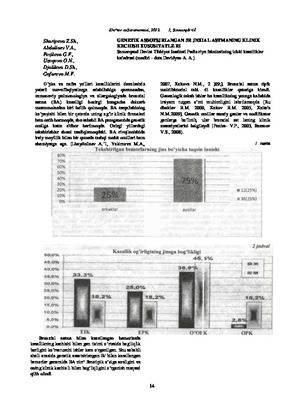 O’pka va nafas yo'llari kasalliklarini davolashda yetarli muvaffaqiyatlarga erishilishiga qaramasdan, zamonaviy pulmonologiya va allergologiyada bronxial astma (BA) kasalligi hozirgi kungacha dolzarb muammolardan biri bo'lib qolmoqda. BA tarqalishining ko’payishi bilan bir qatorda uning og’ir klinik formalari ham ortib bormoqda, shu sababli BA patogenezida genetik omilga katta e'tibor berilmoqda. Oxirgi yillardagi tekshirishlar shuni tasdiqlamoqdaki. BA rivojlanishida irsiy moyillik bilan bir qatorda tashqi muhit omillari ham ahamiyatga ega.