Library search
Search Results
-
OCT CHANGES IN MACULAR RETINAL THICKNESS DURING A-VEGF THERAPY
The American Journal of Medical Sciences and Pharmaceutical ResearchMacular edema is the leading cause of vision loss in the increasingly frequent occurrence of diabetes mellitus, a true epidemic in developed countries. Diabetic macular edema is closely related to lifestyle, obesity, smoking or high blood pressure. Macular oedema is the leading cause of vision loss in people with diabetes in developed countries, and its prevalence is directly related to the duration of diabetes. Its incidence varies from publication to publication, ranging between 7.5% and 15.2%, and is more common in patients with type 2 diabetes receiving insulin.The significance of the incidence of diabetic macular oedema is directly related to the patient's metabolic control and the presence of risk factors, as we will see below.
-
THE POSSIBILITIES OF ULTRASOUND IMAGING IN THE DIAGNOSIS OF ACUTE IDIOPATHIC SCROTAL EDEMA IN CHILDREN
The American Journal of Medical Sciences and Pharmaceutical ResearchDespite many years of experience in the use of ultrasound in the diagnosis of acute testicular ischemia (AISE) in children, the full potential of this technique remains insufficiently studied. As part of a prospective controlled study involving 142 patients with emergency genital disorders, ultrasound was performed for Acute idiopathic scrotal edema. Ultrasound showed differences in the image of the scrotum organs in boys with AISE, depending on the duration of the disease. When using color Dopplerography, changes in perfusion were detected, such as the absence or decrease in blood flow. The localization of the inflammatory process was determined solely by the results of ultrasound of the scrotum organs. The results obtained suggest that with a decrease in blood flow in the testicles with a more pronounced degree or with a longer period of torsion, pronounced changes on the part of the scrotum were observed.
-
 Possibilities of application of kinesio tape and its facial and jaw area in inflammatory diseasesSubject A review of the literature on the topical problem of maxillofacial surgery and surgical dentistry is presented - increasing the efficiency of rehabilitation о f patients with mandibular fractures using kinesio taping. The goal is to study the materials of publications devoted to kinesio taping in inflammatory diseases of the maxillofacial region. Methodology. The mechanisms of action of kinesiological tape applied in the form of applications to the skin, which lead to the creation of favorable conditions for sanogeneticprocesses, which are realized in the normalization о f microcirculation, decrease in edema and the severity of pain, are described in detail. An increase in the number of publications on the use of this method in the prevention and treatment of injuries of the musculoskeletal system in athletes has been noted. Moreover, at present, kinesio taping is also used in clinical medicine, for example, in the practice о f neurology and orthopedics. According to modem scientific research, the use of kinesio tapes in patients with chronic back pain, subacromial impingement syndrome, acute whiplash of the cervical spine can significantly reduce the severity ofpain syndrome.Results. Despue me ramer wiaespreaa use oj me kinesio taping method in sports and clinical medicine, the available literature contains a small number of works devoted to its use in maxillofacial surgery, in particular, for fractures of the mandible. The use of the kinesio taping method after the osteosynthesis of the fragments of the lower jaw made it possible to significantly reduce the level о f inflammatory edema and the intensity of the pain syndrome.Conclusions. The presented results of the literature review indicate that kinesio taping is a promising, simple, поп-traumatic method of rehabilitation after surgical treatment of mandibular fractures, -which does not have side effects and complications and significantly improves the quality of life of patients. However, scientific research devoted to the analysis of the use of kinesio taping for traumatic injuries о f the maxillofacial region is insufficient for this period.
Medicine and innovations -
DIFFERENTIATED HYPEROSMOLARY THERAPY IN CEREBRAL EDEMA IN PATIENTS WITH CRANIOCEREBRAL INJURY
The American Journal of Medical Sciences and Pharmaceutical Researchto study the pathophysiological aspects of cerebral edema and compare the effectiveness of using 15% mannitol solution and hypertonic 3.5%, 7%, 10% sodium chloride solution in the complex treatment of patients with head injury. Material and methods: 90 patients from 18 years old to 68 years old with various traumatic brain injuries and inhibition of consciousness level from 4 to 13 points on the Glasgow coma scale were examined.
Results: infusion of mannitol at the indicated dosage reduced ICP after 30 minutes by 42, 3%, and after 120 minutes it remained below the initial data by 23.9%. Infusion of a 3.5% NaCl solution already by the 30th minute led to a decrease of ICP by 48.6%, and by the end of 120 minutes the ICP remained below the initial data by 35.9%. Infusion of a 7% NaCl solution already by the 30th minute led to a decrease in ICP by 55.4%, and by the end of 120 minutes the ICP remained below the initial data by 39.9%. Infusion of a 10% NaCl solution already by the 30th minute led to a decrease in ICP by 58.4%, and by the end of 120 minutes the ICP remained below the initial data by 45.9%.
Conclusions: the decrease in ICP within 30 and 120 minutes after the introduction of hyperosmolar solutions is more pronounced with iv administration of 3.5%, 7%, 10% NaCl solution relative to 15% Mannitol in calculated dosages, which should be borne in mind in patients with concomitant cardiac and renal pathology.
-
Purpose — to determine the values of central corneal thickness (CCT) in children depending on the level of intraocular pressure
(IOP) and the stage of congenital glaucoma (CG).
Material and methods. Clinical studies were carried out in the eye department of the clinic at the Tashkent Pediatric Medical In-
stitute. The study involved 18 patients (36 eyes) aged 9 to 11 years (mean age 9.3±1.6 years) with confirmed diagnosis of CG.
All patients underwent basic ophthalmologic examination prior to surgical and conservative treatment. In addition to basic meth-
ods, axial eye length and CCT were determined using an automatic non-contact tonometer/pachymeter manufactured by NIDEK
(USA).
Results. Analysis of the obtained data showed that in initial, moderate and advanced stages of glaucoma, the CCT values were sig-
nificantly lower than the age norm values. This indicates stretching of the fibrous capsule and thinning of the cornea in glaucoma.
In terminal stage CG, the CCT values practically did not differ from the age norm, but were higher than in initial, moderate and ad-
vanced stages of the disease. The noted thickening of the corneal membrane in terminal stage may be explained by edema of the cor-
neal tissue as a result of elevated IOP.
Conclusion. The age norm values of CCT should be taken into account when characterizing the severity of glaucomatous process
in children. Compared to the age norm, the cornea is significantly thinner in children aged 9 to 11 years with initial, moderate
and advanced stages of CG, and becomes significantly thicker in terminal stage, which is associated with edema caused by ele-
vated IOP. -
 Assessment of efficiency of complex treatment of diabetic makulopatiya with use of the peptide bioregulator (retinalamin) and lazerkoagulyationResults of the laser treatment in patients with diabetic maculopathy complicated by macular edema have showed that the combined using lasercoagulation and peptid bioregulator Retinalamin contributed the resolu-tion of macular edema and allow to enhance of the visual functions. At application of traditional conservative therapy at DM only at 11,4% visual acuity, in 6 months after treatment doubtfully improved, visual acuity was authentically below initial. As a result of the combined treatment of DM by means of laser photocoagu-lation and Retinalamin's preparation visual acuity improved, with 0,140 to 0,333, changes of the area of rela-tive and absolute scotomas at DM, during treatment wasn't noted, the reached effect remains more than 6 months that speaks about stabilization of function of a retina.
Journal problems of biology and medicine -
 The degree of perivascular and pericellular edema In the cerebral hemispheres of persons who died from acute massive blood lossThe analysis of the state of perivascular and pericellular edema in the brain of people upon death from massive blood loss was carried out. It is established that the thanatogenesis of blood loss varies depending on the type of damaged vessels, as well as the frequency of injuries. With massive blood loss caused by single and especially multiple damage to the heart and great vessels, as well as multiple damage to peripheral vessels in the thanatogenesis. neuronal damage predominates. With a single lesion of peripheral vessels in thanatogenesis with massive blood loss, the role of the vascular component of the brain increases.
Doctor's Herald -
Effectiveness Of Domestic Preparation Kromoviz In Treatment Of Allergic Conjunctivites
The American Journal of Medical Sciences and Pharmaceutical ResearchPurpose: to study the efficacy and tolerability of the domestic drug cromoviz in the treatment of allergic conjunctivitis.
The state of the organ of vision in 60 patients (120 eyes) with allergic conjunctivitis was studied. Depending on the therapy, the patients were divided into two groups of homogeneous clinical manifestations. At the same time, the patients of the main group (30 patients) were instilled with the drug Cromoviz (Uzbekistan), 2 drops 4 times a day for 4 weeks. Patients of the control group (30 patients) were instilled with Aycrol according to the same scheme.
The obtained research results showed that the use of the domestic drug cromoviz against the background of basic treatment is expressed in a decrease in subjective complaints of patients and a significant clinical effect in 95.9% of cases. The revealed economic efficiency of the drug action indicates the achievement of the maximum level of therapeutic result at an acceptable price for the patient and therapeutic-prophylactic institution. Cases of side effects and intolerance to the domestic drug cromoviz were not identified in our studies.
-
 Electronic microscopic image of the myocard in diligated cardiomyopathy
Journal of Biomedicine and PracticeThis paper examines the level of electron microscopy of structural changes that develop in the myocardium as a result of dilated cardiomyopathy (DK.MP). A patient myocardial fragment from DKMP was prepared in a specific order for electron microscopy. The results show that DKMP cardiac-myocardial infarction is manifested by destructive changes in the circulatory and connective tissue, circulatory, edema, dystrophy, destruction submicroscopic structures. Dystrophy, atrophy, and destruction of organelles in the sarcoplasm of cardiomyocytcs led to the destruction of all organelles, including mitochondrial ultrastructure shrinkage, deformation and disintegration of crystals, vacuolation of the matrix, lipidization, and deposition of calcium salts. Myofibrils are characterized by deformation, tearing of sarcomeres, disordered arrangement of actin and myosin structures, separation from each other, in one of which only myosin, in the other actin is stored.
-
 To identify the causes of chronic chronic gingivitis (HCG) and the study of its clinical treatment in children of primary and secondary school age. Material and methods: under observation were 52 schoolchildren with CNG, out of 2 5 - children of younger middle age 7-11 years old - prepubertal period and middle school age 11-14 years old. Results: during a clinical examination of children with CCG found the presence of abundant dental Treatments along the edges of the gums, especially on the contact surfaces of the teeth, in hard-to-reach for cleaning. Edema, hyperemia, bleeding, dental deposits are determined. The severity of HCG occurs with the age of the child: in pre-pubertal disease, it mainly occurs at puberty, a ralized form is observed, which is explained by the hormonal organism Активация Windows Чтобы akthrhoorath Windows, ner
 -
 Anthropometric parameters of the head and maxillofacial part in children with diabetes and their connection with the principle of the golden ratioStudies have shown that morphometric parameter of the heads of the children with diabetes is larger than healthy ones. In our opinion this is due to the constant changes in the level of insulin (hormone status) in a young body, which affects the volume of the brain (cerebral edema). Parameter of the face of healthy children is larger than children with diabetes. This demonstrates the backwardness of the developing bones of the face and dental system in diabetes. And the fullness of the face is due to the accumulation of fat tissue and swelling in the area for a given pathology. Anthropometric parameters of the head and jaw face area in girls is bigger than boys in both groups. This demonstrates the backwardness of the morphometric parame-ters of the head and maxillofacial region in boys in comparison with the girls in the same age. The ratio of the upper, middle and lower parts of the face in girls of the both groups is closer to the law of the golden ra-tio, compared to boys. In the first group relationship between the parameters of face parts is more appropriate to the number of parameters or Fibonacci golden ratio compared with the second group. In the second group the size of the upper segment of the face is more than the lower segment. The transverse dimensions of the face (malar and mandibular diameter) is greater in children of the second group while the longitudinal is in the first.
Journal problems of biology and medicine -
 CHRONIC HEART FAILURE IN PATIENTS WITH EARLY RHEUMATOID ARTHRITISRheumatoid arthritis is a systemic inflammatory disease of connective tissue with a predominant lesion of small joints by the type of erosive- destructive polyarthritis of unclear etiology with a complex autoimmune pathogenesis. The disease is characterized by high disability (70%), which occurs quite early. The main causes of death from the disease are infectious complications and renal failure. Rheumatoid arthritis is widespread all over the world and all ethnic groups are susceptible to it. Chronic heart failure is a clinical syndrome in some diseases, accompanied by characteristic symptoms (shortness of breath, decreased physical activity, fatigue, edema, etc.) associated with inadequate perfusion of organs and tissues at rest or during exercise, accompanied by fluid retention in the body and its accumulation in soft tissues.
Journal of Cardiorespiratory Research -
 Особенности морфофуикциональных изменений в сосудах и тканях легких крыс при реперфузии тонкой кишки после предварительного моделирования ее острой непроходимостиAn important problem in acute intestinal obstruction and its surgical treatment is multiple organ failure, which is based on the phenomenon of "double blow" (double blow) - hyperactive response previously injured body to I more aggression. Inhibition of hepatic enteral and barriers that arise in this case is important in the initiation of lung I dysfunction. The incidence of pneumonia in these cases correlated with the severity of enteric disease. The lungs are the [ target organ, including in terms of mechanisms of bacterial translocation. The purpose of this study was to establish the | particular dynamics and sequence of morphological changes in blood vessels and lung parenchyma when restoring pa- j tency of the small intestine after a preliminary modeling of low acute obstruction. In experiments on rats it was established that the pulmonary artery with intestinal obstruction respond compensatory increase resistance in the form of | increased tone walls with narrowing of the lumen , which may evolve in order to maintain system pressure in response I to hypovolemic , which naturally occurs in such cases. Bronchial arteries respond with an increase in throughput, which I could also have a compensatory value, given the functionality of these vessels. Restoring patency of the alimentary canal after the previous 48 -hour occlusion at first does not lead to improved 1 blood circulation in the lungs and even, on the contrary, found previously frustration deepened slightly. Dilated bronchial arteries alter reactions on spasm, which maybe indicative of similar mechanisms of influence on the vascular wall I. of pulmonary and bronchial arteries. The reason for this may be just "double blow" - local hemodynamic and oxygen [ double blow in the vessels of the small intestine. Changes in the blood vessels of the lungs are secondary, and perhaps already under the influence of hemodynamic factors as well as additional toxic effects.
Doctor's Herald -
 MORPHOFUNCTIONAL STATE ADRENALS WHEN INJECTING DISTILLED WATERThe adrenal glands of intact rats and animals that received distilled water once or repeatedly through a tube did not differ significantly in weight. With a single administration of distilled water, the morphometric and histochemical parameters of the adrenal glands of rats do not significantly differ from those in intact animals. Daily administration of distilled water to control animals leads to changes in their adrenal glands in comparison with intact animals, which are expressed only in the last periods of the experiment (60, 90 days) and are manifested by the presence of mild edema of the interstitium, plethora of capillaries, as well as some thickening and coarsening of argyrophilic fibers. Consequently, the above changes in the morphological and histochemical nature may indicate a weak reaction of the adrenal glands in response to repeated administration of distilled water by the type of stress reaction.
Journal of hepato-gastroenterological research -
ASSISTANCE IN EMERGENCY SITUATIONS (DROWNING, CONNECTION AND POISONIN)
The American Journal of Applied sciencesThe article discusses the author's views on providing assistance in dangerous situations (drowning, suffocation and poisoning). Secondary drowning can also be accompanied by chest pain or exacerbation, shortness of breath, bruising of the skin and mucous membranes and the development of pulmonary edema, and the appearance of ringing in the breath, excessive foamy sputum, wheezing on the entire surface of the lungs, bronchospasm.
-
Бош мия шиши – бош мия шикастининг оғир неспецефик синдроми бўлиб, ҳушдан кетиш ва талваса хуружлари билан тавсифланади. Болаларда бош мия шиши барча инфекциялари; токсик ва гипоксик ҳолатлар; ўткир нейроинфекциялар; бош мия жароҳатлари; эпилептик статус; бош мия қон айланишининг бузилишлари; соматик касалликларда ривожланади.
-
 Clinical and morphological changes in periodontal tissues in patients with chronic glomerulonephritisThe character of clinical morphologic changes in periodontium at chronic glomerulonephritis is not enough researched till present days. Researchers have examined 47 patients with CGN. 10 patients have passed morphologic investigation, which showed that atrophic changes along with appearing of aphtaes and ulcers on mucous membrane prevailed in periodontium tissues at patients with lingering clinical course of CGN. Whereas edema and gingival hemorrhage prevailed at patients with clinical course of 1-2 years.
Stomatologiya -
 In recent years, all over the world, the problem of impaired physical development of adolescents has acquired special medical and social significance, due to the steady increase in morbidity. The spectrum of pathology of physical development is a violation of cardiac activity, accompanied by edema of the extremities, hypothalamic obesity, pathology of the reproductive system with hormonal disorders. Particular caution is caused by impaired physical development with delayed sexual development in obese adolescents - this is a functional, temporal delay in the appearance of signs of puberty by more than two standard deviations compared to the average period.
 -
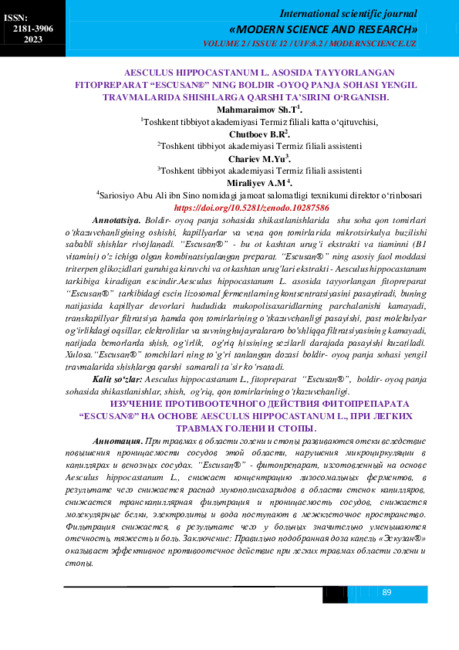 STUDYING THE ANTI-EDEMA EFFECT OF “ESCUSAN®”, A PHYTOPREPARATION PREPARED ON THE BASIS OF AESCULUS HIPPOCASTANUM L., IN MILD TRAUMAS OF THE CALF AND FOOT.In the case of injuries in the calf-paw area, edema develops due to increased permeability of blood vessels in this area, disruption of microcirculation in capillaries and venous blood vessels. Escusan, a phytopreparation made on the basis of Aesculus hippocastanum L., reduces the concentration of lysosomal enzymes, as a result of which the breakdown of mucopolysaccharides in the area of capillary walls decreases, transcapillary filtration and permeability of blood vessels decrease, low molecular weight proteins, electrolytes and water enter the intercellular space. filtration decreases, as a result of which patients experience a significant decrease in swelling, heaviness, and pain. Conclusion. Correctly chosen dose of "Escusan®" drops has an effective anti-edema effect in minor traumas of the leg and foot area.
Modern Science and Research -
 Akilov Н.А., Hakkulov Е.В., Baybekov I.M. Scanning electron microscopy of ureters in ureterohydronephrosis in children.With the aim of scanning electron microscopy different parts of ureter in reflux and obturate ureterohydronephrosis in the children has been studied. The differentials consist in preserve of chitectonics of distal parts of urelres in reflux ureterohydronephrosis wer e revealed. It was shown that a common structure of ureter is remained in this pathology. The differences in structures of distal and proximal parts of ureter consist in the changes of inflammatory character and violation of safety epithelial layer, edema, inflammatory infiltration of lower layers, especially tunica muscularis. In the proximal part of ureter the wall became thin, because all layers became more thin, especially tunica mucosa and tunica muscularis
Doctor's Herald -
 Clinical evaluation of biomechanical features of the fibrous membrane in children with congenital glaucoma
in LibraryThe clinical, functional and biomechanical properties of the fibrous membrane of the eye in children with
primary congenital glaucoma were studied using the method of elastotonometry. The results of a study of the
clinical, functional and biomechanical properties of the eyes in 57 children with primary congenital glaucoma and
in 11 healthy children aged 8 days to 7 years in the eye department of the clinic of the Tashkent Pediatric Medical
Institute are analyzed. Research methods included clinical and functional methods and special (to determine the
rigidity of the membranes of the eye). The results of the study showed that with an increase in elastopod values,
the indicators of corneal deformation decrease as a result of pronounced corneal edema; the level of IOP in the
advanced and terminal stages indicates the weak rigid properties of the fibrous membrane of the eye. Thus, the
elastotonometry method is an objective quantitative diagnostic criterion for assessing the biomechanical proper-
ties of the fibrous membrane of the eye in children with congenital glaucoma. -
 CRYSTALLOGRAPHIC EVALUATION OF EFFICIENCY IN XENOSCLEROPLASTY PROGRESSIVE MYOPIAThe problem of diagnosis and treatment of myopia continues to be one of the urgent problems of modern ophthalmology. One of the factors in the pathogenesis of progressive myopia is malnutrition and hemodynamics, as a result of a lack of blood supply to the inner membranes. Crystallographic signs of tear fluid in diabetic macular edema. Along with all kinds of conservative treatment methods, xenoscleroplastic surgeries occupy a worthy place in progressive myopia.
Journal of hepato-gastroenterological research -
GALL BLADDER POLYP IN A BODYBUILDER WITH SYMPTOMATIC UREMIA: AN EDUCATIONAL ULTRASOUND IMAGE
The American Journal of Applied sciencesBackground: Gallbladder polyps are generally small benign lesions that do not enlarge for years. However, follow-up with ultrasound examination is initially recommended to detect unexpected malignancy. The diagnosis of a gallbladder polyp is generally made with ultrasound examination, and in many instances the polyp is detected while performing the ultrasound examination in the diagnostic work-up of a condition not related to the gallbladder.
Patients and methods: The occurrence of a gallbladder polyp in a bodybuilder with symptomatic uremia is described, and an educational ultrasound image is provided.
Results: At about the age of 50 years, a professional bodybuilder presented with progressive symptomatic uremia associated with nausea, vomiting, pruritus, and mild anemia. He was not havening reduction in urine output, edema or hypertension. Renal ultrasound confirmed the chronicity of renal failure and showed small kidneys. Abdominal ultrasound also showed small polyp in the gall bladder.
Conclusion: The rare association of a gallbladder polyp in a bodybuilder with symptomatic uremia is reported.
-
 Rational use of nonsteroidal anti-inflammatory drugs
Актуальные проблемы педиатрической фармакологииThe article deals with the study effective and safe use NAID (non-steroidal and anti-inflammatory drugs at patients’ treatment). The rational use NAID will help to guarantee the safety of pharmacotherapy.
-
 Abstract. According OCT and visometry data evaluation of the effectiveness of laser photocoagulation at central serous chorioretinopathy .Three patients with central chorioretinopathy have dynamic monitoring of the retina using OCT during medical treatment and after laser coagulation of leakage zones.The use of laser photocoagulation in patients with central serous chorioretinopathy in the absence of therapeutic treatment for 1-2 months has led to the rapid recovery of the architectonics of retina, and made possible to avoid irreversible changes in the neuroepithelium, improve the quality of vision.