

American Journal of Science and
Learning for Development
ISSN 2835-2157
Volume 2 | No 5 | May -2023
Published by
inter-publishing.com | All rights reserved. © 2023
Journal Homepage:
https://inter-publishing.com/index.php/
AJSLD
Page
67
Fish Saprolegniosis (Dermatomycosis) Treatment and Prevention
Measures
Sh. Q. Baliyev
1
, R. M. Urakova
2
, N. A. Sulaymonova
3
, R. Saidalimov
4
1
Director of the Laboratory of Poultry, Rabbit, Fish and Bee Diseases v.f.f.d.
2
Junior researcher, Scientific Research Institute of Veterinary Medicine
3
Basic doctoral student, Scientific Research Institute of Veterinary Medicine
4
Intern researcher, Scientific Research Institute of Veterinary Medicine
Abstract:
The article studied the epizootic course of the disease of fish with saprolegniosis
(dermatomycosis) and explained that it was determined on the basis of clinical signs, pathological
changes and bacteriological examination. In addition, it is emphasized that in the fight against this
disease, preventive measures should be taken on the basis of veterinary and sanitary rules to prevent
the death of fish.
Keywords:
fish, water div, pathogen, Saprolegnia, suslo agar saprolegniosis, dermatomycosis,
ichthyopathology, fungus, disease.
Relevance of the topic:
Wide-scale reforms have been implemented in all areas of agriculture of our
country, especially in the fishing sector, and a number of decisions and assignments of the head of
our state serve to a certain extent in the implementation of the specified tasks in the fishing sector.
The fishing network is one of the strategic areas of ensuring food security. But in the development of
fisheries, the occurrence of fish diseases is hindering the development of the sector.
In particular, fish saprolegniosis (dermatomycosis) causes great economic damage to fisheries.
Saprolegniosis (dermatomycosis) is a disease caused by fungi, the causative agent of which is
Saprolegnia. caused by fungi that have settled in the div or internal organs of fish. In some cases,
fungi develop very quickly, like lightning. Fungal diseases are mainly found in neglected water
bodies. The disease is manifested by the appearance of white threads on the div, fins and wounds
of the fish, and then with characteristic clinical symptoms that cover a large part of the skin of the
fish like cotton.
The purpose of the study
. Diagnosing Saprolegniosis (dermatomycosis) in fish, taking measures for
their treatment and prevention, and providing practical assistance to fish farms in preventing various
fish diseases.
Research materials and methods.
Our researches were carried out in some fisheries water basins,
and laboratory testing procedures were carried out in the Laboratory of Poultry, Rabbit, Fish and Bee
Diseases of VITI. It was found that there are cases of disease and death among the fish kept in some
private households belonging to the fisheries of Payariq and Kattakurgan districts. It was found that
5 types of fish (carp, carp, white carp, white carp, African carp) are kept in these fisheries. It turned
out that the disease occurs only in carp and white bream. During our observations, clinical signs such
as the presence of thin thready white spots on the div of the fish, and the presence of white cotton-

American Journal of Science and Learning for Development
For more information contact:
mailto:editor@inter-publishing.com
Volume 2, No 5|
May - 2023
Published by
inter-publishing.com | All rights reserved. © 2023
Journal Homepage:
http://inter-publishing.com/index.php/AJSLD
Page
68
like spots on the part of the div near the injury were detected, and primary fungal disease was
suspected
.
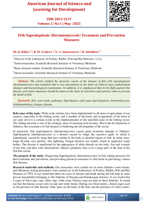
Appearance of fish infected with saprolegniosis (dermatomycosis) in 1-2 ponds and aquariums
Then, 10 live diseased fish weighing between 850 grams and 1 kg were taken from this reservoir,
filled with water from this reservoir, and brought to the laboratory. These imported fish samples
were subjected to bacteriological examination following aseptic-antiseptic rules. Initially, when the
fish were examined pathologically, the blood vessels were hyperemic, filled with fungal hyphae, the
blood vessels in the respiratory layers were tubular-like, and their walls and epithelial tissue were
ruptured. The tissue of parenchymatous organs is filled with blood, the layer of fat and glycogen is
thin. After that, for bacteriological examination of these fish, pathological samples (skin scrapings,
fins, wounds) were taken, and inoculations were planted in Glucose Saburo medium and placed in a
thermostat of 25-30 0C for growth. Planted seedlings grew as white fluffy colonies composed of
elastic undivided hyphae. In order to determine the type of causative agent, thin smears were
prepared from them on sterile, degreased glass slides, stained by the Gram method and subjected to
microscopy. For this purpose, a sterile physiological solution was dripped onto the glass of the
object, a fungal colony was removed with a bacteriological hook and mixed with a light movement,
then fixed. A 40% aqueous solution of glycerol or a liquid consisting of 1 l of 0.85% sodium
chloride solution and 0.5 l of glycerol was used as a preservative, which was titrated to rN 8.0 with
20% sodium phosphate solution and sterilized in an autoclave at 112 °C for 10 minutes. . In
bacteriological examination, isolation of pure culture of microbes, study of their morphocultural,
biochemical characteristics, determination of virulence and sensitivity to antibiotics, and accurate
diagnosis of the disease became the basis.
A reliable diagnostic method in fish mycoses is microscopic examination of the pathological
material. A 50% aqueous solution of glycerin, 0.9% sodium chloride solution, or a few drops of tap
water were used to detect the presence of fungi from swabs prepared from affected organs, based on
microscopic examination. During the bacteriological examination, the used equipment, table,
disinfectants were disinfected, pathological samples of used infected fish, dead fish in the pond were
burned and destroyed. The diseased fish were separated separately and bath methods were applied to
them as follows.
Table 1. Method of treatment of fish infected with saprolegniosis (dermatomycosis)
Т/р
The time is day
Application
to fish
time
Table
salt in
%
Methylin
cookie in
g/l
Formalin
ratio is
equal to
g/l
Copper
sulfate
ratio is
equal to g/l
The ratio of
potassium
permanganat
e is equal to
g/l
1.
2nd time 3 days apart
5-8 minutes
5
2
1 time after 3 days
12 hours
0,05
3.
1 time after 3 days
15 minutes
1:1000
4.
1 time after 3 days
60 minutes
1:200000
5
1 time
15 minutes
1:100000

American Journal of Science and Learning for Development
For more information contact:
mailto:editor@inter-publishing.com
Volume 2, No 5|
May - 2023
Published by
inter-publishing.com | All rights reserved. © 2023
Journal Homepage:
http://inter-publishing.com/index.php/AJSLD
Page
69
For this, baths were used in a 5% solution of common rock salt for 5 minutes or 0.05 g/l methylene
blue for 12 hours. In addition, formalin solutions 1:500 and 1:1000 with exposure for 15 minutes,
copper sulfate 1:200000 -60 minutes; potassium permanganate 1:100000 - for 15 minutes, 5-day
treatment courses were carried out. After that, the condition of the fish was monitored.
In addition, the following effective drugs "Griseofulvin" and "Trichopol" against fish fungal diseases
produced by "Tetra" and "Sera" companies were recommended to fish farmers. When saprolegniosis
appears, it is necessary to create optimal zoohygienic conditions in ponds, exclude damage to fish
and eggs, and check eggs in incubators. Recommendations such as disinfection of inventory and
equipment were made. In addition, once every 4-5 years, the reservoir should be left without water
until the fall of the following year, during which the sediments at the bottom of the reservoir are not
used, and if the height of the sediments in the lower part is more than 20 cm, all the lower sediments
should be cleaned mechanically. The bottom of the pool in winter and drying in summer destroys
pathogenic bacteria, helminths and other pathogenic microorganisms, intermediate hosts of parasites,
especially mollusks that remain on the bottom surface after draining the water. Veterinary-sanitary
measures should be carried out in a timely manner, providing the fish with a complete and necessary
amount of food, diagnosing the condition of the fish, treating fish ponds with quicklime (25 ts/ha) or
bleaching (3-5 ts/ha) lime, calcium hypochlorite (1 ,5-2.5 ts/ha) was recommended as disinfection.
Research results.
It was found that the fishing ponds we conducted research on are one-to-three-
year-old carp, carp, white carp, white dungpeshona, and African carp species. In the course of our
research, it was found that the fungal disease observed in carp and whitehead fish is more common
in winter and early spring, and the rate of fish mortality is high at this time. In addition, it was
observed that saprolegniosis affects fish in artificial ponds, where sanitary standards are neglected.
During the studies, it was found that Saprolegnia fungi develop in the injured areas of the fish and
have a mechanical and toxic effect on the fish. At first, thin white threads appeared on the skin, fins,
and wounds of the fish, and then it was observed that a cottony coating was formed, and this coating
was the mycelium of the fungus, and its color was white, light yellow, and purple. It was observed
that the hyphae grow first in the fish's head, mouth, nose, and eye area, then spread throughout the
div and occupy the lower parts of the skin. As a result of the increased number of zambrugs
covering the injuries, it was observed that the oxygen exchange in the div of the affected fish is
disturbed and due to the lack of oxygen, they swim to the surface of the water and take oxygen
directly from the air. Diseased fish are pale, their response to external stimuli is sharply reduced,
some are completely indifferent. It turned out that sick fish sometimes float on the surface of the
water in a prone position for some time, and they can be easily caught by hand. In the process of
pathologo-anatomical changes, clinical signs and laboratory examination of infected fish, it was
determined that Saprolegniosis is the causative disease, and treatment bath methods were carried out
.
Summary.
1. In the prevention and fight against fish diseases, it would be expedient if fish farms
work in consultation with veterinary ichthyologists.
2. Laboratory tests must be carried out to identify fish diseases. In addition, timely disinfection of
veterinary and sanitary measures in water bodies is important for the prevention of various diseases.
List of used literature
1.
Ф.Э.Сафарова, Д.А.Азимов, Ф.Д.Акрамова, Э.Б.Шакарбоев, Б.А.Қаҳрамонов Балиқлар
касалликлари Тошкент – 2019 й.
2.
Гончаров Г.Д. – Лабораторная диагностика болезней рыб. – М.: Колос, 1973. – 119 с.
3.
Э.Б.Шакарбоев, Ф.Э.Сафарова, Д.А.Азимов, Ф.Д.Акрамова Распространение лигулидозов
карпообразных рыб в водоемах северо-востока Узбекистана // Ветеринария. – Москва,
2016. - № 9. – С. 32-34.
4.
Э.Б.Шакарбоев, Ф.Э.Сафарова, Д.А.Азимов, Ф.Д.Акрамова, У.Т.Мирзаев, В.И.Голованов
Оқ амур ва оқ дўнгпешона балиқларининг гельминтозлари ва инвазияга қарши кураш чора
тадбирлари (Тавсиянома). – Тошкент, 2017. – 52 с.

American Journal of Science and Learning for Development
For more information contact:
mailto:editor@inter-publishing.com
Volume 2, No 5|
May - 2023
Published by
inter-publishing.com | All rights reserved. © 2023
Journal Homepage:
http://inter-publishing.com/index.php/AJSLD
Page
70
5.
Ларцева Л.В., Дудка И.А. Зависимость развития сапролегниевых грибов от
рыбопродуктивного качества осетровых и белорыбицы // Микология и фитопатология.
1990. Т. 24, вып. 2. С.
6.
Ларцева Л.В., Алтуфьев Ю.В. Патогенность сапролегниевых грибов для икры севрюги при
искусственном ее разведении // Гидробиол. журн. 1987.Т. 23, № 2. С.
7.
Abdig’ulomovich, M. E., & Babaqulovich, D. N. (2022, April). Dynamics of triglitsrin in blood
in different conditions. In
E Conference Zone
(pp. 202-204).


