Library search
Search Results
-
 Clinical and functional indicators of the eyes in children with pseudophakia and their mothers
in LibraryAIM: To analyze clinicofunctional and echobiometric indicators of the eyes in children with target refraction, pseudofacial myopia, and their mothers.
MATERIAL AND METHODS: In the eye department of the clinic of the Tashkent Pediatric Medical Institute, a correlation analysis of optical and echobiometric indicators was conducted in 30 children (30 eyes) with artifakia and their mothers (60 eyes). Visiometry, keratorefractometry, and ultrasound examination (A/В scan of the eyeball) were conducted. Children were examined 12-14 months after CC extraction with intraocular lens (I0L) implantation.
RESULTS: A strong direct correlation was determined between the optical power of lOLs in children and their mothers who were theoretically planned for I0L implantation of lOLs in the group that has achieved target refraction. This may indicate the possibility that the child has the same optical power as the mother and the optical power of lOLs in a child is the same as that in adults. No correlation was found between the optical power of the I0L in the eyes of children with pseudophakic myopia and maternal artificial lenses theoretically planned for implantation.
CONCLUSION: The direct strong correlations between the optical power of the I0L of children and the lenses of their mothers in the group with the target refraction achieved by this age make it possible to use the optical power of maternal lenses as a “guideline" when calculating the power of the I0L implanted in children to achieve the target refraction. The lack of correlation between the refractive powers of the I0L in children with pseudophakic myopia and the lenses of mothers may indicate that the SRKII formula with age-related hypocorrection is not adapted to calculate the I0L power in children at risk of excessive refractive enhancement after surgery.
-
 Early indicators of visiometry and refraction in children after intraocular lens implantation
in LibraryThe article presents the result of a survey of 35 patients after extracapsular extraction cataract with IOL implantation at the age of 8 months to 13 years.Patients underwent: viziometry, biomicroscopy, keratorefractometry, skiascopy, A, B-scan ultrasound, ophthalmoscopy, consultation of related specialists. The calculation of the optical power of IOL was performed using the SRK II formula. Refraction in the early postoperative period in children from 8 months to 6 years corresponded to the age range, from 6 to 13 years was presented in the form of ametropia.According to the authors , ametropia is a consequence of post-traumatic scarring of the cornea, tension of the stitches , swelling of the cornea and mistakes made at the calculation of the power of the IOL according to the formula SRK II.
-
 RESULTS OF LENSECTOMY WITH IMPLANTATION OF TWO BACK-TO-BACK IOLS IN HIGH-GRADE MYOPIAThe aim of the study was to evaluate the effectiveness of lensectomy (LE) with the implantation of two intraocular lenses (IOL) by the “back-to-back” method for high myopia using the operating navigation system “VERION. Material and methods. The study included 68 patients (114 eyes) diagnosed with high myopia, which were divided into two groups: the main group included 40 patients (58 eyes) who underwent LE with implantation of two IOLs, the control group consisted of 28 patients (56 eyes), with which the LE was performed by the traditional method. Results and discussion.During implantation of 2 IOLs, an increase in the volume of the lens bag to 1.15 ± 0.05 mm and a lesser displacement of the vitreous body occur, in contrast to a lensectomy with one IOL (LT 0.52 ± 0.02). Conclusion. Analysis of the research results allows us to recommend the removal of a clear crystalline lens with the implantation of two lOLs using the back-to-back method with high myopia, with the aim of creating stability of the anatomical and topographic relative position of intraocular structures and the possibility of reducing the risk of retinal detachment, in contrast to the traditional method of surgical treatment.
Medicine and innovations -
This review presents a variety of views on cochlear implantation. The review showed that promising areas are the introduction of fully implantable cochlear implantation systems; development of new strategies for speech coding, improvement of sound processing technologies with microphones, as well as the search for new surgical approaches when installing a cochlear implant.
-
Recent studies indicate the prevalence of myopic refraction in children with pseudophakia, which significantly reduces the functional results of treatment and may be an indication for replacing the intraocular lens (IOL). Therefore, studies conducted to achieve the target refraction in children with pseudophakia are relevant.
Purpose — to determine the risk factors for the prognosis of myopic refraction in children after extraction of congenital cataract and IOL implantation.
Material and methods. The study presents the results of refraction examination in 69 (110 eyes) children aged 1 to 12 years 36 months after extraction of congenital cataract with implantation of soft IOL.
Results. The obtained data of anamnesis, results of ophthalmological, echobiometric, clinical and laboratory studies were subjected to statistical processing assessing the significance of differences in outcomes depending on the impact of possible risk factors for the development of pseudophakic myopia; a regression logistic model and a ROC-curve were constructed.
Conclusion. According to the authors, reliable risk factors for the development of pseudophakic myopia in children can be such indicators as axial eye length at the time of IOL implantation exceeding the age norm by more than 0.2 mm; the child from the first pregnancy; the AL/CR ratio of 23.0; myopia on the paired eye; strabismus of more than 4 prism diopters; hereditary load; tension of eye's fibrous capsule: pressure of SI 80 mm Hg at the time of IOL implantation. The presented reliable factors, as well as a combination of less significant signs (the child being outside for less than 1 hour per day, intermarriage of the patients parents, near-sight visual loads ot more than 3 hours per day, the blood Ca level of less than 1.8 mmol/L) can be used for prognosis of the development of pseudophakic myopia and to help make adjustments in the management tactics for patients to achieve target refraction.
-
 Clinical efficacy of individual intraocular lens calculation in children with congenital cataract at risk of abnormal refraction
in LibraryTo assess the clinical efficacy of the SRK II formula with a correction factor Rm in children with congenital cataracts who are at risk of pseudophakic myopia. Material and methods. A complex examination of 48 children (86 eyes) with congenital cataracts involved visometrics, tonometry, tonography, biomicroscopy, keratorefractometry, ophthalmoscopy, ultrasonography, and pachymetry. To determine the IOL power, we used the SRK II formula supplemented with the individual correction factor Rm, proposed by the authors. The examined children were divided into 2 groups. The main group 1 included 22 patients (42 eyes), for which the IOL power was calculated with the Rm factor. The control group 2 consisted of 26 patients (44 eyes) for which the IOL power was calculated according to the traditional SRK II formula using age-related hypocorrection of refraction but without the Rm coefficient. Results. The correction factor Rm, allowed us to achieve the targeted refraction in children who were at risk of developing pseudophakic myopia in 83.3 % of cases of the main group (versus 45.4 % of the control group cases) and reduce the development of high age-related refraction) by 37.9 %. In children of the main group, visual acuity reached, on average, 0.5 ± 0.001, while in the control group it was also higher but only reached 0.200 ± 0.001. Conclusion.The method of calculating the IOL optical power involving an individual correction factor Rm, according to the formula: SRK II – R – Rm can be recommended for clinical practice focused on children at risk of abnormal refractogenesis.
-
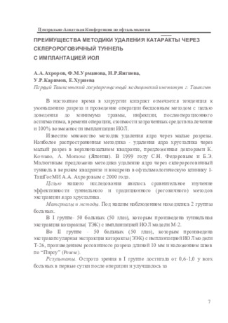 Преимущества методики удаления катаракты через склеро роговичный тоннель с имплантацией иол
in LibraryВ настоящее время в хирургии катаракт отмечается тенденция к уменьшению разреза и проведение операции бесшовным методом с целью доведения до минимума травмы, инфекции, послеоперационного астигматизма, времени операции, стоимости затраченных средств на лечение и 100% возможности имплантации ИОЛ. Известно множество методик удаления ядра через малые разрезы. Наиболее распространенная методика - удаления ядра хрусталика через малый разрез в верхненазальном квадранте, предложенная докторами К. Kowano, A. Momose (Япония). В 1999 году С.Н. Федоровым и Б.Э. Малюгиным предложена методика удаление ядра через склеророговичный туннель в верхнем квадранте и внедрена в офтальмологическую клинику I-ТашГосМИ А.А. Ахроровым с 2000 года.
-
 ASSESSMENT OF THE POSSIBLE RISK OF DENTAL IMPLANTATION ACCORDING TO MORPHOLOGICAL CRITERIA IN PATIENTS WITH BACKGROUND SOMATIC PATHOLOGYAmong dental patients, more often than others, people over 55-60 years old need prosthetics, and they have a history of somatic diseases. Correction of immune homeostasis have an important place in the complex treatment of dental implant sin patients of a specified age. On the material of 24 patients aged 50 to 65 years, morphological methods of the study analyzed the results of dental implantation in patients with diabetes mellitus. It was revealed that immunocompetent mucosal immunocytes reflect the success of dental implantation measures and can be used to predict the effectiveness of therapeutic measures.
Journal of oral medicine and craniofacial research -
 The main rules and principles of intraoperative direct prosthetics, the key to success in immediate implantation
in LibraryAccording to the Protocol of intraoperative direct prosthetics (IDP), the immediate functional load is provided by temporary orthopedic structures. The Protocol significantly reduces the treatment time and the need for preliminary pre-prosthetic reconstructive surgery-bone transplantation methods and allows you to bypass areas where transplants are ineffective. The orthopedic Protocol is simplified as much as possible. The possibility of prosthetics without the use of cement fixation of prostheses is realized. Protocols of intraoperative direct prosthetics for implant-prosthetic rehabilitation of patients require prototyping and designing of treatment results.
-
Имплантация зубов – самое актуальное, перспективное и востребованное направление в современной стоматологии и протезировании. По данным статистических исследований, распространенность дефектов зубных рядов достигает 80% у населения трудоспособного возраста Узбекистана. Данные показатели свидетельствуют о высокой нуждаемости населения в ортопедической и хирургической помощи. Именно поэтому дентальная имплантация столь актуальна в настоящее время. Несмотря на множество преимуществ, быстрое развитие и совершенствование технологий, современная имплантация имеет существенный недостаток – осложнения в послеоперационном периоде. В результате чего, вопрос о разработке профилактических мероприятий по снижению риска осложнений в послеоперационный период при проведении зубной имплантации имеет большое значение.
-
 Peculiarities of the course of the intra- and postoperative period in conditionally frequently ill children after iol implantation
in LibraryTo study the results of IOL implantation in children with frequent respiratory diseases - conditionally frequently ill children (FCCI) taking into account the parameters of the composition of the chamber moisture of the eye. Material and methods: A retrospective and prospective analysis of the case histories of 50 children (50 eyes) aged from 1 to 5 years, who were treated in the eye department of the TashPMI clinic, was carried out. All patients underwent ophthalmological, clinical and laboratory studies: biochemical studies of blood and chamber moisture of the eye (EC). Results: The children were divided into 2 groups: the 1st group - 28 UCBD, the 2nd group (control) - 22 patients with no pathology from the somatic status. In patients of the 1st group, intraoperative complications occurred 1.8 times more often than in patients of the 2nd group, postoperative complications - 2.5 times more often. Of the late postoperative complications in patients of the 1st group, there was fibrosis of the posterior lens capsule (61%), poste-rior synechia (18%), and IOL dislocation (14%), which were indications for repeated surgical interventions. Conclu-sions: UCBD has a higher percentage of early postoperative inflammatory and late proliferative reactions. In patients of the 1st group, a significant increase in the protein content in the chamber moisture and a significant decrease in the protein level in the blood before cataract extraction were also revealed.
-
 Features of the course of the post-operative period in frequently affect children after extraction of congenital cataract
in LibraryThe main link in the treatment of congenital cataracts (VK) is the extraction of the lens with the
subsequent correction of ametropia by various methods, one of which is the implantation of an
intraocular lens (IOL). The aim of the study was to study the features of the course of the postoperative period in frequently ill children (FBI) after VC extraction. We examined 35 children
(59 eyes) with VK who are undergoing surgical treatment in the eye department of the clinic of the Tashkent Pediatric Medical Institute. A comparative analysis showed significant changes in the hydrodynamic parameters of the eyes and a high percentage of intra- (hyphema in 71%, fibrin
reaction in 59%, prolapse of the vitreous in 47% of cases) and postoperative complications (corneal
edema in 36 %, inflammatory exudative reaction in the anterior Chambers: 1+, 2+ in 64% of cases) in FWB compared to children who are not in the category of frequently ill. The revealed feature
indicates the need for adequate management of the preoperative and postoperative periods in
Frequently ill children -
 HEMATOLOGICAL INDICATORS OF EXPERIMENTAL STUDY OF CHRONIC TOXICITY OF DOMESTIC PASTE-LIKE COMPOSITEThe emergence of osteoplastic materials and new methods of bone regeneration not only solve the problem of the volume and density of the jaw bones, but also significantly improve the results of implantation. The ami of the research is search and development of new domestic osteoplastic materials remains relevant. The results of acute toxicity detection allow us to classify the domestic pasty composite material as a group of practically non-toxic materials according to the International classification of material toxicity.
Stomatologiya -
 In patients with diabetes, any operation of a particularly open sinus lifting with single-step dental implantation is dangerous complications. In patients with diabetes mellitus taking into account risk factors, the actuality of the problem is to prevent purulent-inflammatory complications and optimize the conditions of reparative regeneration. We have examined 46 patients with diabetes mellitus at the age of 20 to 65 years old with defects in dentition and atrophy of the alveolar process of different degrees, which were performed dental implantation with bone grafting using the closed sinus lifting method. Tactics of preoperative pharmacological preparation of the patient and development of less traumatic operating approach with aheightening of the alveolar process of the upper jaw by the method of subantral augmentation (closed sinus lifting) with single-step installation of the implant is a solution to the above problems.
 -
 MORPHOLOGICAL FEATURES OF THE PROCESS OF DECIDUALIZATION AFTER SPONTANEOUS ABORTION IN WOMEN WITH ANTIPHOSPHOLIPID SYNDROMEIn the structure of obstetric diseases, a violation of the process of endometrial implantation occurs due to many diseases. The endometrium is a rare functional and morphological target structure that leads to disruption of the decidual process during pregnancy as a result of the interaction of hormones, autoimmune processes caused by its damage.
Doctor's Herald -
The paper presents the results of extraction of congenital cataract with IOL implantation in 50 (86 eyes) children aged 7 months to 12 years. In the examined patients, intraoperative complications were noted, such as posterior capsule rupture (8%), hyphema (6%), postoperative: fibrin effusion on the IOL (7%) and corneal edema (4.6%). With congenital bilateral cataracts after treatment, visual acuity increased on average from pr.l.certae to 0.1; with unilateral - from pr.l.certae to 0.01.
-
 Factors influencing target refraction in children with pseudophakia after extraction of congenital cataract
in LibraryThe article describes the factors affecting the target refraction of pseudophakic eyes of children after extraction of congenital cat-
aracts. The factors include features of the echobiometric parameters of the eye, refraction, comorbidity of congenital cataracts and
ocular pathologies, margins of error in calculating strength of the intraocular lens, localization and structure of the artificial lens,
as well as correction of obscure or refractive amblyopia in pseudophakic eyes. Development of the algorithm for correction of re-
sidual refraction of pseudophakic eyes in children both before and after IOL implantation with consideration of each of those fac-
tors currently remains a relevant problem. -
 Functional results low-invasive by energy industry with implantation of IOL at 40 patients with senile cataract are studied. Visual acuity at all patients at receipt fluctuated from the correct projection of light to 0,05. Before operative measure of 30 patients (75%) distinguished only light with the correct svetoproyektion, at 10 - visual acuity has made 0,02-0,05 (25%). Within the first week after operation visual acuity has increased to 0,3 at 21 patients (52,5%), 0,4 - at 12 (30%). The maximum visual acuity without correction which managed to achieve as a result of operation, has made 0,8-0,9 at 7 patients - 17,5%.
 -
Каждый год более чем одному миллиону человек выполняют хирургическое лечение по поводу катаракты (помутнения хрусталика, в норме прозрачной линзы, расположенной внутри глаза). В настоящее время в большинстве случаев, после проведения операции по поводу удаления катаракты, для восстановления зрения используются искусственные хрусталики (ИОЛ) или искусственные внутриглазные линзы. Имплантация факичной линзы производится через разрез роговицы длиной 3.03.5 мм. Настоятельно рекомендуется проведение интраоперационной или предварительной иридэктомии. Для того, чтобы убедиться, что не произошло повышение внутриглазного давления, пациента осматривают через несколько часов после операции. Обязательно наготове должен быть набор для экстракции катаракты, если произойдет интраоперационное повреждение хрусталика.
-
Cataract in children, which is one of the leading causes of primary blindness, is a clouding of the lens. The prevalence of cataracts in developed countries, as well as in Russia, is 1.6–2.4 per 100,000 children. Intraocular lens (IOL) implantation has become widespread in recent decades and is considered the most optimal method for correcting aphakia. Despite the introduction of new high-tech methods of surgical treatment of congenital cataracts, there is currently a fairly high percentage of complications. All of the above does not reduce the urgency of the problem of treating children with congenital cataract (CC) and requires further research.
-
Причины нарушения слуха у ребенка могут быть врожденными или приобретенными, возможны и сочетания различных факторов. Устойчивые снижения слуха приводят к критическим нарушениям психоречевого развития, вплоть до полного отсутствия речи с нарушениями интеллекта. Распространенными причинами потери слуха являются генетические дефекты у новорожденных, ушные инфекции и серная пробка у детей. Генетической предрасположенностью вызваны около 40% случаев тугоухости у детей. Если в семье были слабослышащие люди, есть вероятность того, что заболевание передастся на уровне генов.Ослабленные болезнью малыши чаще других страдают от порока развития уха или слухового нерва. Поэтому здоровью детей, которые родились недоношенными или с маленьким весом, либо переболели желтухой в раннем возрасте, нужно уделить пристальное внимание.
-
Purpose: comparative evaluation of the clinical effectiveness of dental implantation (EMS, Switzerland) in the complex treatment of patients with peri-implantitis and peri-implant mucositis and traditional methods. Material and methods: the results of treatment of para-implantation inflammatory diseases in 17 patients aged 24-56 years, including 9 men and 8 women (main group), with terms of using implants from 1 to 7 years, were studied. Results: a comparative analysis of the two methods in terms of PIC depth reduction revealed a statistically significantly higher efficiency of the research method compared to the control (p<0.01). After 3 and 6 months from the start of treatment, the research method led to a more significant reduction in the depth of PIC compared to the method used in the control group, which we explain by the better restoration of the biocompatibility of the tissues of the peri-implant pocket during aquakinetic treatment due to the destruction of the biofilm compared to the traditional method. Conclusions: a schedule of dynamic monitoring of patients with dental implants: one month after implantation, then every 3-4 months, with stable remission - 2-3 times a year. If necessary, light probing is carried out, and radiography is used to assess the level.
-
 MORPHOMETRIC CHARACTERISTICS OF THE ULTRASONIC ANATOMY OF THE YELLOW SACKS AS A FACTOR OF FETAL LOSSThe article provides data on the study of the size of the yolk sac of pregnant women with antiphospholipid syndrome up to 8 weeks of pregnancy. Studied ultrasound anatomy, the nature of development in the early gestational period. In case of a violation of pregnancy, the yolk sac in the first trimester is characterized either by other sizes, or not determined. The diameter of this extraembryonic organ is one of the criteria that allows you to assess the state of the developing fetus.
Doctor's Herald -
 Increasing the height of the alveolar process of the upper jaw when performing a sinus lift operation with simultaneous implantationOne of the factors prevented from the placement of dental implants is the severe form of atrophy. After extraction of teeth in lateral part of upper jaw bone resorption is caused by pneumatization of maxilla sinus. The placement of dental implants in this part is possible by combination of operation sinus-lifting and bone tissue placement.
Stomatologiya -
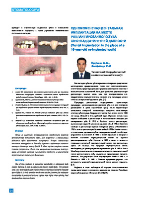 Dental Implantation In the place of a 16-year-old re-implanted tooth Tooth replacement Is о promising tooth-preserving operation, with Inconclusive conservative, endodontic treatment. Although Infrequent resort to re-Implantatlon, this possibility must be considered among other treatment options. Most cases of resorption of the Implanted tooth are diagnosed In the first 2-3 years after the re-Implantatlon, however, resorption can occur even after 5 or 10 years or more. According to our results and reviews, the literature shows that re-Implantatlon Is a reliable and predictable procedure used to preserve the natural dentition. This operation can be carried out much more often than at the present time. The tooth replacement not only preserves the tooth, but also the bone tissue of the alveolar process, preventing atrophy and deformation. Also, the reconstructed root part can serve as a bone support for dental Implants. Considering the possibility of dental Implantation on the site of Implanted teeth, this tactic Is recommended for wide Introduction Into practical dental Implantology.