Library search
Search Results
-
 Сlinical and functional results of the primary intraocular lens implantation in children with cataracts of various etiologies
in LibraryThe study showed that 66 chi/dren aged from 8
months to 13 years with cataracts of different etiology
showed high efficiency in the primary implantation of
soft spherica/ monohlock intraoci1lar /ensf!S. Тhе high-
est rates of viziometry after treatment were observed in
chi/dren with congenital bl/ateral and traumatic cata-
racts. After _treatment, children with uveal and uni/at-
era/ congenital cataracts had relative/y /ow visua/ acu-
ity va/ues. -
 Features of the course of the post-operative period in frequently affect children after extraction of congenital cataract
in LibraryThe main link in the treatment of congenital cataracts (VK) is the extraction of the lens with the
subsequent correction of ametropia by various methods, one of which is the implantation of an
intraocular lens (IOL). The aim of the study was to study the features of the course of the postoperative period in frequently ill children (FBI) after VC extraction. We examined 35 children
(59 eyes) with VK who are undergoing surgical treatment in the eye department of the clinic of the Tashkent Pediatric Medical Institute. A comparative analysis showed significant changes in the hydrodynamic parameters of the eyes and a high percentage of intra- (hyphema in 71%, fibrin
reaction in 59%, prolapse of the vitreous in 47% of cases) and postoperative complications (corneal
edema in 36 %, inflammatory exudative reaction in the anterior Chambers: 1+, 2+ in 64% of cases) in FWB compared to children who are not in the category of frequently ill. The revealed feature
indicates the need for adequate management of the preoperative and postoperative periods in
Frequently ill children -
 Crystallographytearfluidin patients with age-related cataractA crystallographic study of tear fluid was performed in 20 healthy and 24 patients with age-related cata-ract. Crystallization of the tear fluid of patients with age-related cataract occurs in the same way as in healthy individuals, and is characterized by the formation of continuous figures in the form of snowflakes, daisies or fern branches
Journal problems of biology and medicine -
 The comparative characteristic of novocaine and lidocaine in the conductive anesthesia in ophthalmologyWe examined 49 patients with a diagnosis of cataract (acquired) were operated under local anesthesia. All patients used the retrobulbar blockade and siege of the facial nerve. Used local anesthetics lidocaine and procaine. The duration of anesthesia in patients with lidocaine is 2-3 times greater than in group 1 patients with novocaine
Journal problems of biology and medicine -
 The effectiveness of the operation of phacoemulsification with intra ocular lenses implantation with mature senile cataractFunctional outcomes were studied surgery phacoemulsification with IOL implantation in patients with senile cataract. We performed an analysis of surgical treatment of 22 patients (22 eyes) with this eye disease. Visual acuity at delivery ranged from the correct projection of light to 0.01. By the degree of core density of 54.5% (12 eyes) - had grade 2, 36.4% (8 eyes) grade 3 and 9.1 % (2 eyes) - 4 degree very dense cataract are drilling core. Discharge of patients from hospital acuity of 0.6-1.0 was observed in 45.4% cases (10 eyes), 0.4-0.5- in 31.8% cases (7 eyes). 0.2-0.3- in 22.7% cases (5 eyes). Poor eyesight in the postoperative comorbidity explained by the retina.
Doctor's Herald -
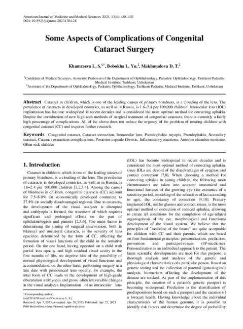
Cataract in children, which is one of the leading causes of primary blindness, is a clouding of the lens. The prevalence of cataracts in developed countries, as well as in Russia, is 1.6–2.4 per 100,000 children. Intraocular lens (IOL) implantation has become widespread in recent decades and is considered the most optimal method for correcting aphakia. Despite the introduction of new high-tech methods of surgical treatment of congenital cataracts, there is currently a fairly high percentage of complications. All of the above does not reduce the urgency of the problem of treating children with congenital cataract (CC) and requires further research.
-
 Clinical and functional aspects of correction of ametropia in children after extraction of congenital cataracts
in LibraryCongenital cataracts occupy a significant place in the structure of blindness and low vision and are one of the main causes of visual disability since childhood. Currently , cataract in children is one of the urgent problems of pediatric ophthalmology , given its fairly high prevalence and significant role in the structure of blindness and low vision . Due to clouding of the lens, the development of the visual analyzer is disrupted and amblyopia is formed, the treatment of which requires significant and lengthy efforts on the part of ophthalmologists and parents. Among the causes of blindness in children, the share of congenital cataracts varies from 7.5% (in developed countries) to 27.4% (in socially disadvantaged regions). The prevalence of cataracts in developed countries, as well as in Russia, is 1.6-2.4 per 100,000 children [25.].
-
 Medical protection of psychoemotional stress in elderly patients with accompanying hypertensive disease in optalmosurgeryAmong them were 38 women (46.3%) and 44 men (53.7%). The age of patients was 60 to 75 years, the duration of arterial hypertension (AH) was 10 to 23 years. Preoperative preparation of patients with concomi-tant GB, as well as subsequent hemodynamic monitoring is described. The results of the study suggest that ad-equate antihypertensive therapy in combination with sedation and neuroleptanalgesia in the wards of waiting allows achieving stabilization of the clinical state of patients, clearly correlating with improvement of hemody-namic parameters
Journal problems of biology and medicine -
 Clinical and functional indicators of the eyes in children with pseudophakia and their mothers
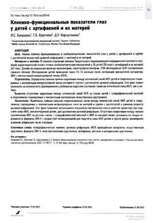
in LibraryAIM: To analyze clinicofunctional and echobiometric indicators of the eyes in children with target refraction, pseudofacial myopia, and their mothers.
MATERIAL AND METHODS: In the eye department of the clinic of the Tashkent Pediatric Medical Institute, a correlation analysis of optical and echobiometric indicators was conducted in 30 children (30 eyes) with artifakia and their mothers (60 eyes). Visiometry, keratorefractometry, and ultrasound examination (A/В scan of the eyeball) were conducted. Children were examined 12-14 months after CC extraction with intraocular lens (I0L) implantation.
RESULTS: A strong direct correlation was determined between the optical power of lOLs in children and their mothers who were theoretically planned for I0L implantation of lOLs in the group that has achieved target refraction. This may indicate the possibility that the child has the same optical power as the mother and the optical power of lOLs in a child is the same as that in adults. No correlation was found between the optical power of the I0L in the eyes of children with pseudophakic myopia and maternal artificial lenses theoretically planned for implantation.
CONCLUSION: The direct strong correlations between the optical power of the I0L of children and the lenses of their mothers in the group with the target refraction achieved by this age make it possible to use the optical power of maternal lenses as a “guideline" when calculating the power of the I0L implanted in children to achieve the target refraction. The lack of correlation between the refractive powers of the I0L in children with pseudophakic myopia and the lenses of mothers may indicate that the SRKII formula with age-related hypocorrection is not adapted to calculate the I0L power in children at risk of excessive refractive enhancement after surgery.
-
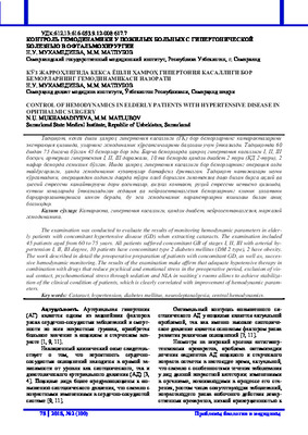 Сontrol of hemodynamics in elderly patients with hypertensive disease in ophthalmic surgeryThe examination was conducted to evaluate the results of monitoring hemodynamic parameters in elderly patients with concomitant hypertensive disease (GD) when extracting cataracts. The examination included 45 patients aged from 60 to 75 years. All patients suffered concomitant GB of stages I, II, III with arterial hy-pertension I, II, III degree, 10 patients have concomitant type 2 diabetes mellitus (DM 2 type), 2 have obesity. The work described in detail the preoperative preparation of patients with concomitant GD, as well as, succes-sive hemodynamic monitoring. The results of the examination make affirm that adequate hypotensive therapy in combination with drugs that reduce psychical and emotional stress in the preoperative period, exclusion of vis-ual contact, psychoemotional stress through sedation and NLA in waiting`s rooms allows to achieve stabiliza-tion of the clinical condition of patients, which is clearly correlated with improvement of hemodynamic param-eters
Journal problems of biology and medicine -
For parents, a child of any age seems vulnerable, so adults take care of him, they want protection from all difficulties. Unfortunately, a person is not strong and some diseases are very dangerous for the life of children. Some pathologies pass quickly and affect the future life, otherwise others will significantly affect the future life of the child. In order to reduce the impact of pathologies on the child's body, it is possible to diagnose the disease in the early stages, it is necessary to identify and immediately begin treatment. Among these diseases are ophthalmic we can not do without the introduction of diseases. If a child has vision problems from an early age, it can lead to a delay in the development of the child in the future. Ophthalmic diseases the main part: eye injuries, glaucoma, cataracts, glaucoma, retinopathy, myopia, cataract diseases, etc. More than 20% of diseases in ophthalmological practice, depending on the injury, damage the orbit and the eyeball. From an eye injury, then in 13% of cases, subatrophy of the eye develops, in 25% anophthalmos may occur. As for the characteristics of injuries, 10% of children suffer from damage to the organ of vision. This leads to various pathologies of the eye, in 30-60% of cases it can lead to one- or two-sided blindness. The most important traumatic factors in children are: knives, bullets, stones and clubs, hockey sticks, spears, nails, wire, etc. Glaucoma is one of these diseases. The disease also requires special attention. Reason: Prevalence of glaucoma in children Occurs in 1:10,000-1:12,000 cases. Its share in eye pathology is 0.1%. enough. More than 75% of glaucoma cases are bilateral. In parallel, there was glaucoma in 5 to 15% among children, blind and non-blind schoolchildren. Blindness in children, the proportion of this pathology ranges from 2 to 15%. in the Russian Federation Congenital glaucoma accounts for 10.1% of childhood blindness.
-
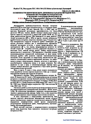 Особенности хирургического лечения катаракты методом тоннельной экстракции катаракты (тэк) у больных сахарным диабетом (сд)Количество больных сахарным диабетом (СД) во всем мире неуклонно растет и в настоящее время насчитывается около 160 млн. больных СД, а к 2025 году, по прогнозу Всемирной организации здравоохранения, это число достигнет 380 млн. человек. Диабет занимает первое место среди причин слепоты у пациентов в возрастной группе 30-60 лет [2], среди диабетических причин 70% приходится на долю диабетической ретинопатии (ДР) и 30% на другие глазные заболевания (диабетическая катаракта, вторичная глаукома и др.). У пациентов с СД своевременная хирургия катаракты крайне необходима для оценки состояния глазного дна и своевременного проведения лазерной коагуляции сетчатки, с целью предотвращения прогрессирования ДР и, тем самым, предупреждения развития конечной стадии ДР - отслойки сетчатки, рубеозной глаукомы и, в конечном счете, полной потери зрения. Любые хирургические вмешательства у пациентов с СД сопряжены с большим риском послеоперационных осложнений, возникающих на фоне декомпенсации диабета либо развития тяжелых гипогликемических состояний. Ответ организма на оперативное вмешательство проявляется активизацией симпато-адреналовой системы, что может вызвать резкую гипергликемию, липолиз, кетогенез и протеолиз, усугубляющиеся голоданием перед и во время операции. Воздержание от еды в пред- и послеоперационном периоде представляет особую опасность для пациентов с СД в плане развития гипогликемических состояний.
Doctor's Herald -
 Indicators of the development of inflammatory reactions in the moisture of the eye cameras in children after extraction of a connected cataract with implantation of an intraocular lens
in LibraryBiochemical studies of blood and chamber moisture in children with congenital cataract
are of great clinical importance. Chamber moisture is an ultrafiltrate of blood plasma and can be
an indicator of various pathological conditions of the body as a whole, and the eye in particular.
The study of the level of protein, glucose in the blood and chamber moisture of all the examined
children was carried out. As an indicator of inflammatory complications in the postoperative period,
the threshold value of total blood protein of 62.2±1.3 g/l and an increase protein content in chamber
moisture to 3.5±0.09 g/l can be used. -
 Propophol is a sedative with large therapeutic activity and 1OP depressed effect. Propophol in combination with retrobulbar anesthetization is a perfect method for short-term sedation management in cataract surgery, which satisfies patient and surgeons.
 -
A clinical case of familial congenital cataract in two patients is presented. born from consanguineous marriage. The studies were carried out in the department of ophthalmology clinics of the Tashkent Pediatric Medical Institute. Patients underwent standard laboratory and instrumental studies, as well as ophthalmological examination , including visometry , biomicroscopy , ophthalmoscopy . The a clinical case indicates the risk of having children from a closely related marriage with congenital cataract, optic nerve hypoplasia, which can further lead to visual impairment.
-
On the basis of the eye department of the TashPMI clinic, a retrospective study of the case
histories of 42 (84 eyes) patients with congenital cataracts was carried out. The age of the patients
ranged from 1 month to 13 years. All patients underwent a comprehensive clinical, ophthalmological, laboratory and instrumental examination and consultations of related specialists. The reasons for the late surgical treatment of children with congenital cataracts are: late diagnosis associated with clinical
forms of cataracts that are difficult to detect during external examination, the presence of concomitant somatic pathology, which is a contraindication to surgical treatment. -
 Factors influencing target refraction in children with pseudophakia after extraction of congenital cataract
in LibraryThe article describes the factors affecting the target refraction of pseudophakic eyes of children after extraction of congenital cat-
aracts. The factors include features of the echobiometric parameters of the eye, refraction, comorbidity of congenital cataracts and
ocular pathologies, margins of error in calculating strength of the intraocular lens, localization and structure of the artificial lens,
as well as correction of obscure or refractive amblyopia in pseudophakic eyes. Development of the algorithm for correction of re-
sidual refraction of pseudophakic eyes in children both before and after IOL implantation with consideration of each of those fac-
tors currently remains a relevant problem. -
 Functional results low-invasive by energy industry with implantation of IOL at 40 patients with senile cataract are studied. Visual acuity at all patients at receipt fluctuated from the correct projection of light to 0,05. Before operative measure of 30 patients (75%) distinguished only light with the correct svetoproyektion, at 10 - visual acuity has made 0,02-0,05 (25%). Within the first week after operation visual acuity has increased to 0,3 at 21 patients (52,5%), 0,4 - at 12 (30%). The maximum visual acuity without correction which managed to achieve as a result of operation, has made 0,8-0,9 at 7 patients - 17,5%.
 -
Features of calculating the optical power of an intraocular lens in children with congenital cataracts at the risk of developing pseudomyopic myopia.
in LibraryThe article presents the results of the clinical efficacy of the calculation formula with the correction factor of the optical force of the intraocular lens in children with congenital cataracts at risk of development of pseudophakic myopia. Personalized Rm corrective coefficient in the formula for calculating the force of IOL in children with risk of development of pseudophakic myopia makes it possible to achieve target refraction in 83.3% cases and reduce the development of reduce myopic refraction.
-
 INTRАOCULAR AMMETROPIA CORRECTION WITH PHAKIC LENSESThe analysis of literature sources (database PubMed and Google scholar) concerning modern views on the use of Phakic intraocular lenses in order to correct the refractive error of the eye. The current models of these lenses are quite sophisticated and many times increase the possibilities of their use than before. With the help of PIOL it is possible to correct any degree of myopia and hypermetropia, as well as astigmatism. There are examples of using such lenses to correct presbyopia. Currently, lenses are predominantly used in the posterior chamber of the eye. In the long term (on average after 8-10 years) complications are possible in some (up to 10%) patients. When the PIOL is inserted into the anterior chamber, the development of endothelial dystrophy of the cornea or the appearance of glaucoma is possible; when the PIOL is inserted into the posterior chamber, cataracts may develop.
Journal of oral medicine and craniofacial research -
 The structure of ophthalmopathology in infants based on the materials of the clinic of the Tashkent Pediatric Medical Institute
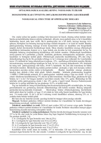
in LibraryPrevention of visual disability should begin in childhood. To plan high-tech specialized ophthalmic care for young patients, it is necessary to monitor the structure of ophthalmopathology in infants in leading domestic children's medical institutions. Purpose of the study. The study of the structure of eye diseases in infants based on the materials of the department of ophthalmology of the clinic of the Tashkent Pediatric Medical Institute (TashPMI). Material and methods. A retrospective analysis of statistical coupons of case histories of 685 patients aged 0 to 1 years who were hospitalized in the ophthalmological department of the TashPMI clinic for 2018-2021 was carried out. Results and conclusion. The spectrum of nosological units revealed the predominance of glaucoma (41.3%) and lens diseases (30.4%). At the same time, it was noted that eye injuries (10.5%), diseases of the eyelids, lacrimal ducts (phlegmon of the lacrimal sac) and orbit (9.2%), although they did not occupy a leading position, nevertheless represented severe acquired lesions, which could have been prevented. Age-related aspects of nosologies are due to the timing of clinical manifestations, diagnosis and treatment of pathologies, the anatomical and physiological characteristics of the child's body, and a decrease in parental control over children.
-
Recent studies indicate the prevalence of myopic refraction in children with pseudophakia, which significantly reduces the functional results of treatment and may be an indication for replacing the intraocular lens (IOL). Therefore, studies conducted to achieve the target refraction in children with pseudophakia are relevant.
Purpose — to determine the risk factors for the prognosis of myopic refraction in children after extraction of congenital cataract and IOL implantation.
Material and methods. The study presents the results of refraction examination in 69 (110 eyes) children aged 1 to 12 years 36 months after extraction of congenital cataract with implantation of soft IOL.
Results. The obtained data of anamnesis, results of ophthalmological, echobiometric, clinical and laboratory studies were subjected to statistical processing assessing the significance of differences in outcomes depending on the impact of possible risk factors for the development of pseudophakic myopia; a regression logistic model and a ROC-curve were constructed.
Conclusion. According to the authors, reliable risk factors for the development of pseudophakic myopia in children can be such indicators as axial eye length at the time of IOL implantation exceeding the age norm by more than 0.2 mm; the child from the first pregnancy; the AL/CR ratio of 23.0; myopia on the paired eye; strabismus of more than 4 prism diopters; hereditary load; tension of eye's fibrous capsule: pressure of SI 80 mm Hg at the time of IOL implantation. The presented reliable factors, as well as a combination of less significant signs (the child being outside for less than 1 hour per day, intermarriage of the patients parents, near-sight visual loads ot more than 3 hours per day, the blood Ca level of less than 1.8 mmol/L) can be used for prognosis of the development of pseudophakic myopia and to help make adjustments in the management tactics for patients to achieve target refraction.
-
 Comparative age-related assessment of the anterio-posterior axis length in children with normal eyes and with unilateral congenital cataract or congenital glaucoma
in LibraryThe length parameter of anterior-posterior axis is compared for children’s hyperopic eyes (302 eyes, hyperopia of 0.5 to 3.0 D). 109 eyes (109 children) with unilateral congenital cataract (UCC), and 132 eyes (90 children) with congenital glaucoma, aged 1 month to 15 years. Extended observation of anterior-posterior axis growth and refraction in eyes with artifakia showed a partial tendency to myopisation (3— 7%), which requires additional research of the pathogenetic process and should be taken into account when calculating the optical power of 1OL. The analysis of data on age-related growth of the anterio-posterior axis of eyes with congenital glaucoma showed the agreement with average figures for the developed and advanced stages of the condition. In contrast, terminal and advanced glaucoma (in children aged 2 to 3) the obtained data showed a reliable increase as compared to previously published values
-
 Clinical efficacy of individual intraocular lens calculation in children with congenital cataract at risk of abnormal refraction
in LibraryTo assess the clinical efficacy of the SRK II formula with a correction factor Rm in children with congenital cataracts who are at risk of pseudophakic myopia. Material and methods. A complex examination of 48 children (86 eyes) with congenital cataracts involved visometrics, tonometry, tonography, biomicroscopy, keratorefractometry, ophthalmoscopy, ultrasonography, and pachymetry. To determine the IOL power, we used the SRK II formula supplemented with the individual correction factor Rm, proposed by the authors. The examined children were divided into 2 groups. The main group 1 included 22 patients (42 eyes), for which the IOL power was calculated with the Rm factor. The control group 2 consisted of 26 patients (44 eyes) for which the IOL power was calculated according to the traditional SRK II formula using age-related hypocorrection of refraction but without the Rm coefficient. Results. The correction factor Rm, allowed us to achieve the targeted refraction in children who were at risk of developing pseudophakic myopia in 83.3 % of cases of the main group (versus 45.4 % of the control group cases) and reduce the development of high age-related refraction) by 37.9 %. In children of the main group, visual acuity reached, on average, 0.5 ± 0.001, while in the control group it was also higher but only reached 0.200 ± 0.001. Conclusion.The method of calculating the IOL optical power involving an individual correction factor Rm, according to the formula: SRK II – R – Rm can be recommended for clinical practice focused on children at risk of abnormal refractogenesis.
-
Purpose. An analysis of complications in case of penetrating injuries of visual organ in children and methods of their elimination.
Material and methods. There were in the follow-up 36 patients (36 eyes) aged from 6 to 14 years with various ocular injuries: boys - 70% of cases, girls - 30% of cases. All children underwent ophthalmic and clinical examinations.
Results. The primary surgical treatment (PST) was performed in all patients at the place of residence in different regions of the Republic. The secondary surgical treatment (SST) was carried out according to the following indications: swelling cataract, filtration of corneal and scleral injuries, local endophthalmitis, iris incarceration, hypotension, secondary glaucoma, sutures failure, torpid uveitis, deformation of eyelids, abruption of lacrimal canaliculi, non-sutured scleral wound, suspect intraocular foreign body, the destruction of the eyeball, suspected intraocular foreign body, the destruction of the eyeball. Causes of PST complications were: the severity of injuries (47.2%), an unskilled first aid (41.7%), a late appeal of patient (47.2%).
Conclusion. As a SST result the inflammatory response was stopped and the eyes were maintained as an anatomical organ in 99.6% of cases, visual functions were preserved in 67.4% of cases. It is necessary to intensify educational work among the population, to improve the quality of emergency eye care and the adequate rehabilitation of patients.