Library search
Search Results
-
 Апикальное позиционирование ортопедической платформы имплантата и толщина мягких тканей
Actual problems of dentistry and maxillofacial surgery 4Достижение стабильности костной ткани в области дентальных имплантатов по-прежнему является наиболее актуальной и до конца нерешенной проблемой в имплантологической практике. При наличии тонких мягких тканей гребня многие клиницисты предпочитают более глубокую установку имплантата, что позволяет в ряде случаев добиться стабильного уровня кости и здорового состояния мягких тканей. Однако существует ряд мнений о несостоятельности данного подхода: невозможность прогнозирования реакции прилегающей костной ткани - опасность ремоделирования, трудность контроля глубины препарирования костного ложа, возможность развития периимплантита за счет избыточной площади контакта поверхности имплантата с эпителием, сложность выполнения клинических этапов изготовления протезов [1].
-
 SIMULTANEOUS IMPLANTATION IS A MODERN SOLUTION IN DIFFERENT CLINICAL SITUATIONS IN THE PRACTICE OF AN IMPLANTOLOGISTIn clinical situations when it is planned to install dental implants immediately after tooth extraction, various surgical protocols of implantation and postoperative management are possible. Achieving primary stability of dental implants is a key factor for further osscointegration and the success of the entire treatment. Another problem with immediate implantation is the correct positioning of the implant, due to the discrepancy between the shape and size of the well, the size and shape of the implant. Considering the above, it is important to have a clear algorithm for direct implantation, which will be optimal for ensuring osseointegration and subsequent functional load and achieving aesthetic results. This review article analyzes modem approaches for simultaneous dental implantation into the hole of a removed tooth, describes methods for solving these problems
Medicine and innovations -
 COMPARATIVE ANALYSIS OF THE INFLUENCE OF THE SURFACE OF THE DENTAL IMPLANT "IMPLANT.UZ" FOR THE PROCESS OF OSTEOINTEGRATIONComparative study of the surface characteristics and the value of the untwisting torque of the dental implant “Iinplant.uz” subjected to sandblasting with 125 pmA12O3 particles, followed by immersion in a biomimetic liquid and the mechanically treated surface of the same implant without immersion in this liquid. Material and methods: In an experiment on dogs, the surface characteristics of the dental implant “Iinplant.uz” were studied. Results: The mechanically treated implants showed 49.5± 10.3 cN m removal torque values after a 6-week healing period, but for immersed in biomimic fluid and sandblasted implants the same parameter was 72.7±15.98 cN m. After a 16-week recovery period, these values increased to 77.5±15.16 and 89.7±11.83 cN m, respectively. Conclusions: For biomimetically incubated sandblasted dental implants, faster recovery was observed compared to mechanically treated implants. Key words: dental implant “Implant, uz”, mechanical surface treatment, sandblasting with A12O3 particles.
Stomatologiya -
 Предоперационная конусно-лучевая компьютерная томография качество кости оценка для предсказуемого немедленного размещения имплантата и реставрация в эстетической зоне.
in LibraryИспользование управляемого хирургического подхода с помощью компьютерного моделирования позволяет обеспечить установку имплантата с точностью около 98%, управляемая хирургия выгодна для обычного размещения имплантата, немедленного имплантата размещение и потенциальная немедленная провизионализация.
-
 Practical recommendations on application of the developed biocompatible implant "geprotsel" for the prevention and treatment of disorders of aero - and hemostasis in surgery of the lungsVSU "RPMZ named Akad. V. Vakhidov street" together with NYSAFP Academy of Sciences of Uzbeki-stan developed a new aerial and hemostatic implant "Gabriel" which has passed preclinical testing. The article presents the results of clinical studies of the developed implant. Studies have shown that the use of a biological implant "Geprotsel" has reduced the need for additional single stitching lung tissue to ensure adequate Aero-and hemostasis from 38.2% to 11.4% and multiple strengthening seams from 29.4% to 5.7% (χ2=7,706; Df=2; P=0.021). The introduction of the domestic biological implant into lung surgery allowed to reduce the period to achieve Aero-and hemostasis from 32.8±2.5 to 12.5±1.2 minutes (P<0.001), the total duration of the operation from 135.6±6.1 to 107.2±4.7 minutes (P<0.001), as well as to reduce the duration of pleural drainage from 3.38±0.31 to 2.09±0.06 (P<0.001) and the entire period of hospitalization from 12.1±0.4 to 10.7±0.2 days (P<0.01)
Journal problems of biology and medicine -
 Assessment of the effectiveness of using the bioactive layer for the intraosseous part of domestic dental implants. Results: bioactive coatings formed on the surface of titanium plates under optimal conditions and electrolysis conditions have a nanoscale density m the range of 10-300 nm. Conclusions: substantiated positive dynamics of osseomtegration at the implant border - bone during implantation.
 -
 On the basis of the conducted researches a new design of a dental implant was developed. It provides the increased reliability of fixing, convenience in use. Creation of the implant in one-stage and two-stage forms expands the range of instrumentations of implantation depending on medical indications and desire of the patient.
 -
 Bilateral ophthalmohypertension after the installation of an intravascular implant - a flow stent in the left internal carotid artery in the projection of the neck of the aneurysm
in Libraryassessment of risk factors for the occurrence of ophthalmohypertension in a patient after the installation of an intravascular implant - a flow stent in the left internal carotid artery (ICA) in the projection of the aneurysm neck. Material and Methods: Patient A., 69 years old, with the development of bilateral ophthalmohypertension after the installation of an intravascular implant - a flow stent in the left ICA in the projection of the aneurysm neck. Results: Based on the examination of the patient and the study of all possible risk factors for the occurrence of ophthalmohypertension, confirmed by the data of modern literature, we can assume a multifactorial cause of bilateral ophthalmohypertension after the installation of an intravascular implant - a flow stent in the left internal carotid artery in the projection of the aneurysm neck. Conclusions: An interdisciplinary approach to the surgical treatment of an aneurysm of the supracune-shaped part of the internal carotid artery is important.
-
 Digital method of radiation diagnostics in the assessment of bone structure during dental implantation in patients with diabetes mellitusResults digital method of radiodiagnosis in assessment of bone structure at the dental implantation at patients with diabetes and of intrabone dental miplants osteointegration after restoration of an atrophy of an alveolar bone and dental implantation are shown on a material of 220 patients. Patients with the second type of diabetes at a stage of compensation are in comparative group. For the analysis of results of osteointegration and stability miplants we were used the device «Osstell ISQ». it was manufactured by firm Integration Diagnostics (Sweden), it defining frequency-resonant analysis and stability factor of implants by method RFA (Resonance Frequency Analysis), which registries of resonant electromagnetic fluctuations of implants and a surrounding bone influences on them with electromagnetic field. ISQ (Implant stability Quotient) is expressed on a scale from one to hundred. The obtained data allows assuming influence of objective methods of research of osteointegration dental implants with the subsequent reduction of terms of orthopedic treatment and choice of optimum term of prosthetics. The general average value of implants stability is about 70 units ISQ.
Stomatologiya -
 One of the fundamental factors that ensure a high effect during implantation is the correct determination of indications for this method of treatment. The mam requirements for materials for dental implants are: clinico-biological. determmed by the features of the interaction of living tissues with the material of the implant: biological, associated with toxicological, carcinogenic, corrosive properties of the implant material. However, the possibility of immediate implant placement in the alveolus of the tooth after its removal is an actual and economically justified way of improving dental care. A number of methods for the immediate restoration of included dental flaws in the removal of teeth from various indications, including elements of bone plasty with auto- and allogeneic bone or preparations based on calcium hydroxyapatite, are known [Grigoriants LA with co-workers, 2006; Zuev Yu.A. with co-workers, 2003: Pavlyuchenko I I. with co-authors. 2004: Sirac SV with co-workers., 2010; 2011].
 -
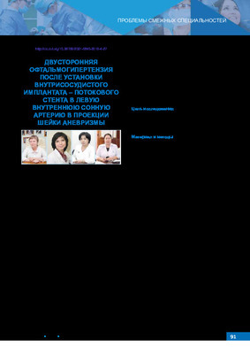 BILATERAL OPHTHALMOHYPERTENSION AFTER INSTALLATION OF AN INTRAVASCULAR IMPLANT – FLOW STENT INTO THE LEFT INTERNAL CAROTID ARTERY IN THE PROJECTION OF THE NECK OF THE ANEURYSMA clinical case of the development of bilateral ophthalmic hypertension after the installation of an intravascular implant - a flow stent in the left internal carotid artery in the projection of the aneurysm neck is described, which may be of interest not only to ophthalmologists, but also confirms the importance of an interdisciplinary approach in the surgical treatment of supraclinical aneuiysm (internal carotid artery aneurysm), which implies die mandatory awareness of neurosurgeons, vascular surgeons, ophthalmologists about possible sensations from tire organ of vision.
Stomatologiya -
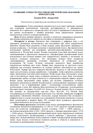 Сравнение точности трех типов хирургических шаблонов имплантатов
Topical issues of surgical dentistry and dental implantologyТочное размещение имплантатов в кости имеет решающее значение для сохранения анатомические структуры, реставрируемость и приживаемость имплантата. В то время как в литературе много об успехе и выживаемости имплантатов, нет данных исследований о влиянии различных типов хирургических шаблонов имплантатов на точность установки имплантата
-
 Оценка эффективности способов фиксации мостовидных протезов на дентальные имплантаты.
Actual problems of dentistry and maxillofacial surgery 4Этап конструирования и изготовления зубных протезов имеет особое значение в обеспечении долгосрочной эффективности протезирования на дентальных имплантатах. К прогрессирующей резорбции периимплантатной костной ткан и может привести не только функциональная перегрузка имплантата, но и недостаточная прецизионность припасовки и ненадежная фиксация протеза к абатменту имплантата.
-
 New technologies for the prevention of adhesions in thoraco-abdominal surgery
Catalog of dissertations and abstractsThe aim of the research work is determination of the prospects for the use of a domestic agent for the prevention of adhesion formation in thoraco-abdominal surgery on the basis of experimental and morphological studies.
Research objectives were white outbred rats in the amount of 62 individuals, in two experimental studies on the abdominal and pleural cavities, in each series of experiments the studies were carried out in 2 comparative groups, control and main. Experiments on the formation of adhesions in the abdominal and pleural cavities were carried out on the basis of the Republican Specialized Scientific and Practical Medical Center of Surgery named after acad. V.Vakhidov in the Department of Experimental Surgery for the period from 2019 to 2020.
The scientific novelty of the research consists of the followings: it is proved according to the data of experimental research that when modeling the adhesion process in the abdominal cavity, the local application of an anti-adhesion coating made of cellulose derivatives reduces the processes of adhesiogenesis and the development of changes in architectonics, bends and narrowings of the intestinal lumen; it was found in an experimental study that when modeling the adhesion process in the chest cavity, the local use of an anti-adhesion implant provides a significant decrease in the risk of adhesiogenesis in the form of the formation of coarse adhesions or planar adhesions; it was determined that when blood serum was applied over a powder implant, the quality of adhesion and the uniformity of its distribution on the surface of the experimental defect of the peritoneum or lung did not change, but, in contrast to activation by blood (to ensure a hemostatic effect), it was not accompanied by the development of cellular inflammation due to the resorption of thrombotic masses; it was found that the formation of a gel film over the area of damage to the peritoneum in the absence of cellular elements of blood makes it possible to achieve biodegradation of the coating without a pronounced cellular-inflammatory reaction, providing cicatricial replacement of defects with a significant reduction in the risk of developing a massive adhesive process; the morphostructural features of the formation of the adhesive process when using an anti-adhesive coating, characterized by regression in the dynamics of the number of connective tissue cells of the inflammatory infiltrate with scarring of the defect zone without the development of adhesive conglomerates with the surrounding tissues, have been determined.
Introduction of the research results. According to the results of a scientific study on a comparative analysis of the use of a domestic agent for the prevention of adhesion formation in thoraco-abdominal surgery: methodological recommendations were developed: "New technologies for the prevention of adhesions in thoraco-abdominal surgery" (certificate of the Ministry of Health No. 08-09/10055 of August 12, 2021). The proposed recommendations for performing surgical interventions on the organs of the abdominal and thoracic cavities will allow for sparing local hemostasis, as well as prevent the formation of a coarse adhesive process in the abdominal cavity.
The obtained scientific results on a comparative analysis of the use of the domestic remedy for the prevention of adhesion formation in thoraco-abdominal surgery have been introduced into the practical activities of health care, including in the Republican Specialized Scientific and Practical Medical Center for Surgery named after V.I. Academician V. Vakhidov, surgical departments of the clinics of the Andijan and Samarkand State Medical Institutes (certificate of the Ministry of Health No. 08-09/10055 of August 12, 2021). Based on the proposed results of experimental studies, it was shown that the use of an anti-adhesive coating made of cellulose derivatives made it possible to reduce the risk of adhesion formation from 60% to 20%, bowel deformation without manifestations of obstruction from 33.3% to 13.3% and the possibility of acute adhesive intestinal obstruction from 6.7% to 0%.
Structure and scope of the dissertation. The dissertation consists of an introduction, four chapters, conclusions, practical recommendations and a list of cited literature. The volume of work is 113 pages.
-
 Objective assessment of osseointegration of dental implants by resonance frequency analysis in patients with diabetes mellitusResults of intrabone dental implants osteointegration after restoration of an atrophy of an alveolar bone and dental implantation are shown on a material of 220 patients. Patients with the second type of diabetes at a stage of compensation are in comparative group. For the analysis of results of osteointegration and stability implants we were used the device «Osstell ISQ», it was manufactured by firm Integration Diagnostics (Sweden), it defining frequency-resonant analysis and stability factor of implants by method RFA (Resonance Frequency Analysis), which registries of resonant electromagnetic fluctuations of implants and a surrounding bone influences on them with electromagnetic field. ISQ (Implant stability Quotient) is expressed on a scale from one to hundred. The obtained data allows assuming influence of objective methods of research of osteointegration dental miplants with the subsequent reduction of terms of orthopedic treatment and choice of optimum term of prosthetics. The general average value of implants stability is about 70 units ISQ.
Stomatologiya -
 OPTIMIZATION OF THE SURGICAL STAGE OF THE DENTAL IMPLANTATIONSA reduction in the number of complications following tooth implantation remains of critical importance to dentistry. All risk factors can be divided into general, including smoking, systemic diseases, patient’s condition following radiotherapy, etc. and local ones comprising poor oral hygiene, periodontal disease, iatrogenic disorders, design and quality of the transgingival implant. In order to better understand inflammatory degenerative processes around dental implants researchers and dental implant manufacturers are forced to improve currently used conservative and surgical methods of treating and preventing these diseases, focusing more on risk factors for the development of peri-implantitis. The development of new surgical and prosthetic techniques, new dental implant systems will contribute greatly to increasing the life span of implant-supported dentures and improving the patient’s quality of life.
Journal of oral medicine and craniofacial research -
 Влияние скорости установки дентальных имплантатов на первичную стабилизацию
Topical issues of surgical dentistry and dental implantologyКариес, заболевания пародонта, инфекции и травмы могут вызвать резорбцию корней и альвеол постоянных зубов. Эти условия могут привести к отрыву зуба или даже к необходимости удаления зуба. Одним из способов лечения потери зубов является хирургическая имплантация зубов. Остеоинтеграция является ключевым фактором, определяющим успех имплантации зубов, а начальная стабильность имплантата является хорошим показателем эффективности остеоинтеграции. Остеоинтеграция относится к естественной связи между имплантатом и альвеолярной костью. Другими словами, первичная стабилизация имплантата способствует успеху последующей остеоинтеграции, тем самым способствуя долгосрочному успеху. Перед проведением операции по имплантации зубов также необходимо учитывать биомеханические факторы, чтобы повысить первичную стабилизацию после установки.
-
 Assessment of changes in the quality of life of patients with dentition defects before and after prosthetics and dental implantation using an implant Implant.uz
StomatologiyaComparative evaluation of the results of defect replacement with the installation of a domestic Implant.uz implant.
-
Reducing the number of complications after dental implantation remains an urgent problem in dentistry. All risk factors can be divided into general (comorbidities, bad habits, systemic pathology, condition after radiation therapy, etc.,) and local (unsatisfactory dentures, poor oral hygiene, periodontal disease, iatrogenic conditions, defects in the transgingival part of the implant and etc). Understanding the course of inflammatory and destructive processes around a dental implant after its installation directs scientists and implant manufacturers to improve the already used conservative and surgical methods of treating these diseases, as well as to pay more attention to the prevention of this pathology, and, accordingly, risk factors for its development. The development of new methods of surgical interventions and prosthetics, the creation of new implant systems will help to increase the service life of dental prostheses on implants, improve the quality of life of patients
-
 Факторы подавляющие процесс остеоинтеграции имплантата в костную ткань.
Actual problems of dentistry and maxillofacial surgery 4Ключом успешной дентальной имплантации является остеоинтеграция, то есть образование прямой структурной и функциональной связи между живой костной тканью и поверхностью вживленного в нее имплантата на молекулярном уровне.
-
 Воздействия биоактивных покрытий на остинтеграцию имплантов
Topical issues of surgical dentistry and dental implantologyДизайн поверхности имплантатов эволюционировал для решения проблем реабилитации полости рта как в здоровой, так и в нарушенной кости. Например, чтобы победить наиболее распространенные осложнения, связанные с зубными имплантатами, периимплантит и последующую потерю имплантата, поверхности имплантатов были модифицированы, чтобы придать зубному имплантату желаемые свойства и тем самым повысить процент успешного приживления имплантата и расширить показания к их применению. Биоактивные покрытия имеют потенциал для улучшения костной интеграции механически нагруженных ортопедических керамических имплантатов. Есть гипотеза о том, что биоактивное покрытие будет способствовать интеграции отменной кости. Биоактивное покрытие замедлить скорость коррозии сплавов и ускорить процесс заживления кости. Основная Цель биоактивных покрытий - усилить прямое прикрепление живых тканей и тем самым способствовать остеокондукции.
-
 Delivery device for the application of hemostatic implant geprocel in minimum-invasive surgeryDespite the achievement of modern surgery in nowadays, there is no special delivery device for laparo-scopic surgery in the minimally invasive surgery. This kind of device must meet the following characteristics: the ability to dispense a certain amount of powder in the target zone, safety and efficiency at the high pressure in the abdominal cavity. A new delivery device for the use of hemostatic powder Heprocel in minimally invasive surgery was developed by the developers of SI “RSSPMCS named after acad. V.Vakhidov”. The effectiveness of the delivery device was evaluated experimentally on 8 mongrel dogs weighing up to 7.5 kg. The developed de-livery device ensures an uniform distribution of the hemostatic implant Heprocel over the wound surface of the liver. The proposed device is an effective delivery system capable of transport a dosed amount of hemostatic powder to the target bleeding area during manipulations in laparoscopic surgery
Journal problems of biology and medicine -
 Повышение эффективности дентальной имплантации при сохранении зубо-альвеолярного сегмента верхней челюсти с помощью метода “root membrane”
in LibraryПротезирование вторичных адентий, которые длительное время присутствуют в верхней и нижней челюстях, является одной из актуальных проблем, стоящих перед стоматологами. Одним из современных методов устранения этой проблемы, является дентальная имплантология. Одним из сдерживающих факторов для широкого распространения имплантации является недостаточный объем костной ткани для установки имплантата. Процесс дентальной имплантации во фронтальную часть верхней челюсти, которая на высоком уровне атрофирована или сопровождается вертикальным переломом, зубов, требует дополнительных костных изделий и длительного реабилитационного периода, причина этого в том, что вестибулярная пластинка фронтальной части верхней челюсти тонкая и он характеризуется переломом во время удаления зуба. Атрофия костной ткани после удаления зубов является одним из важнейших вопросов современной стоматологии, так как значительная атрофия костной ткани челюстей делает невозможным выполнение внутрикостной имплантации, а также создает серьезные трудности при ортопедическом лечении пациентов.
-
 Определение стабильности дентальных имплантатов в эксперименте методом голографической интерферометрии
Actual problems of dentistry and maxillofacial surgery 4Стабильность дентальных имплантатов в процессе их остеоинтеграции делится на первичную и вторичную. Первичная или механическая стабильность достигается за счёт механического соединения конструкции дентального имплантата с костной тканью.
-
 ESTIMATE OF THE EFFECTIVENESS OF OSTEOPLASTIC MATERIAL OSTEON COLLAGEN 3 AFTER THE SOCKET PRESERVATION TECHNIQUE USING A DPTFE MEMBRANEMechanism of irreversible changes is started in the alveolar ridge, when the tooth is removed from its socket, in particular the alveolar ridge itself in the region of this extraction well begins to decrease in volume and morphologically transform. [5] These changes subsequently create unfavorable conditions for prosthetics in the extraction site and sometimes the impossibility of installing a dental implant. [6,7] Preventive procedures of the dental surgeon by mean specific manipulations with the tooth socket will be preservation of its volume and help not only to carry out the implantation procedure in this place, but also to achieve an excellent aesthetic and functional result during prosthetics on the implant. This article open the possibility of using a non-resorbable membrane dPTFE in well socket preservation techniques for multiple tooth removal using sole a single xenogenic bone-plastic material Osteon collagen 3
Medicine and innovations