Library search
Search Results
-
Cytogenetic Study Of Peripheral Blood Leukocytes In Patients With Uterine Mesenchymal Tumors
The American Journal of Medical Sciences and Pharmaceutical ResearchAccording to the World Health Organization, uterine tumours rank second in the structure of oncological morbidity in women. Epithelial tumours account for 95% of all genital tumours and only 5% are mesenchymal tumours. Malignant mesenchymal tumours include uterine sarcomas. Annually, 10 cases of uterine sarcomas are diagnosed in 1 million women worldwide. The problems of early diagnosis and screening of malignant uterine mesenchymal tumours (sarcomas) have not yet been solved. Due to the fact that this disease is rare and the treatment of uterine sarcomas remains a pressing problem.
-
EVALUATING THE EFFICACY OF TARGET PXT IN METASTATIC LIVER TUMORS
European International Journal of Multidisciplinary Research and Management StudiesThe goal of assessing tumour response on imaging is to identify patients who are likely to benefit — or not — from anticancer treatment, especially in relation to survival. The World Health Organization was the first to develop assessment criteria. This early score, which assessed tumour burden by standardising lesion size measurements, laid the groundwork for many of the criteria that followed. This was then improved by the Response Evaluation Criteria in Solid Tumours (RECIST) which was quickly adopted by the oncology community. At the same time, many interventional oncology treatments were developed to target specific features of liver tumours that result in significant changes in tumours but have little effect on tumour size. New criteria focusing on the viable part of tumours were therefore designed to provide more appropriate feedback to guide patient management. Targeted therapy has resulted in a breakthrough that challenges conventional response criteria due to the non-linear relationship between response and tumour size, requiring the development of methods that emphasize the appearance of tumours. More recently, research into functional and quantitative imaging has created new opportunities in liver imaging. These results have suggested that certain parameters could serve as early predictors of response or could predict later tumour response at baseline. These approaches have now been extended by machine learning and deep learning. This clinical review focuses on the progress made in the evaluation of liver tumours on imaging, discussing the rationale for this approach, addressing challenges and controversies in the field, and suggesting possible future developments.
-
 Echographic criteria for the choice of tactics of management of women with failure of the seam on the uterine after cesarian section
Journal of Biomedicine and PracticeWe studied the echographic and elastographic parameters and features of hemodynamics in the uterine vessels at 79 women, including 59 women by cesarean section and 20 women after vaginal birth. In the case of complications with endometritis after cesarean section, an increase in uterine body volume by 25.5%, an increase in the rate of end diastolic blood flow in the arch arteries of the uterine body was found. Symptoms of the inflammatory process in the myometrium were combined with the manifestation of its anatomical inconsistency, which was manifested by the formation of a wall defect in the form of an irregular triangular “thorn” by the uterine cavity and a clear thinning. its distal part. On day 3 after cesarean delivery, the myometrium in the uterine scar area had a higher index of elasticity than the control.
-
REGULARITY OF PATHOMORPHOLOGICAL CHANGES OF ENDO AND MYOMETRY IN THE DEVELOPMENT OF ABNORMAL UTERINE BLEEDING IN WOMEN IN PERIMENOPAUSE
The American Journal of Medical Sciences and Pharmaceutical ResearchA special place in the problem of abnormal uterine bleeding is occupied by the issues of qualitative diagnosis of patients with abnormal uterine bleeding, the cause of which is pathomorphological changes in the endomyometrium, such as: endometrial hyperplasia, uterine myoma, adenomyosis, a combination of fibroids and endometrial hyperplasia, a combination of adenomyosis and endometrial hyperplasia. Abnormal uterine bleeding is observed in 10-30% of women of reproductive age, in perimenopause - their frequency increases to 50%.
-
 Features of ultrasound and dopplerometric changes in women with uterine fibrous and / or adenomiosis and their role in the choice of treatment method
in LibraryUterine fibroids and adenomyosis are an urgent problem in modern gynecology. In recent years, there has been an increase in the combined form of uterine fibroids and adenomyosis. Improvement of non-invasive methods of ultrasound diagnostics with color Doppler hysterosonography and magnetic resonance imaging (MRI) can improve the accuracy of diagnosing the combined pathology of uterine fibroids and adenomyosis. The effectiveness of treatment of patients with a combined form of uterine fibroids and adenomyosis depends on determining the severity of a particular disease.
-
 Vexed and unsolved questions of treatmentand prophylaxis of hysteromyoma for patientsof reproductive periodUterine fibroids are less common in women aged 20-30; but the risk of getting sick increases after 35-40 years. Often the problem of maintaining reproductive function in women with uterine fibroids occurs at the end of reproductive age. The most optimal treatment for uterine fibroids in the reproductive period is conservative myomectomy. This article describes the manifestations and methods of conservative myomectomy, as well as methods of adjuvant drug treatment.
Journal problems of biology and medicine -
 Standards for the management of patients with uterine myomaIntroduction. According to various authors, the frequency of detection of uterine fibroids among the modern female population ranges from 20 to 77% [5, 6, 8]. At the same time, there is a noticeable increase in the incidence among women of reproductive age [1, 2, 8]. Considering that in modern conditions women often realize their reproductive function after 30 years, the issues of management of uterine fibroids in patients of reproductive age have grown into an independent topical problem. Fragmentation of works, contradictions, lack of protocol schemes for treatment and rehabilitation, creates a lot of questions about the management of this category of women. A number of researchers see a solution to this problem in the introduction of clinical protocols, which should become the basis for the formation of the medical State Standard [8].
Journal problems of biology and medicine -
 Acyclic uterine bleeding is a formidable complication of the menopause, and remains relevant to this day. The data of world literature shows that there is no consensus on the data on the pathological condition of the endometrium. In this connection, the attention of world researchers is drawn to a further study of the state of the endometrium, depending on the cyclic changes in the endometrium, in order to identify preventive measures for acyclic uterine bleeding.
 -
 Uterine myoma is relatively rare in women aged 20-30, but disease risk increases significantly after the age of 35-40. The need to preserve fertility occurs in women of late reproductive age with uterine myoma also increases. Conservative myomectomy is considered to be the method of choice for surgical treatment of uterine myomas in reproductive period. The article describes types and techniques of conservative myomectomy.
 -
 The role of magnetic resonance imaging in the comprehensive radial diagnosis of volumetric masses of the eye organ
Catalog of abstractsRelevance of the problem. The difficulties of diagnostics of orbital diseases are well known. Especially difficult is intraspecies differentiation among the multitude of tumour, pseudotumour, inflammatory, vascular, endocrine and other diseases occurring here, manifested by the symptom complex of unilateral exophthalmos [Beradze I.N., 1978; Brovkina A.F., 1993].
Malignant intraocular neoplasms are the main cause of death of patients with diseases of the organ of vision, with 45-48% of patients dying from metastases in the first 5 years after enucleation [Alekseeva I.B., 1990, Barkhash S.A.1978, Brovkina A.F..1991, 1997; Keizer R.W.. Viclvoyc G.L.,1986],
Retinoblastoma is the most frequent malignant neoplasm in children. According to different authors, the frequency of its occurrence is 1 case per 14000 - 35000 newborns. [Bobrova N.F. and Vit V.V., 1993; Brovkina A.F., 1997; Provenzale J.M., et al., 1995; Skulski M., et al., 1997; Weber A.L., Mafee M.F, 1992; Wilms G., et al., 1989]. The frequency of patients with the most malignant intraocular tumour in adults - uveal melanoma has recently reached 7-9 people per 1 million population [Brovkina A.F., 1997; Kotslyansky E.O., 1989; Yushko N.A., Peskova L.I., Kalenich L.A., 1989; Peyster R.G., Augsburger J..I., Shields J.A., 1988; Romani A.. Baldeschi L., ct al 1998; Scott I.U., 1998].
The fundamental difference in treatment tactics, depending on the stage of development, size and topography of the tumour, as well as the seriousness of the prognosis in retinoblastomas and melanomas sharply increase the requirements for the accuracy of their differential diagnosis. At the same time, the number of diagnostic errors in ocular tumours continues to be 10-30% even when complex clinical and instrumental examination is applied in specialised ophthalmological centres [Ternovoy S.K., Panfilova G.V., Rogozhin V.A., 1979; Friedman F.E., Malyuta G.D., Kodzov M.V., 1995; Song G.X., 1991].
Widely used in ophthalmological practice traditional diagnostic methods (ophthalmoscopy, gonioscopy, diaphanoscopy, fluorescence angiography, laboratory tests) are insufficient to obtain comprehensive information about the localisation, nature of growth and prevalence of volumetric pathological formations of the eye and orbit. This circumstance and not quite satisfactory results of surgical treatment are the causes of high mortality of patients [Muratova T.T., Nigmanova N.H., Kozlovskaya G.M.. 1989, Naches A.I., 1980; Cheremisin V.M., Trufanov G.E., Kholin A.V., 1991]. Untimely or erroneous recognition of pathological processes of the orbit leads to a sharp deterioration of visual functions, up to blindness, and in some cases to the death of the patient [Yuzhakov A.M., Travkin A.G., Kiseleva O.A., 1991]. All this determines the importance of timely and accurate diagnosis of diseases of the orbit, on the one hand, and the difficulty of such diagnosis - on the other [Gabunia R.I., Kolesnikova E.K., Tumanov L.B., 1982].
The fact that the orbit is closed from direct inspection and palpation by bone walls and the eyeball, indicates the advantage of radial diagnostics in comparison with other methods of examination. In the arsenal of clinicians there is a great variety of methods of clinical-radial diagnostics of orbital pathology, however, at present the information in the literature about their resolving capabilities and significance in comparative aspect is incomplete and not fully studied. The priority of using one or another instrumental investigation, their sequence and expedient combination have not been determined yet. This makes it difficult to choose the optimal standardised approach for diagnosis and adequate treatment [Cheremisin V.M., Trufanov G.E., 1993, Weber A.L., Sabates N.R., 1996; Wenig V.M., Mafee M.F., 1998].
Thus, the study of these and other questions, contributing to the improvement of diagnostics and treatment of patients with neoplasms of the eye and ocular cavity, should be recognised as urgent urgent.
Purpose of the study. Comparative evaluation of magnetic resonance tomography capabilities and development of algorithms for complex radial diagnostics of volumetric formations of the visual organ. To solve this goal we set the following tasks.
1. To study the normal picture of the magnetic resonance image of the visual organ in comparison with other methods of visualisation.
2. To find out the possibilities of magnetic resonance tomography, ultrasound and computed tomography in detection and evaluation of intraocular neoplasms.
3. To determine the role and place of magnetic resonance tomography in differential diagnostics of volumetric pathological formations of the eye cavity in comparison with other radial methods of research.
4. To determine the indications and to develop an algorithm for the complex application of radiography, ultrasound, computer and magnetic resonance tomography for diagnostics of volumetric formations of the eye organ.
Scientific novelty.
The present work is the first to give a detailed and detailed description of the complex clinical and radiation examination, with generalisation and standardisation of magnetic resonance, computer and ultrasound semiotics of volumetric pathological formations of the eye and eye cavity. The conducted clinical and instrumental investigations allowed to determine the diagnostic value and resolving capabilities of each of the applied methods. The ultrasound, CT and MRI signs of volumetric formations of the eye organ were studied, clarified and supplemented taking into account the use of low-field magnetic field and general-purpose ultrasound apparatus. The developed standardised diagnostic algorithm of examination of patients with this pathology is new, thanks to which the pre-oppositional diagnosis of tumour and other diseases of the visual organ is improved and the total radiation load on the patient is reduced.
Conclusions
1. MPT will provide an opportunity to study the weight of the soft tissue and anatomical components of the ocular cavity, up to the optic nerve sheath and perineural liquor space, the orbital apex and chiasmal-sellar region, as well as to assess the condition of adjacent structures of the brain and facial skull. The method is limited in the evaluation of changes in the bony walls of the orbital cavity.
2. MRI is inferior in detecting characteristic signs of retinoblastoma (presence of calcification). The sensitivity of MRI was 66.6%, while for ultrasound and CT these values were 96.1 and 100%, respectively. But when the tumour spreads rstrobulbarly outside the eyeball (at 3-4 stages) the informativeness of MRI increases significantly. In uveal melanoma the sensitivity and specificity of MRI reaches 100%.
3. Both MRI and CT have a high detection rate (98.1% and 95.8% respectively) of benign orbital tumours of both primary and secondary origin. However, MRI is the preferred method of investigation. MRI is especially informative when a cranioorbital tumour and pseudotumour are suspected. The sensitivity of the method is 90.9% and 91.6%, respectively
4. In some cases ultrasound can be used to differentiate between encapsulated and diffuse neoplasms, which facilitates the diagnosis. However, when the pathological process is localised near the orbital apex, the diagnostic value of ultrasound decreases. In such cases it is advisable to use MRI.
5. In detection of primary and secondary malignant tumours of the orbital cavity both MRI and CT are quite informative (sensitivity 97,2% and 95,4% respectively), but the most comprehensive information about the state of bone walls will be provided by CT. When the process spreads intracranially, the value of MRI increases significantly, especially with the use of contrast enhancement.
6. The developed algorithm of complex clinical and radiation examination of patients with the use of ultrasound, CT and MRI is the most effective in the diagnosis of volumetric pathological formations of the eye and eye cavity, allowing to reduce to an adequate minimum the total radiation load on the patient and diagnostic period, excluding duplication of research techniques and choosing the most informative in each case, which in turn allows to develop appropriate treatment tactics and reduce the level of disability of the patient. -
 The frequency of caesarean section is the main reason for the increase in the number of women with a uterine scar. The choice of tactics of pregnancy, the optimal timing and methods of delivery in such patients presents considerable difficulties in each specific case and should be addressed individually.
 -
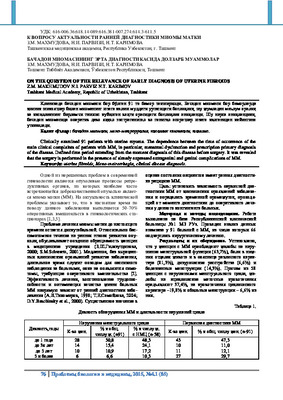 On the question of the relevance of early diagnosis of uterine fibroidsClinically examined 91 patients with uterine myoma. The dependence between the time of occurrence of the main clinical complaints of patients with MM, in particular, menstrual dysfunction and prescription primary diagnosis of the disease. Defined time period extending from the moment diagnosis of this disease before surgery. It was revealed that the surgery is performed in the presence of already expressed extragenital and genital complications of MM.
Journal problems of biology and medicine -
 The greatest value in the diagnosis of nondeveloping pregnancy represents an ultrasound. Diagnosting of anembrioniya strated in earlier periods more frequently at 5- 6 weeks and the death of the embryo often after 7 weeks of gestation. Length of stay empty' chorion sac or dead embryos in the uterine cavity coincide with the time of diagnosing non-developing pregnancy. The greatest danger to the health of patients tested current is non-developing pregnancies by type of death of the embryo, as it is diagnosed later, and in most cases the duration of stay of the dead embryo in the uterine cavity is more than 4 weeks
 -
 The method of ligation of the uterine vessels with myomectomy in pregnant womenA case of successful myomectomy large intramuscular ischemia myoma over, was node in fist pregnant women in the period of 9 weeks. For decreasing the stage of blood losing before enucleating of myoma staging was using homelier bandaging uterine vessels on the side of tumor localization. A complication isn’t present now, pregnant is progressing and now the term of pregnant period going good of 14 weeks.
Journal problems of biology and medicine -
 Changes in pro- and anti-inflammatory cytokines before and after treatment in women with combined form of uterine fibrous and adenomyosis
in LibraryThe aim of the study was to study the immunological cytokine status in women with combined forms of fibroids and adenomyosis to determine their role in the course of the disease. Methods. A total of 165 women with uterine myoma and / or adenomyosis were examined and the patients were divided into 3 groups. The research methods were dynamic study of blood flow of nodes by ultrasound with Doppler and immunological blood tests. Results: determination of the level of proinflammatory cytokines in patients before treatment showed the following results: the level of IL-1, IL-6 and TNF-α was higher in all three groups than in the control group. IL-6 and TNF-α, as well as VEGF, which proved the effectiveness of therapy. Discussion: an increase in the level of cytokines in the blood is most pronounced in combined forms of fibroids and adenomyosis compared with isolated forms of fibroids and adenomyosis. This indicates the participation of the immune system in the development and progression of such hyperplastic processes of the uterus as myoma and adenomyosis.
-
 The role of dopplerometry in the differential diagnosis of adenomyosis and uterine fibroids in young women
in LibraryIn gynecological practice, fibroids and adenomyosis are among the most common diseases among women of reproductive age, since these diseases are often the cause of radical operations. Ultrasound examinations are used in the world as screening for the detection of fibroids and adenomyosis. Conducting ultrasound on expert-class devices with blood flow Dopplerometry allows you to differentiate the type of myomatous node, determine the degree of adenomyosis and offer a comprehensive treatment depending on the activity of the process.
-
 This article presents the results of research on women with uterine myoma. Studies show the positive efficiency of the drug T-lifc used as an immunocorrcctivc treatment for uterine myomas.
 -
 The analysis of observations of 68 patients with relapses of soft tissue sarcoma of extremities and trunk is assumed as a basis of this work. The relapses of soft tissue sarcoma of the head and neck arc not included into the work. There were 38 women and 30 men among the observed. The localization of the relapses was various: 22 patients had them on the upper extremity, 13- on the trunk, 33- on the lower extremity. There were 6 patients at the age of before 19, 14 - from 20 tO 29, 19- from 30 to 39, 12- from 40 to 49, 11- from 50 to 59, and 6 patients at the age of 60 and older. There were the most patients with relapses at the age of 30 to 39, as sarcomata occur more often at this age group. Two patients had relapsing tumours with the structure of rhabdomyosarcoma; 4 patients- with angiosarcoma, 20- with fibrosarcoma, 13- with synovial sarcoma, 3- with malignant schivanoma, 4- with liposarcoma, 2- with leiomyosarcoma. Twenty patients had unclassified tumours.
 -
 Topographic-anatomic features of the development of the pancreas in the prenatal period of human ontogenesisAim of investigation: to detect the topographic-anatomical relationship of the pancreas in the embry-onic period of human ontogenesis. The study was made on 5 step by step series of the histological sections of human embryos using the microscopic method and the method of plastic reconstruction. The study found that at the end of 5th week of the embryonic period caudally from stomach the laying of pancreas develops. It is located tightly to the laying of duodenum. During this period, when the dorsal pancreas active develops the blood islets a forming, within the mesenchy me which surrounds them, the mesenchymal cordsand fi-brous structures are visible. In the dorsal mesentery the dense structure that is made of mesenchymal cells. It is embryo spleen. In embryosat the end of 6th weekthe dorsal pancreas and ventral pancreas have not grouped each other, but most likely under the influence of the growth of organs that surround them: the liver, stomach, duodenum, and also as a result of growth of these germs the ventral pancreas displays shifts to the left and dorsally, coming toward the dorsal laying of the pancreas.
Journal problems of biology and medicine -
 The value of predicting postpartum hemorrhage in the syndrome of uterine overstretching in the personification of preventive measures
in LibraryThe problem of the development of polyhydramnios is relevant in connection with the complications of pregnancy and childbirth, both for the mother and for the fetus. One of the most dangerous complications is hypotonic bleeding in the postpartum period, which leads to an increase in maternal morbidity and mortality. Among the causes of bleeding, polyhydramnios, the frequency of which reaches 1–8% of the total pool of pregnant women, occupies one of the leading positions. The aim of the study was to study the features of the clinic and the morphological structure of the uterine wall in polyhydramnios of varying severity. All this dictates the need to improve ways to prevent hypotonic postpartum bleeding in women with polyhydramnios.
-
 Premature labor (PL) continues to be at the epicenter of obstetricians and gynecologists attention around the world. The nature of the relationship between the synthesis of placenta-specific proteins and uterine hemodynamics in threatening preterm labor was studied. The study included 150 women with a clinic of threatened preterm birth with gestational terms from 22 to 36.6 weeks (main group), who were treated in the Perinatal Center and City Maternity Complex in Bukhara. The control group consisted of 50 pregnant women with the physiological course of gestation at the same time. The revealed character of correlations between the content of gravidar proteins and the parameter of uterine hemodynamics in threatening preterm labor indicates the presence of progressive disorders in the motherplacenta-fetus system. The results of the study dictate the need for further study of the pathogenetic mechanisms of miscarriage in order to improve the existing principles of treatment.
 -
 Along with surgical diseases, patients with an increasingly common cardiovascular disease (20%). arterial hypertension (60%), vascular dystonia (70%), cerebral circulatory disorders, or a history of stroke are more common. The importance of preoperative preparation of patients has increased, in particular, the demand for therapeutic premedication has increased [1-3]. In operative gynecology, a similar situation occurs in women aged 40-55 years with menopausal uterine fibroids. The aim of the study was to determine the effectiveness of the use of climadynon and grandaxine as part of therapeutic premedication.
 -
 Today a modern woman wishes to give birth at a sufficiently mature age, pre-planning the number of children, unfortunately often at this age is found out uterine myoma. Established fact, negative affection of both submucous and intramural nodes on the ability to conceive and safe bearing of pregnancy. In our study were women of different ages symptomatic uterine fibroids which was recommended for hysterectomy surgery and which was previously as pre-opcrative preparation to prescribe the drug ulipristal acctat in the from of a course of treatment.
 -
 Uterine prolapsed is ver}' common in patients of any age, but is determined by the 10% of women in the age if 30 years of pathology are concerned 40% over the age of thirty years old and younger than forty uterine prolapsed disease over 50 years old suffer 50 % of patients. Patients held in gynecology women. Of all he operations that the genitalis, 15% were operations at the omission or loss. High frequency occurrence and often unfavorable outcome division hair loss uterus do her important medica-social problem.
 -
 The morphological feature proliferativnoy activities celluar element endometriya under uterinas mioma on background of the viral infection
Doctor's HeraldThe morphological study to activities of proliferative cellular endometrial element under uterine of miami has shown that viral
infection aggravates the development an mioma’s process and brings about widespread proliferation process both histiotitar, and muscular of the hutches with appearance new miomatosis centre