


Биология ва тиббиёт муаммолари, 2015, №4.1 (85) 121
УДК: 616-003.7+616.62-089
BLADDER CALCULUS RESULTING FROM THE MIGRATION OF AN INTRAUTERINE
CONTRACEPTIVE DEVICE
A.K. ZAKIROV, O.I. KASIMOV
Republic specialized center of Urology, Republic of Uzbekistan, Tashkent
БАЧАДОН ИЧКИ ВОСИТАСИНИНГ МЕГРАЦИЯСИ НАТИЖАСИДА КЕЛИБ ЧИҚГАН ҚОВУҚ
ТОШИ
А.К. ЗАКИРОВ, О.И. КАСИМОВ
Республика ихтисослашган урология маркази. Ўзбекистон Республикаси, Тошкент
КАМЕНЬ МОЧЕВОГО ПУЗЫРЯ В РЕЗУЛЬТАТЕ МИГРАЦИИ ВНУТРИМАТОЧНОГО СРЕДСТВА
А.К. ЗАКИРОВ, О.И. КАСИМОВ
Республиканский Специализированный Центр Урологии, Республика Узбекистан, г. Ташкент
Бачадон ички воситаси (БИВ) билан бачадоннинг перфарацияси кам учрамайдиган ҳолатдир, қовуқ
ичига миграцияси ва иккиламчи тошнинг ҳосил бўлиши жуда кам учрайдигин асоратдир. БИВнинг бачадондан
қовуқга миграцияси қовуқда тош ҳосил бўлишига олиб келади. Ўртача ёш 42,6 ёш (33-59). Пастки сийдик
йўллари симптоми деярли ҳамма ҳолатларда асосий шикоят бўлди. БИВ ни қўйилиши ва симптомларнинг
юзага келишигача бўлган вақт оралиғи 2 йилдан 12 йилгача ташкил этди. 59 та холатда цистоскопия БИВнинг
қисман қовуқ ичида жойлашганини кўрсатди. Хамма беморларда тошни эндоскопик литотрипсия йўли билан
майдалаб бир вақтнинг ўзида БИВ олиб ташланган. Амалиётлар яхши ўтди, асоратлар кўзатилмади.
Калит сўзлар:
қовуқ тошлари, бачадон ичи воситаси, бачадон перфорацияси, эндоскопия.
Перфорация матки внутриматочным средством (ВМС) не является редкостью, внутрипузырное миграция
и формирование вторичных камней очень редкое осложнение. ВМС мигрировал из матки в мочевой пузырь и
привел к образованию камней. Средний возраст составил 42,6 лет (33-59). Симптомы нижних мочевых путей
были основной жалобой почти во всех случаях. Интервал между введением ВМС и появления симптомов ко-
леблется от 2 до 12 лет. Цистоскопия показала частичное внутрипузырное положение ВМС в 59 случаях. Все
пациенты прошли эндоскопическую литотрипсию камня с извлечением внутриматочного контрацептива. Про-
цедуры прошли хорошо, без осложнений.
Ключевые слова:
камень мочевого пузыря, внутриматочное средство, перфорация матки, эндоскопия.
Introduction. Currently, IUD is the most widely
used method of reversible contraception and worldwide,
over 100 million women use it (1). It is a widely accepted
contraception method among women because of it low-
complication rates. There has been concomitant large
number of reported complications (2), the spectrum of
which varies greatly from slight discomfort at time of
insertion to death (3). Perforation of the uterus by an IUD
with migration into the bladder is very uncommon. Most
of these cases have only been published as abstracts and
case reports. Stones can form as a result of complete mi-
gration of the IUD. To date, approximately 110 cases of
IUD migration to the bladder have been reported in the
scientific literature, and about half of them resulted in
stone formation, with established stone sizes varying
from 1 cm to 8 cm (4, 5). To the best of our knowledge
no large series of intravesical IUD resulting in stone for-
mation have been reported. On review of reported cases,
there was no general consensus about the diagnostic tools
and proper management. In this study, we report ten cases
of IUD type copper-T migrating to the bladder compli-
cated by bladder stone formation. Our aim is to define the
proper investigations as well as management.
Materials and Methods. Between September 2010
and July 2015, sixty women were endoscopically treated
for bladder stones resulting from migration of IUD to the
bladder. The mean age at the time of diagnosis was 42.6
years (range 33–59). Only 3 of these patients have had
ultrasonography immediately after the insertion of IUD to
verify the device location. Medical history of recurrent
urinary infections was reported by three patients. Almost
all patients (n=59) reported that gynecologist or the nurse
was unable to locate the device and assumed that it had
fallen out. Additionally, all of the patients failed to have
their device medically controlled on regular basis. Urine
analysis and culture were performed for all the cases.
Initial radiological investigations were requested by the
treating doctor before referral to us. They included US
and/or plain KUB film in all the cases. In three cases,
IVU was carried out for evaluation of the upper urinary
tract. Cystoscopy was performed at the time of surgical
intervention in all the cases.
Results. In our department, over the last 6 years,
there have been 337 female patients with the diagnosis of
primary bladder lithiasis. Siхty of them had an intravesi-
cal IUD complicated by bladder stone (17.8 %).
In spite of the presence of the IUD, ten patients
became pregnant within 5 months to 2 yrs. Persistent
LUTS (such as dysuria, frequency, and suprapubic pain)
were the main complaint in all cases, while 12 had micro-
scopic hematuria of variable duration, five patient suf-
fered from urinary pseudo-incontinence and another one
had acute urinary retention. The time interval between
insertion of the IUD and appearance of urinary tract
symptoms is variable and ranges between 2 to 12 yrs.
Clinical examination was unremarkable in all the pa-
tients. Positive urine cultures were present in three cases;
they were treated with proper antibiotics and a sterile

Bladder calculus resulting from the migration of an intrauterine contraceptive device
122 Проблемы биологии и медицины, 2015, №4.1 (85)
culture was obtained before intervention. KUB plain ra-
diographs showed bladder stone on IUD in all the cases
with variable sizes (1-4 cm) (Fig. 1). The stone size was
greater than 2 cm in 19 patients. The US revealed normal
upper tracts. IVU confirmed the diagnosis of intravesical
IUD (Fig. 2).
The migrated IUD was partially inside the vesical
lumen with calculus formation on top in 51 cases (Fig. 3).
Three patient was found to have a bladder stone mobile in
the bladder with intact bladder mucosa. The stone was
fragmented endoscopically using a ballistic lithotripter
(Swiss Lithoclast, Le Sentier, Switzerland). Both frag-
mented calculus and IUD were removed cystoscopically
by a grasping forceps without any complication (Fig. 4).
A Foley catheter was left for 2 days. All patients did well
and were discharged to their homes with no complica-
tions. Recovery was uneventful. Four weeks after, pa-
tients remained clinically asymptomatic. Follow-up ultra-
sound and urine culture performed at 6 months were
normal. Later on, 10 patients became pregnant and they
had delivered their babies without any problem. After a
mean follow-up of 3.5 years (2-7 years) none of our pa-
tients have presented new recurrences oftilithiasis or uri-
nary tract infections and all of them were sexually active.
Recurrences of bladder lithiasis or urinary tract infections
and all of them were sexually active.
Discussion. IUD is the most popular method of
reversible contraception (6) due to its high efficacy for
fertility regulation, low risk and low-cost (7). It has been
used for over 30 years and is a widely accepted world-
wide contraceptive instrument especially in the develop-
ing countries (8). However, its use has been associated
with some complications, e.g. pelvic inflammatory dis-
ease, infertility due to upper genital infections, spontane-
ous and septic abortion, bowel perforation and
vesicouterine fistula and endometrial adenocarcinoma.
Other reported complications include dysmenorrhea, hy-
permenorrhea, pain, pelvic infections, ectopic pregnancy,
uterine rupture and migration into adjacent organs (2, 3,
9-12). The mechanism of uterine perforation by IUD may
be primarily at the time of insertion (13). It is closely
related to the time and technique of insertion, the type of
IUD, the skill of the physician, and the anatomy of the
cervix and uterus (3). Undetected extreme posterior uter-
ine position is the most common reason for perforation at
the time of insertion. This risk increases especially during
the puerperium or out of the menstruation, when the uter-
us is small and its wall is thin predisposing to IUD migra-
tion. Inept insertion and position, fragile uterine wall,
multiparity, recent abortion or pregnancy, following ce-
sarean section and sepsis are some of the factors associat-
ed with uterine perforation and subsequent transvesical
migration (14, 15). Patients may be asymptomatic or may
present with abdominal or pelvic pain and lower urinary
tract voiding symptoms like recurrent urinary tract infec-
tion. These cases underline the need for a closer meticu-
lous post-insertion follow up and a high index of suspi-
cion (15). Secondary perforation can occur by slow mi-
gration through the muscular wall of the uterus which can
be augmented by spontaneous uterine contractions, uri-
nary bladder contractions (14). Maskey et al (13) reported
a case of intravesical migration of an IUD one month
after its insertion. Dietrick et al (11) reported a case in
which the device migrated into the pelvis 3 years after its
placement, and remained there for an additional 13 years
before migrating into the bladder. In our series, the blad-
der perforations presented long time after IUD insertion,
suggesting slow migration. It has been suggested that
pregnancy helps in erosion of the uterine wall with IUD
and therefore, secondary perforation is considered to be
the most likelihood mechanism (2). Our data support this
hypothesis because pregnancy had occurred in four cases
(about half of patients) after IUD insertion. Upon review-
ing the literature, there were no reported cases of preg-
nancy except 1 report from Turkey with the IUD perforat-
ing into the bladder (1). Experience of the practitioner is a
crucial element in determining the risk of uterine perfora-
tion. It was shown in a large-scale study that doctors who
reported inserting less than ten devices (in a study period
of 7 years) reported significantly more perforations than
those who reported inserting between 10 and 100 devices
(15).
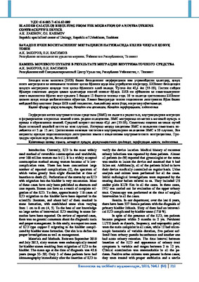
Figure 1.
KUB x-ray. A stone forming on the long arm of
the copper-T IUD is observed in the bladder.
Figure 2.
Post void x-ray during IV pyelogram: bladder
stone (arrowheads) formed on a partially intra-vesical mi-
grating contraceptive device and adhered to the blood
wall.

A.K. Zakirov, O.I. Kasimov
Биология ва тиббиёт муаммолари, 2015, №4.1 (85) 123
Figure 3.
Cystoscopy: Intravesical calcified IUD. Note
the thread of the IUD (arrow)
Figure 4.
Cystoscopy: Endoscopic view after stone frag-
mentation.
These findings stress the fact that placing an IUD
is an invasive procedure and should be performed by ex-
perienced doctors. In developing countries, the device is
often inserted by paramedics with variable skills (in fami-
ly planning facilities, and in rural areas), and follow-up
evaluations are irregular or absent that explain the im-
portance of our series. An IUD in the bladder can also be
the consequence of inserting it erroneously in the bladder
through the urethra (16). In our tenth case, cystoscopy
showed a totally mobile T-shaped bladder stone covering
the IUD with no mucosal lesions. These findings can be
consistent either with an early bladder perforation during
insertion of the device or an erroneous placement of the
IUD directly in the bladder by an inexperienced paramed-
ic lacking basic anatomical knowledge. In a literature
review by Kassab and Audra (17), a total of 165 cases of
migrating IUDs were collected, and only 23 were in the
bladder (14%). The incidence of uterine perforation was
reported to be 1.6 for 1,000 insertions (18). The true inci-
dence of perforation is most likely higher because of the
frequently asymptomatic nature of perforation (3). Migra-
tion into the bladder and secondary bladder stone for-
mation is very uncommon (2, 3, 9, 16, 18). It has been
reported in fewer than 70 cases in the literature. However,
less than half of these cases have resulted in bladder cal-
culus formation (4). Only 31 cases of complete or incom-
plete migration of IUD into the bladder and calculus for-
mation have been reported in the literature by 2006 (8).
From a review of the literature, it appears that most cases
of intravesical migration of IUDs have been associated
with the Copper T. However, we did not find any scien-
tific evidence to suggest that Copper T IUD is more
prone to such complications. It seems that hormone re-
leasing IUDs may also cause bladder perforation (19). To
reduce the incidence of such complications of IUD use,
new improved devices have become available during the
past few years. However, in many parts of the world like
Tunisia, Copper T devices are still frequently used. They
result in more severe inflammatory reaction and adhesion
(20). From a review of the literature it appears that any
foreign div placed in the proximity of the bladder has
the potential to migrate into bladder, e.g. vaginal dia-
phragm (21), cerclages (22), surgical clips used in hernia
repair (23), prosthetic slings (24) etc. Once an IUD has
eroded in to the bladder, it plays the role of matrix (24)
and the deposition of urinary sediments leads to calculus
formation on the device. However, the degree of encrus-
tation is variable and independent of the duration of the
device in the bladder (11). Thus, the device can either be
partially or completely encrusted with calculi. In only one
patient, there was complete encrustation of the device and
the stone measured 4 cm. The migrated IUD may remain
silent for a long period (25) and not be discovered until it
is found to be missing. Nine of our patients were noted to
have lost their IUD years before the development of uri-
nary tract symptomatology and, instead of carrying out
radiological investigations, they were told that the IUD
must have fallen out. Total or partial migration into the
bladder usually presents with LUTS as urinary frequency,
tenesmus, suprapubic pain, dysuria, hematuria, urinary
tract infection, urinary tract obstruction secondary to li-
thiasis, and urinary incontinence (2-4, 12). Persistent or
recurrent urinary tract infections are the most frequent
presentation, being the diagnosis of intravesical IUD a
finding during diagnostic workup (4, 16). Recurrent uri-
nary tract infections after appropriate antibiotic therapy
should also arouse suspicion of a foreign div in the uri-
nary tract (18). Primary vesical calculi are very unusual
in women and presence of intravesical stones should raise
suspicion of the presence of a foreign div (11). A care-
ful search for the lost device must be pre-formed with the
hope of preventing dangerous squeal. All IUDs are radio-
opaque; therefore, plane pelvic radiography may be used
for detection of the IUD (16) as well as US and Comput-
ed Tomography Scan. The main function of the plain film
is to show whether it is present within the patient (16).
Transvaginal US provides the best view for locat-
ing the IUD, but it restricts the space for itssimultaneous
removal (20). From our experience, we found that US can
be the investigation of choice for the diagnosis of in-
travesical migrated IUD. Moreover, the extent of myome-
trial and bladder wall perforation could be precisely de-
picted without the need for other invasive technique. For
other authors (27), noncontrast Computed Tomography
for detection of the site of the IUD and diagnosis of asso-
ciated complications such as stone or fistula is mandato-

Bladder calculus resulting from the migration of an intrauterine contraceptive device
124 Проблемы биологии и медицины, 2015, №4.1 (85)
ry. Cystoscopy is another method to detect the intravesi-
cal IUD and can help in more effectively planning the
optimal approach for removing the IUD. The adherence
of the IUD to the bladder wall, as well as the degree of
intravesical protrusion, can readily be identified (26) Cys-
toscopy will confirm the presence of an IUD in the blad-
der and, it might be possible to retrieve the IUD endo-
scopically (28). Although the management of the migrat-
ing IUD in asymptomatic patients remains controversial,
no controversy exists about the management of the IUD
that migrates into the bladder. All migrated IUDs in the
bladder must be removed. Even if the IUD migration is
asymptomatic, it should be removed for the prevention of
complications such as pelvic abscess, bladder rupture,
and adhesions. A migrant IUD in the bladder can be re-
moved by cystoscopy, as reported in some cases (2, 12,
16, 18). It can also be removed by suprapubic cystotomy
such as was used in other reports (3, 9). Open surgery
was generally used for the removal of the big stones
around IUD (17). However, open surgery has definitive
morbidity over the patient. We opted for endoscopic
management in all our patients. This was done because of
minimal invasiveness concern and for the reason that the
endoscopic management does not prevent conversion to
open surgery should it be a failure. Endocorporeal
lithoripsy and IUD extraction were easily performed in
our cases. Because the partially migrating IUD was either
under the bladder mucosa or within the bladder wall, gen-
tle traction on it allowed its complete extraction. The
punctuate bladder perforation caused by pulling the IUD
out of the bladder wall was insignificant and healed simp-
ly by prolonged urinary drainage. The most effective
treatment remains prevention. The IUD should be cor-
rectly inserted by an experienced person. A proper selec-
tion of patient and a thorough history and physical exam-
ination is crucial. If uterine rupture is suspected, US
should be performed to determine the probable location
of the rupture. Women should be informed of the poten-
tial complications and should be suggested to check the
device string regularly. If the string is not found, ab-
dominal radiography is required even in asymptomatic
patients. In any woman who has an IUD in situ and who
presents with LUTS, with recurrent urinary tract infec-
tions in spite of appropriate antibiotic therapy, the possi-
bility of intravesical migration of the device should be
included in the differential diagnosis.
Conclusions. Migration of an IUD into the bladder
is a low frequency complication. Persistent LUTS, recur-
rent or persistent urinary tract infections, and moreover,
bladder lithiasis, in women with IUD should raise the
suspicion of intravesical migration. Ultrasonography is
generally the first test in which suspicion is raised, and it
should be confirmed by cystoscopy. Endoscopic retrieval
is a feasible and safe procedure to achieve complete ex-
traction of the stone and IUD with very low morbidity for
the patient. To the best of our knowledge, we have re-
ported the largest series of bladder calculus resulting
from the migration of an intrauterine contraceptive device
managed endoscopically with excellent outcome.
References:
1. Thomalla JV. Perforation of urinary bladder by intrau-
terine device. Urology. 2000. 27:260-4.
2. Shokeir AA, el-Gharib MS, et al. Bladder stone: a
complication of intravesical migration of Lippes
loop. Scand J Urol Nephrol. 2013;27:279-80.
4. Serin IS, Basbug M, et al. Differential diagnosis of
intra uterine device migrating to bladder using radio-
graphic image of calculus formation and review of litera-
ture. Eur J Obstet Gynecol Reprod Biol. 2013;108:94-6.
5. Istanbulluoglu MO, Ozcimen EE, Ozturk B, et al.
Bladder perforation related to intrauterine device. J Chin
Med Assoc. 2010;71:207-9.
6. Mosher WD, Pratt WF. Contraceptive use in the Unit-
ed States, 1973-88. Patient Educ Couns. 2011;16:163-72.
7. Cheng D. The intrauterine device: still misunderstood
after all these years. South Med J. 2010;93:859-64.
8. Demirci D, Ekmekзioğlu O, Demirtaş A, et al. Big
bladder stones around an intravesical migrated intrauter-
ine device. Int Urol Nephrol. 2013;35:495-6.
9. Lu HF, Chen JH, Chen WC, et al. Vesicle calculus
caused by migrant intrauterine device. AJR Am J Roent-
genol. 2009;173:504-5.
10. Grimes DA. Intrauterine device and upper-genital-
tract infection. Lancet. 2010;356:1013-9.
BLADDER CALCULUS RESULTING FROM THE
MIGRATION OF AN INTRAUTERINE
CONTRACEPTIVE DEVICE
A.K. ZAKIROV, O.I. KASIMOV
Perforation of the uterus by an intrauterine contra-
ceptive device is not rare event, intravesical migration
and secondary stone formation is a very rare complica-
tion. We report a series of 60 women in whom an intrau-
terine contraceptive Copper-T device migrated from the
uterus to the bladder and resulted in formation of a stone.
Methods: Between September 2010 and July
2015, sixty women were treated for bladder stones be-
cause of migrated intrauterine contraceptive device. Di-
agnosis was established after performing pelvic ultraso-
nography and/or intravenous urogram. We describe histo-
ry, clinical course, diagnostic workup and treatment data
obtained from the hospital charts. Results: The mean age
was 42.6 years (33-59). Persistent lower urinary tract
symptoms were the main complaint in almost all the cas-
es, while 26 patients presented with macroscopic hematu-
ria. The interval between insertion of intrauterine contra-
ceptive device and onset of symptoms ranged from 2 to
12 yrs. Cystoscopy revealed partial intravesical position
of the intrauterine contraceptive device in 59 cases and an
entire intravesical intrauterine contraceptive device in one
case with calculus formation in all the cases. All patients
underwent endoscopic lithotripsy of the stone with ex-
traction of intrauterine contraceptive device. Procedures
went well with no complications. Patients received uri-
nary drainage for 10 days. Postoperative course was une-
ventful with a 2 years follow-up. Conclusions: Intrauter-
ine contraceptive device perforation to the bladder with
stone formation is a rare event. Persistent lower urinary
tract symptoms in women with intrauterine contraceptive
device should raise the suspicion of intravesical migra-
tion. Ultrasonography permits excellent depiction of in-
travesical migrated intrauterine contraceptive device.
Endoscopic retrieval is a feasible and safe procedure.
Key words:
Intrauterine Devices, Uterine Perfo-
ration, Urinary Bladder Calculi, Endoscopy.