Library search
Search Results
-
 The role of magnetic resonance imaging in the comprehensive radial diagnosis of volumetric masses of the eye organ
Catalog of abstractsRelevance of the problem. The difficulties of diagnostics of orbital diseases are well known. Especially difficult is intraspecies differentiation among the multitude of tumour, pseudotumour, inflammatory, vascular, endocrine and other diseases occurring here, manifested by the symptom complex of unilateral exophthalmos [Beradze I.N., 1978; Brovkina A.F., 1993].
Malignant intraocular neoplasms are the main cause of death of patients with diseases of the organ of vision, with 45-48% of patients dying from metastases in the first 5 years after enucleation [Alekseeva I.B., 1990, Barkhash S.A.1978, Brovkina A.F..1991, 1997; Keizer R.W.. Viclvoyc G.L.,1986],
Retinoblastoma is the most frequent malignant neoplasm in children. According to different authors, the frequency of its occurrence is 1 case per 14000 - 35000 newborns. [Bobrova N.F. and Vit V.V., 1993; Brovkina A.F., 1997; Provenzale J.M., et al., 1995; Skulski M., et al., 1997; Weber A.L., Mafee M.F, 1992; Wilms G., et al., 1989]. The frequency of patients with the most malignant intraocular tumour in adults - uveal melanoma has recently reached 7-9 people per 1 million population [Brovkina A.F., 1997; Kotslyansky E.O., 1989; Yushko N.A., Peskova L.I., Kalenich L.A., 1989; Peyster R.G., Augsburger J..I., Shields J.A., 1988; Romani A.. Baldeschi L., ct al 1998; Scott I.U., 1998].
The fundamental difference in treatment tactics, depending on the stage of development, size and topography of the tumour, as well as the seriousness of the prognosis in retinoblastomas and melanomas sharply increase the requirements for the accuracy of their differential diagnosis. At the same time, the number of diagnostic errors in ocular tumours continues to be 10-30% even when complex clinical and instrumental examination is applied in specialised ophthalmological centres [Ternovoy S.K., Panfilova G.V., Rogozhin V.A., 1979; Friedman F.E., Malyuta G.D., Kodzov M.V., 1995; Song G.X., 1991].
Widely used in ophthalmological practice traditional diagnostic methods (ophthalmoscopy, gonioscopy, diaphanoscopy, fluorescence angiography, laboratory tests) are insufficient to obtain comprehensive information about the localisation, nature of growth and prevalence of volumetric pathological formations of the eye and orbit. This circumstance and not quite satisfactory results of surgical treatment are the causes of high mortality of patients [Muratova T.T., Nigmanova N.H., Kozlovskaya G.M.. 1989, Naches A.I., 1980; Cheremisin V.M., Trufanov G.E., Kholin A.V., 1991]. Untimely or erroneous recognition of pathological processes of the orbit leads to a sharp deterioration of visual functions, up to blindness, and in some cases to the death of the patient [Yuzhakov A.M., Travkin A.G., Kiseleva O.A., 1991]. All this determines the importance of timely and accurate diagnosis of diseases of the orbit, on the one hand, and the difficulty of such diagnosis - on the other [Gabunia R.I., Kolesnikova E.K., Tumanov L.B., 1982].
The fact that the orbit is closed from direct inspection and palpation by bone walls and the eyeball, indicates the advantage of radial diagnostics in comparison with other methods of examination. In the arsenal of clinicians there is a great variety of methods of clinical-radial diagnostics of orbital pathology, however, at present the information in the literature about their resolving capabilities and significance in comparative aspect is incomplete and not fully studied. The priority of using one or another instrumental investigation, their sequence and expedient combination have not been determined yet. This makes it difficult to choose the optimal standardised approach for diagnosis and adequate treatment [Cheremisin V.M., Trufanov G.E., 1993, Weber A.L., Sabates N.R., 1996; Wenig V.M., Mafee M.F., 1998].
Thus, the study of these and other questions, contributing to the improvement of diagnostics and treatment of patients with neoplasms of the eye and ocular cavity, should be recognised as urgent urgent.
Purpose of the study. Comparative evaluation of magnetic resonance tomography capabilities and development of algorithms for complex radial diagnostics of volumetric formations of the visual organ. To solve this goal we set the following tasks.
1. To study the normal picture of the magnetic resonance image of the visual organ in comparison with other methods of visualisation.
2. To find out the possibilities of magnetic resonance tomography, ultrasound and computed tomography in detection and evaluation of intraocular neoplasms.
3. To determine the role and place of magnetic resonance tomography in differential diagnostics of volumetric pathological formations of the eye cavity in comparison with other radial methods of research.
4. To determine the indications and to develop an algorithm for the complex application of radiography, ultrasound, computer and magnetic resonance tomography for diagnostics of volumetric formations of the eye organ.
Scientific novelty.
The present work is the first to give a detailed and detailed description of the complex clinical and radiation examination, with generalisation and standardisation of magnetic resonance, computer and ultrasound semiotics of volumetric pathological formations of the eye and eye cavity. The conducted clinical and instrumental investigations allowed to determine the diagnostic value and resolving capabilities of each of the applied methods. The ultrasound, CT and MRI signs of volumetric formations of the eye organ were studied, clarified and supplemented taking into account the use of low-field magnetic field and general-purpose ultrasound apparatus. The developed standardised diagnostic algorithm of examination of patients with this pathology is new, thanks to which the pre-oppositional diagnosis of tumour and other diseases of the visual organ is improved and the total radiation load on the patient is reduced.
Conclusions
1. MPT will provide an opportunity to study the weight of the soft tissue and anatomical components of the ocular cavity, up to the optic nerve sheath and perineural liquor space, the orbital apex and chiasmal-sellar region, as well as to assess the condition of adjacent structures of the brain and facial skull. The method is limited in the evaluation of changes in the bony walls of the orbital cavity.
2. MRI is inferior in detecting characteristic signs of retinoblastoma (presence of calcification). The sensitivity of MRI was 66.6%, while for ultrasound and CT these values were 96.1 and 100%, respectively. But when the tumour spreads rstrobulbarly outside the eyeball (at 3-4 stages) the informativeness of MRI increases significantly. In uveal melanoma the sensitivity and specificity of MRI reaches 100%.
3. Both MRI and CT have a high detection rate (98.1% and 95.8% respectively) of benign orbital tumours of both primary and secondary origin. However, MRI is the preferred method of investigation. MRI is especially informative when a cranioorbital tumour and pseudotumour are suspected. The sensitivity of the method is 90.9% and 91.6%, respectively
4. In some cases ultrasound can be used to differentiate between encapsulated and diffuse neoplasms, which facilitates the diagnosis. However, when the pathological process is localised near the orbital apex, the diagnostic value of ultrasound decreases. In such cases it is advisable to use MRI.
5. In detection of primary and secondary malignant tumours of the orbital cavity both MRI and CT are quite informative (sensitivity 97,2% and 95,4% respectively), but the most comprehensive information about the state of bone walls will be provided by CT. When the process spreads intracranially, the value of MRI increases significantly, especially with the use of contrast enhancement.
6. The developed algorithm of complex clinical and radiation examination of patients with the use of ultrasound, CT and MRI is the most effective in the diagnosis of volumetric pathological formations of the eye and eye cavity, allowing to reduce to an adequate minimum the total radiation load on the patient and diagnostic period, excluding duplication of research techniques and choosing the most informative in each case, which in turn allows to develop appropriate treatment tactics and reduce the level of disability of the patient. -
 Improvement of diagnosis and treatment of patients with combined trauma of facial bone
Catalog of abstractsTopicality and demand of the subject of dissertation. In the world lat days chanchcd structures of trauma, increase the number of heavy combined traumas, which resulting in more heavy nature of simultaneous injuries of three , four or more anatomical regions, which creates difficulties in determining of the order of care and surgical tactics in patients with combined traumas of the facial skeleton bones (CTFSB). The syndrome of mutual burdening injuries of various anatomical regions, variety, hcavity and speed of the development of pathological process did difficulty of diagnosis of the CTFSB. Complexity of the clinical picture, features of the progress of post-traumatic shock, the development of traumatic disease cause difficulties which arise in the course of examination of patients and put tasks to the experts to find new ways of developing diagnostic algorithms and early surgical treatment of the CTFSB.
Frequency of CTFSB ranges from 34,8 to 63,3%. Fractures of orbit has been observed with an extremely high frequency (98%) in CTFSB, injury of the orbit is accompanied by damage of the eyeball and its subsidiary bodies has been observed in 66 % of eases. Consequences of eye injuries arc becoming the leading cause of disability and in 50% of eases could cause permanent loss of vision. By reason of death combined trauma take the third part after coronary heart diseases. Frequency of disfiguring defects and deformities of face occurs in 12 and 57%, disability in CTFSB reaches up to 23%. CTFSB, combined with TBI, causes up to 60% of deaths.
The causes of unsufficient results is non-availability of a diagnostic algorithm, which includes the most informative research methods, determining the order of interaction and priority of work of doctors of various specialties in CTFSB.
In some eases, requires specified an indications, character, scope, sequence and timing of surgical interventions, depending of the objective assessment of heaviness of injuries to various anatomical regions, prognosis criteria, the nature and heaviness of life-threatening consequences of combined trauma. The research work earned out within the framework of the achievement of the set by the Decree of the President of Republic of Uzbekistan “About measures on the further deepening reform the health care system” November 28, 2011, № PD-1652, maintenance of high-quality medical aid to the population under modem requirements and standards.In this regard the need for the development of algorithms of diagnosis and early methods of surgical treatment of patients with CTFSB constitute one of the important criteria demand the theme of dissertation.
Purpose of research is improvement of the diagnostic tactics and therapeutic interventions in patients with acute combined injuries of the facial bones according to the severity and location of the injury.
Scientific novelty of disscrtational research consists in the following: revealed the structure and features provide consistent care to patients with combined injuries in Republic of Uzbekistan;
The sequence of diagnostic and therapeutic measures, depending on the patient's general condition with CTFSB first determined by using created CT program "ADIL
developed innovative methods for early reduction and fixation of bone fragments in CTFSB;
identified endogenous factors, affecting on the wound process, disclosed the mechanisms of post-traumatic complications in CTFSB;
proved, that at 2 - 3rd days after the injury occurs the depression of cell and humoral immunity in the blood. Increases the level of proinflammatory cytokines, reduced the level of anti-inflammatory cytokine (in 2,8 at patients with heavy commonl condition. Increased levels of pro - and reducing anti - inflammatory cytokines is a poor prognostic factor in the development of inflammatory complications (bone wound suppuration, osteomyelitis of the jaw bones, soft tissue abscess);
patients with CTFSB at 2 - 3rd days after the injury occurs the depression of the content of protein and micronutrients (calcium, potassium and phosphorus) in the blood, which is a prognostic factor of the development of complications;
a scheme was developed for integrated medical correction of endogenous factors affecting on the development of posttraumatic complications;
1. CTFSB in 100% of cases combined with TBI, in 27.7 % with injuries of skeleton and internal injuries. In the diagnosis and treatment of patients with CTFSB should participate resuscitator, maxillofacial surgeon, neurosurgeon, ophthalmologist, and otolaryngologist. Primary debridement of wounds, reduction and fixation of bone fragments in patients in compensated state should be done within 3 hours after injury, while at subcompensated state - during the first day, and at the decompensated state - within 3 days.
2. With the CT program "ADIL" can determine the overall condition of patients in a short time. The most informative diagnostic criteria arc the general condition of patients, level of consciousness, hemodynamic stability, shock index and temperature gradient. The severity of the general condition of patients is directly dependent on the localization of the fracture of the facial bones. Multiple fractures of the upper and middle areas of the face arc the most serious injury in patients.
3. Patients with CTFSB in compensated and subcompensated state emergency surgical aid and diagnostic procedures should be performed in full volume (maxillofacial surgery, traumatology, neurosurgery, surgery, ophthalmology and otorhinolaryngologist), including the reduction and fixation of bone fragments in the first day. To patients with CTFSB in state decompensated should be performed at least diagnostic procedures, limiting the amount of emergency surgery. Reduction and fixation of bone fragments should be done after the restoration of function of vital organs and systems.
4. The method of choice for the treatment of depressed large bone fragments of facial bones is a titanium distractor, the use of which gives a good clinical and functional outcome.
5. When depressed fracture of the zygomatic arch application of the developed device will allow us to produce reduction and fixation of bone fragments in the early stages (within one day) with a good cosmetic result.
6. At patients with CTFSB in posttraumatic period (7- 14th day.) there arc a deep depression of CD3, CD4 cell composition, humoral factors and secretory immune system, increased necrosis factor CD95, increasing the levels of proin-flammatory (IL-6 ) and a decrease - anti- inflammatory (IL -10) cytokines. On 9-10th day reduced total protein, calcium, potassium and phosphorus in the blood .
7. Reduction of cellular and humoral immunity, increased proinflammatory cytokine and tumor necrosis factor, reducing the anti-inflammatory cytokine , the protein concentration in the blood, calcium, potassium and phosphorus arc predictors of complications.
8. Application of complex drug therapy within the 1-3 days after the injury with the inclusion of immune ( immunomoduline, ribomunil ), enzyme ( Voben-zym ) drugs osteoplastic materials allows to correct the violation of homeostasis, also used to prevent complications. -
 Improvement of surgical aspects in providing help for sufficients wi thcombined pelvic and femoral bone damages
Catalog of dissertations and abstractsThe aim of the research work is to improve the results of treatment of patients with combined injuries of the pelvis and femur, by developing tactical and technical aspects based on the severity of the injury and the severity of the condition.
The object of the study was 130 patients with injuries of the pelvic and hip bones with concomitant trauma, treated at the Republican Scientific Center for Emergency Medical Aid and its Samarkand branch for the period 2016-2021 years.
The scientific novelty of the research work is the following: the structure and frequency of combined injuries of the pelvis and femur in the general structure of injuries, in the structure of injuries to the pelvis and femur separately were evaluated. the risk factors for the development of unsatisfactory results of treatment of concomitant injuries of the pelvis and hip, based on traditional clinical and diagnostic standards, have been determined; a direct relationship has been proven in the dynamics of the condition of the victims and the prognosis, taking into account the type and nature of segmental injuries; the device for external fixation for stable functional minimally invasive osteosynthesis has been improved and the possibility of expanding the indications for surgical treatment for combined injuries of the pelvis and hip in the early period of traumatic disease has been proved; the technical advantages of a complete set of an improved rod device for external fixation have been proved, the pelvic and femoral versions of which make it possible to use them for effective stabilization of the pelvis and hip separately during anti-shock measures, and for the final reposition of bone fragments; the direct dependence of treatment results on the proposed tactics of providing trauma care at an early hospital stage, depending on the type, nature, severity of pelvic and hip injuries, and the severity of the condition has been proved.
The introduction of research results. Based on the results of scientific research to improve the surgical aspects of providing assistance to victims with concomitant injuries of the pelvis and femur: based on the results of the development of a device for the treatment of fractures, a patent for an invention was obtained from the Intellectual Property Agency of the Russian Federation "Apparatus for the treatment of combined fractures of the pelvic and hip bones" (patent No. 2749897 dated 06/18/2020). The results obtained made it possible to improve the tactics of surgical treatment of patients, to shorten the period of hospitalization and the period of postoperative rehabilitation, to ensure the possibility of patients with minimal economic costs; on the basis of the results of scientific research on the diagnosis and treatment of concomitant injuries of the pelvic and femur bones, methodological recommendations were approved "Method for the treatment of victims with concomitant injuries of the pelvis and hip, depending on the severity" (Conclusion of the Ministry of Health of the Republic of Uzbekistan No. 8 n-z / 288 dated August 31, 2021 of the year). The results obtained made it possible to improve the quality of wound diagnosis and rehabilitation of patients with injuries of the pelvic and hip bones in concomitant injury; approved methodological recommendations "Tactics of rendering assistance to victims with combined injuries of the pelvis and hip, taking into account the severity of the condition" (Conclusion of the Ministry of Health of the Republic of Uzbekistan No. 8 n-z / 288 of August 31, 2020). The results obtained made it possible to improve the tactical and technical aspects in the treatment of injuries to the pelvic and hip bones, based on the severity of the injury and the severity of the patient's condition.
Scientific results have been introduced into the practical activities of healthcare, in particular, the Samarkand branch of the Republican Specialized Scientific and Practical Medical Center of Traumatology and Orthopedics, the Jizzakh Branch of the Republican Scientific Center for Emergency Medical Aid, the Samarkand branch of the Republican Scientific Center for Emergency Medical Aid (certificate of the Ministry of Health No. 08-09 / 18979 dated December 02, 2021). The proposed tactics for the treatment of combined injuries of the pelvis and femur made it possible to reduce the incidence of postoperative complications of excellent and good long-term functional results from 66.1% to 92.6%.
The structure and scope of the dissertation. The dissertation consists of an introduction, five chapters, conclusions, practical recommendations, a list of referencesand applications. The volume of the text material of the work is 111 pages.
-
Subjects of the research: The diagnostics of data transmission systems’ (DTS) elements under operation terms, methods of the control of DTA (data transmission apparatus) and diagnostics of digital devises on the base of signature analysis.
Purpose of work: Investigation and development of effective control methods and diagnostic of data transmission systems’ elements.
.Method of research: On solving of given problems analytical and program methods of investigation have been used, including elaborated models and methodic with following processing and analyses of the received results. Analytical methods were based on probability theory, flow - chart theory, reliability theory, algebra logic theory, machine modeling theory.
The results obtained and their novelty: The cascade model of error source of discreet channel and the strategy of diagnostics and restoring the efficiency of DTS elements. The mathematic model of embedded control over DTS elements with and without self control and evaluation of volumes value and efficiency. Evaluation methodic of sample signatures reliability and calculation. The algorithms of despairs detection at signature analysis application, minimizing the time of search. An imitation model for evaluating the methods of compact testing and sample signatures shaping.
Practical value: The elaborated methodic, algorithms and programs are recommended for practical use at designing of control - diagnostic provision at the stage of operation of data transmission systems.
Degree of embed and economic cffcctivitv: The results of dissertation work have been adopted in AK «Uzbektelecom». The theoretical and practical results have been used at TU1T on «Telecommunications» specialty and 5A522205 - «Communication networks and control systems» specialty.
Field of application: The proposed methodic, algorithms and programs could be widely used at operation of data transmission systems, development of control - diagnostic supply of digital systems and devices of telecommunication equipment. -
 РАННЯЯ ДИАГНОСТИКА РАКА МОЛОЧНОЙ ЖЕЛЕЗЫ НА УРОВНЕ ПЕРВИЧНОГО ЗВЕНА ЗДРАВООХРАНЕНИЯРАННЯЯ ДИАГНОСТИКА РАКА МОЛОЧНОЙ ЖЕЛЕЗЫ НА УРОВНЕ ПЕРВИЧНОГО ЗВЕНА ЗДРАВООХРАНЕНИЯ
Modern Science and Research -
 Management of a higher educational institution as an object of economic diagnostics
Economics and innovative technologiesThe article studies the features of higher educational institutions as an object of economic diagnostics. The place of economic diagnostics in ensuring the effective operation of higher educational institutions and the importance of the principles of diagnostics in management are highlighted. The approaches to assessing the quality of higher education from the point of view of expected results in accordance with the interests of the participants in the educational process are substantiated. There are scientific conclusions and practical recommendations on the economic assessment of the quality of higher education.
-
Subject of the inquiry: 120 patients with inflammatory diseases of the maxillary sinuses (177 affected sinuses).
Aim of the inquiry: improve the diagnosis of inflammatory diseases of maxillary sinus diseases using B-modc ultrasonography
Methods of research: Ultrasonogrpahy, X-ray, magnetic resonance imaging.
The results achieved and their novelty: Based on the results of investigation of 120 patients with inflammatory diseases of the maxillary sinuses (177 affected sinuses) the following was found: in acute sinusitis (irrespective of the form) ultrasonography had quite low specificity - 50.0+6.7%. However the sensitivity of the method in catarrhal and purulent forms of acute sinusitis came to 75.0+11.6% and 93.0+3.9% respectively. In all the forms of chronic sinusitis the parameters indicated good informativeness of B-modc ultrasonography: hyperplastic - sensitivity 82.0+5.2%, specificity 95.0+3.0%; purulent - sensitivity 88.0+6.8%, specificity 79.0+8.5%; cysts and polyps - sensitivity 87.0+5.0%, specificity 93.0+3.8%. The research demonstrated possibilities of ultrasonography in the diagnosis of inflammatory diseases of the maxillary sinuses. Diagnostic value of B-modc ultrasonograhy was shown in different forms of maxillary sinusitis. Practically convenient interpretation technique of maxillary sonograms and protocol of examination were developed. Sonographic criteria were defined for differential diagnosis of different inflammatory diseases of the maxillary sinuses as well as the role of B-modc ultrasonography in the algorhithm of radiological investigation of the maxillary sinuses.
Practical value: improvement of maxillary sinusitis, decrease of patient’s exposure to radiation (unjustified use of X-ray in the absence of pathology), avoiding of invasive methods (diagnostic puncture in case of mucosal hyperplasia of cysts) Degree of inculcate: The results of the investigation were introduced in the Otorhynolaryngology and Radiology departments of the 1-st Tashkent Medical Institute Sphere of usage: radiology, otorhynolaryngology. -
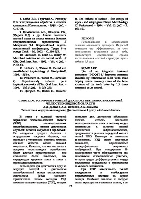
 Methodic of sonoelastography in the time of examination in patients with new formations in salivary glands used in this work.Also.we can see his high informative methodin time of differentiated detecting of bemgn.pre-cancer and malignant tumors.because of different staining of salivary gland tissue depending on degree of its malignancy and deformation coefficient.
 -
 Methodic of sonoelastography in the time of examination in patients with soft tissular new formations of jaw-facial area is used in this work. Also, we can see high information of the method in time of differentiated detecting of benign, pre-cancer and malignant tumors, because of different staining of tissue depending on degree of its malignancy and deformation coefficient.
 -
 Epidemiological-clinical peculiarities of comorbid pathology in hiv infected patients with neurocirculatory dystoniaThe research, which we conducted, is based on an epidemiological analysis of the results of a questionnaire of 341 HIV-infected persons with NCDs. This study was conducted using specially developed questionnaires, as well as instrumental diagnostic methods. The results of the research showed that the clinical diagnosis of NCDs in HIV-infected patients with comorbid pathology is a big challenge. In turn, this indicates the importance of carrying out complex instrumental and special diagnostic methods for patients in this group.
Doctor's Herald -
 Methodic of sonoelastography in the time of examination in patients with soft tissular new formations of jawfacial area is used in this work. Also, we can see high information of the method in time of differentiated detecting of benign, pre-cancer and malignant tumors, because of different staining of tissue depending on degree of its malignancy and deformation coefficient.
 -
 CHAKKA-PASTKI ZHAG BOGIMINING DYSFUNCTION AND DIAGNOSTICS MUTAHASSISLARNING FANLARARO YONDASHUVI ASOSIDA DAVOLASH SAMARADORLIGINI TAKOMILLASHTIRISHObjective: To evaluate the effectiveness of an interdisciplinary approach to the diagnosis and treatment of pathology of the temporomandibular joint. Material and methods: 1197 patients with pathology of the temporomandibular joint, 478 of them women and 719 men, turned to the dental center of the Bukhara State Medical Institute for specialized help and advice from orthopedic dentists, psychotherapists and neurologists of the Institute. Results: 46.1% of patients complained of tingling in the mouth when opening and / or closing it, including 45 (15.84%) on the right, 80 (28.57%) on the left. In the area of the temporomandibular joint. 49.5% of patients felt pain, discomfort, malocclusion was noted in 53.1%. and limited opening of the mouth was felt by 29.6% of the subjects. Conclusions: The diagnosis of patients with temporomandibular jomt dysfunction should be carried out taking mto account the participation of both biological, psychological and neurological factors in the development of the disease.
Stomatologiya -
 The article presents result of diagnostic and surgical treatment of II type of stomach ulcers of 354 pa-tients. II type of stomach ulcers i.e. combined ulcers stomach and duodenal. II type of stomach ulcers have disposition aggressively current, more often of whiplash, high number surgical complication and without result conservative treatment. X - Ray and endoscopy diagnostic have their of identity therefore needle to know some other feature Atypical resections of stomach should be choice operation
 -
 В статье унифицированы общие принципы диагностики острого гематогенного остеомиелита у детей. Описаны особенности клинического течения и дифференциальной диагностики. Обоснована диагностическая и лечебная тактика. Авторы считают, что остит и периостит являются осложнениями начальной стадии остеомиелита (то есть воспаления костного мозга), которые возникают по разным причинам, главная из которых — позднее начало комплексного лечения. Сделан вывод, что ранняя диагностика острого гематогенного остеомиелита у детей способствует снижению числа осложнений и летальности.
 -
 Climate and geographic characteristics of systemic lupus erythematosusSystemic lupus erythematosus (SLE) is one of the most severe systemic diseases of the connective tissue, more common at the age of 20-40 years, about 90% of cases are women [1;2]. Over the past decades, there has been an increase in the incidence of SLE (50-250 cases per 100 thousand population), which is due to the improvement of diagnostic methods, the identification of latent and chronic forms, and in some cases, overdiagnosis. The diagnostic criteria of the American College of Rheumatology (ACR) (1982, 1997) were developed for epidemiological studies and do not always allow one to exclude or confirm the diagnosis of SLE with absolute probability, especially in the early stages and in atypical variants of the disease [3;4] . When studying the medical records of one of the major medical institutions, J. Calyo et al. [5] found that among patients with a diagnosis of SLE, only two-thirds met the criteria for ACR, while about 10% had signs of lupus that were not sufficient to verify a reliable diagnosis, and 25% had a picture of fibromyalgia in combination with positive antinuclear antibodies (ANA), while even with long-term follow-up, no evolution of the disease into reliable SLE was noted. The question of improving the criteria for diagnosing SLE, “chameleon disease”, or “great disease simulator” has been repeatedly raised [6]. In 2012, a group of experts from Systemic Lupus International Collaborating Clinics (SLICC) presented the results of a long-term analysis of a large number of case histories (about 700 patients) with SLE and proposed modified diagnostic criteria for the diagnosis, expanded by introducing a number of dermatological , neurological signs with more accurate indicators [7;8].
Journal problems of biology and medicine -
 Diagnosis of chronic venous insufficiency of lower extremitiesChronic venous insufficiency of the lower extremities (CVI) is of great social and economic importance in society due to the high prevalence, duration of treatment and loss of working capacity. One of the main reasons for its formation is varicose veins. The frequency of varicose veins is 25-33% among women and 10-20% among men [4, 11]. Diagnosis and treatment of the disease in the early stages, before the addition of trophic disorders that require long-term treatment and significant costs, are important. So, in the UK, about six hundred million pounds sterling is spent annually on the treatment of venous ulcers, and in the USA - more than a billion dollars. Currently, in European countries, there has been a tendency to reduce the frequency of common forms of the disease due to the introduction of a program for early diagnosis of the disease [18, 20]
Journal problems of biology and medicine -
 Herpes simplex virus infection is one of the most common oral-facial infections. Despite modern methods of diagnosis and treatment, herpetic stomatitis remains an urgent problem in dentistry. The authors analyzed literature data to assess the clinical and pathological characteristics of HSV-1 infection. Relevant MED LINE studies published since 2006 have been reviewed. This search has been supplemented by a search for all bibliographic references from reference lists of included literature.
 -
 Treatment of fracture of the scaphoid bone and its consequences.(Literature analysis)
Journal of Biomedicine and PracticeThe materials of rendering aid to 78 patients with fractures of the scaphoid bone in different periods after the injury were analyzed. The patients underwent treatment in the Republican Specialized Scientific and Practical Medical Center of Traumatology and Orthopedics in the department "Hand and foot surgery" 2019-2021. In the process of diagnosis, 14 patients were diagnosed between the years 2019-2021 , with modern methods of clinical examination, early diagnostic weight loss and primary medical treatment, and in suspected cases, CSCT and MRT examinations have clarified the diagnosis.
-
 In order to increase the effectiveness of treatment at the modern, complex, market stage of the formation of the state system for the protection of citizens, it is necessary to systematize approaches to the tactics of conducting ballrooms. It is necessary to unify the optimal modes of the diagnostic and treatment process from the point of view of the implementation of standards (protocols) approved by evidence-based medicine. Certification and licensing of medical activities should be carried out through organizational, preventive, diagnostic, therapeutic and rehabilitation standards (protocols). We need a unified, national, standard approach of health authorities and institutions to the International Classification of Diseases and Related Health Problems (ICD X revision). Performers must be aware of accepted medical standards. Ignorance of medical standards is not an excuse for the lack of medical action or inaction of the doctor. To ensure adequate therapy, consultations are needed,substantiating the expediency of using certain diagnostic and treatment methods to ensure the quality of the diagnostic and treatment process in the modern level.
 -
 Leukoplakia is a fairly common pathology’ of the oral mucosa, with a tendency to malignant development in 20-30% of cases. This problem makes dentists think about cancer vigilance. At certain stages of development leukoplakia is reversible, timely diagnostics and treatment can prevent the development of malignancy. This paper includes the review of current data of published literature for the evaluation of clinical and pathological characteristics of leukoplakia as precancerous lesion.
 -
 Ранняя диагностика первичной глаукомы на основании тонометрии
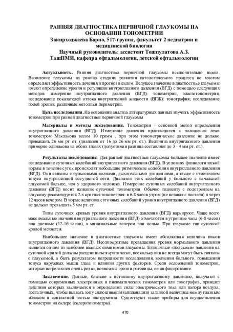
Prospects for the development of medicineРанняя диагностика первичной глаукомы исключительно важна. Выявление глаукомы на ранних стадиях развития патологического процесса во многом определяет эффективность лечения и прогноз в целом. Ведущее значение в диагностике глаукомы имеют определение уровня и регуляции внутриглазного давления (ВГД) с помощью следующих методов: измерение внутриглазного давления (ВГД): тонометрии, эластотонометрия; исследование показателей оттока внутриглазной жидкости (ВГЖ): тонография; исследование полей зрения: различные методики периметрии.
-
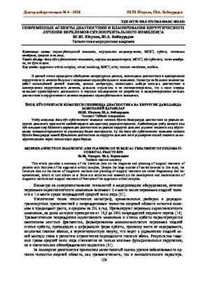 Modern aspects of diagnostic and planning of surgical treatment of zygomati- corbital fracturesThis article provides a summary of the literature data on the diagnosis and planning of surgical treatment of patients with fractures of the zygomatic-orbital complex. Despite the large number of works devoted to this topic, the literature data on the choice of diagnostic methods and planning of surgical treatment are rather fragmentary and not systematized, which in turn allows us to focus our activities and research on the development and modernization of diagnostic methods and surgical treatment of fractures of the zygomatic-orbital complex.
Doctor's Herald -
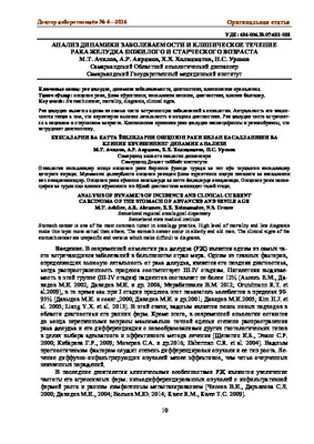 Analysis of dynamics of incidence and clinical current carcinoma of the stomach of advanced and senile ageStomach cancer is one of the most common tumor in oncology practice. High level of mortality and late diagnosis make this topic more actual than others. The stomach cancer occur in elderly and old men. The clinical signs of the stomach cancer are unspecific and various which make difficult to diagnosis.
Doctor's Herald -
 Methodic of sonoelastography in the time of examination in patients with soft tissular new formations of jaw-fecial area is used m this work. Also, we can see high information of the method m time of differentiated detecting of benign, pre-cancer and malignant tumors, because of different staining of tissue depending on degree of its malignancy and deformation coefficient.
 -
 Мультиметрическая ультрозвуковая диагностика нефробластом у детей дошкольного возраста региона Андижанской области
Экспериментальная медицина: сегодня и в будущемСреди всех опухолевых заболеваний у детей нефробластома занимает 4 место, уступая только гемобластозам, новообразованиям ЦНС и саркомам мягких тканей. Частота её составляет от 0,4 до 1 на 100 000 детей. Чаще всего ОВ встречается у детей в возрасте 2-5 лет, редко у новорожденных и еще реже у детей старше 8 лет, как казуистика - у взрослых. Обычно ОВ возникает спорадически, а в 2% случаев она имеет семейный характер. В 10% случаев она развивается у детей с пороками развития (чаще мочеполовой системы) или генетическими синдромами. Частота заболевания среди девочек и мальчиков одинакова. В 6-10% случаев встречается билатеральное поражение почек, преимущественно у детей до 2 лет. В 2% случаев опухоль поражает подковообразную почку. Описаны случаи внеорганного расположения опухоли. Важное значение для этой патологии имеет не просто правильная, но и ранняя диагностика заболевания. Необходимо не только оценивать общее состояние больного, уточнять локализацию опухоли и степень ее распространения, с обязательным морфологическим подтверждением диагноза, но и использовать как можно больше инструментальных методов диагностики. При этом необходимо соблюдение принципа ургентности.