Library search
Search Results
-
 Improvement of diagnosis and treatment of patients with combined trauma of facial bone
Catalog of abstractsTopicality and demand of the subject of dissertation. In the world lat days chanchcd structures of trauma, increase the number of heavy combined traumas, which resulting in more heavy nature of simultaneous injuries of three , four or more anatomical regions, which creates difficulties in determining of the order of care and surgical tactics in patients with combined traumas of the facial skeleton bones (CTFSB). The syndrome of mutual burdening injuries of various anatomical regions, variety, hcavity and speed of the development of pathological process did difficulty of diagnosis of the CTFSB. Complexity of the clinical picture, features of the progress of post-traumatic shock, the development of traumatic disease cause difficulties which arise in the course of examination of patients and put tasks to the experts to find new ways of developing diagnostic algorithms and early surgical treatment of the CTFSB.
Frequency of CTFSB ranges from 34,8 to 63,3%. Fractures of orbit has been observed with an extremely high frequency (98%) in CTFSB, injury of the orbit is accompanied by damage of the eyeball and its subsidiary bodies has been observed in 66 % of eases. Consequences of eye injuries arc becoming the leading cause of disability and in 50% of eases could cause permanent loss of vision. By reason of death combined trauma take the third part after coronary heart diseases. Frequency of disfiguring defects and deformities of face occurs in 12 and 57%, disability in CTFSB reaches up to 23%. CTFSB, combined with TBI, causes up to 60% of deaths.
The causes of unsufficient results is non-availability of a diagnostic algorithm, which includes the most informative research methods, determining the order of interaction and priority of work of doctors of various specialties in CTFSB.
In some eases, requires specified an indications, character, scope, sequence and timing of surgical interventions, depending of the objective assessment of heaviness of injuries to various anatomical regions, prognosis criteria, the nature and heaviness of life-threatening consequences of combined trauma. The research work earned out within the framework of the achievement of the set by the Decree of the President of Republic of Uzbekistan “About measures on the further deepening reform the health care system” November 28, 2011, № PD-1652, maintenance of high-quality medical aid to the population under modem requirements and standards.In this regard the need for the development of algorithms of diagnosis and early methods of surgical treatment of patients with CTFSB constitute one of the important criteria demand the theme of dissertation.
Purpose of research is improvement of the diagnostic tactics and therapeutic interventions in patients with acute combined injuries of the facial bones according to the severity and location of the injury.
Scientific novelty of disscrtational research consists in the following: revealed the structure and features provide consistent care to patients with combined injuries in Republic of Uzbekistan;
The sequence of diagnostic and therapeutic measures, depending on the patient's general condition with CTFSB first determined by using created CT program "ADIL
developed innovative methods for early reduction and fixation of bone fragments in CTFSB;
identified endogenous factors, affecting on the wound process, disclosed the mechanisms of post-traumatic complications in CTFSB;
proved, that at 2 - 3rd days after the injury occurs the depression of cell and humoral immunity in the blood. Increases the level of proinflammatory cytokines, reduced the level of anti-inflammatory cytokine (in 2,8 at patients with heavy commonl condition. Increased levels of pro - and reducing anti - inflammatory cytokines is a poor prognostic factor in the development of inflammatory complications (bone wound suppuration, osteomyelitis of the jaw bones, soft tissue abscess);
patients with CTFSB at 2 - 3rd days after the injury occurs the depression of the content of protein and micronutrients (calcium, potassium and phosphorus) in the blood, which is a prognostic factor of the development of complications;
a scheme was developed for integrated medical correction of endogenous factors affecting on the development of posttraumatic complications;
1. CTFSB in 100% of cases combined with TBI, in 27.7 % with injuries of skeleton and internal injuries. In the diagnosis and treatment of patients with CTFSB should participate resuscitator, maxillofacial surgeon, neurosurgeon, ophthalmologist, and otolaryngologist. Primary debridement of wounds, reduction and fixation of bone fragments in patients in compensated state should be done within 3 hours after injury, while at subcompensated state - during the first day, and at the decompensated state - within 3 days.
2. With the CT program "ADIL" can determine the overall condition of patients in a short time. The most informative diagnostic criteria arc the general condition of patients, level of consciousness, hemodynamic stability, shock index and temperature gradient. The severity of the general condition of patients is directly dependent on the localization of the fracture of the facial bones. Multiple fractures of the upper and middle areas of the face arc the most serious injury in patients.
3. Patients with CTFSB in compensated and subcompensated state emergency surgical aid and diagnostic procedures should be performed in full volume (maxillofacial surgery, traumatology, neurosurgery, surgery, ophthalmology and otorhinolaryngologist), including the reduction and fixation of bone fragments in the first day. To patients with CTFSB in state decompensated should be performed at least diagnostic procedures, limiting the amount of emergency surgery. Reduction and fixation of bone fragments should be done after the restoration of function of vital organs and systems.
4. The method of choice for the treatment of depressed large bone fragments of facial bones is a titanium distractor, the use of which gives a good clinical and functional outcome.
5. When depressed fracture of the zygomatic arch application of the developed device will allow us to produce reduction and fixation of bone fragments in the early stages (within one day) with a good cosmetic result.
6. At patients with CTFSB in posttraumatic period (7- 14th day.) there arc a deep depression of CD3, CD4 cell composition, humoral factors and secretory immune system, increased necrosis factor CD95, increasing the levels of proin-flammatory (IL-6 ) and a decrease - anti- inflammatory (IL -10) cytokines. On 9-10th day reduced total protein, calcium, potassium and phosphorus in the blood .
7. Reduction of cellular and humoral immunity, increased proinflammatory cytokine and tumor necrosis factor, reducing the anti-inflammatory cytokine , the protein concentration in the blood, calcium, potassium and phosphorus arc predictors of complications.
8. Application of complex drug therapy within the 1-3 days after the injury with the inclusion of immune ( immunomoduline, ribomunil ), enzyme ( Voben-zym ) drugs osteoplastic materials allows to correct the violation of homeostasis, also used to prevent complications. -
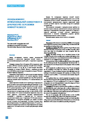 The purpose of the study is to assess the possibilities of sonography in the diagnosis of fractures of the mandible using a functional test with the opening and closing of the mouth. Materials and methods: 96 patients, aged from 6 up to 59 years were examined, 56 of them had fractures of the mandible. X-ray films. MSCT scans were taken for all patients and were compared with the ultrasonographicfindings. Results: All patients showed one or two-sided fracture of the mandible. Fractures of the mandible were combined with injuries of the middle zone of the face in 4 cases Isolated fractures of the mandible were detected in 17. and multiple fractures in 35 patients Among fractures of the mandible prevailed fractures of the angle (in 22). the articular process (in 16). parasymphysis (in 19). the branch (in 11). the corpus (8). the coronoid process (in 3), the symphysis of the mandible (in 6 cases). MSCT revealed 82 mandibular fractures. X-ray 77. and sonography 80, the sensitivity of the latter two methods was 93.9% and 97.5%. respectively. In 13 cases of unbiased fractures, when difficulties arose, the technigue of functional sonography with opening and closing of the mouth was used In addition. 11 more fractures were revealed using this technigue In 34 patients, there was a need for repeated sonography to assess the state of bone fragments after repositioning. The state of the fragments was satisfactory at 19 and unsatisfactory in 15 cases. In 2 patients, sonography was used intraoperatively to assess the adeguacy of bone fragments during reposition of the mandible. Conclusion: On sonograms, fractures of the mandible are manifested by interrupting the outer cortical layer with or without displacement of bone fragments, displacement of bone fragments when using functional loads: dislocation of the head of the articular process by the absence of its contour in the projection of the joint fossa. The use of sonography with functional tests on the mandible increases the sensitivity of the method in the diagnosis of fractures. Ultrasonic monitoring of the adeguacy of open and closed reposition of fragments improves the effectiveness of interventions and allows timely elimination of the causes of unsatisfactory standing of fragments.
 -
 Аннотация. По литературным источникам зарубежных и отечественных авторов изучены факторы стабильности костного аугментата. Исследователями доказано, что, независимо от происхождения аутогенного блока, использование методов направленной костной регенерации способствует лучшей стабилизации костного аугментата. Обеспечение минимального промежутка между ограничивающими структурами и костными наполнителями позволит повысить клиническую эффективность процедуры твердоткаппой аугментации при реабилитации стоматологических пациентов.
 -
 ВЛИЯНИЕ ИНФЛЯЦИИ И КУРСА ВАЛЮТ НА ФОНДОВЫЙ РЫНОКОбеспечение социально-экономического развития требует от государства проведения эффективной налогово-бюджетной, денежно-кредитной и структурной политики. Любая экономика устойчиво развивается только в условиях макроэкономической стабильности, элементами которой являются сбалансированный бюджет, низкая инфляция и стабильность внешнего сектора экономики. Во всем мире задача по обеспечению стабильности цен традиционно возлагается на центральный банк, так как воздействие на совокупный спрос в экономике более всего оказывает денежно-кредитная политика.
Priority directions, modern trends and prospects of the development of the financial market -
 This article describes the author's thoughts and opinions about the impact of women's gender enlightenment on social stability.
 -
 The article describes the issues of ensuring economic stability in industrial enterprises, especially in the textile industry, in particular, the content, essence and importance of the concept of "economic stability" and the factors influencing it
 -
 The role of magnetic resonance imaging in the comprehensive radial diagnosis of volumetric masses of the eye organ
Catalog of abstractsRelevance of the problem. The difficulties of diagnostics of orbital diseases are well known. Especially difficult is intraspecies differentiation among the multitude of tumour, pseudotumour, inflammatory, vascular, endocrine and other diseases occurring here, manifested by the symptom complex of unilateral exophthalmos [Beradze I.N., 1978; Brovkina A.F., 1993].
Malignant intraocular neoplasms are the main cause of death of patients with diseases of the organ of vision, with 45-48% of patients dying from metastases in the first 5 years after enucleation [Alekseeva I.B., 1990, Barkhash S.A.1978, Brovkina A.F..1991, 1997; Keizer R.W.. Viclvoyc G.L.,1986],
Retinoblastoma is the most frequent malignant neoplasm in children. According to different authors, the frequency of its occurrence is 1 case per 14000 - 35000 newborns. [Bobrova N.F. and Vit V.V., 1993; Brovkina A.F., 1997; Provenzale J.M., et al., 1995; Skulski M., et al., 1997; Weber A.L., Mafee M.F, 1992; Wilms G., et al., 1989]. The frequency of patients with the most malignant intraocular tumour in adults - uveal melanoma has recently reached 7-9 people per 1 million population [Brovkina A.F., 1997; Kotslyansky E.O., 1989; Yushko N.A., Peskova L.I., Kalenich L.A., 1989; Peyster R.G., Augsburger J..I., Shields J.A., 1988; Romani A.. Baldeschi L., ct al 1998; Scott I.U., 1998].
The fundamental difference in treatment tactics, depending on the stage of development, size and topography of the tumour, as well as the seriousness of the prognosis in retinoblastomas and melanomas sharply increase the requirements for the accuracy of their differential diagnosis. At the same time, the number of diagnostic errors in ocular tumours continues to be 10-30% even when complex clinical and instrumental examination is applied in specialised ophthalmological centres [Ternovoy S.K., Panfilova G.V., Rogozhin V.A., 1979; Friedman F.E., Malyuta G.D., Kodzov M.V., 1995; Song G.X., 1991].
Widely used in ophthalmological practice traditional diagnostic methods (ophthalmoscopy, gonioscopy, diaphanoscopy, fluorescence angiography, laboratory tests) are insufficient to obtain comprehensive information about the localisation, nature of growth and prevalence of volumetric pathological formations of the eye and orbit. This circumstance and not quite satisfactory results of surgical treatment are the causes of high mortality of patients [Muratova T.T., Nigmanova N.H., Kozlovskaya G.M.. 1989, Naches A.I., 1980; Cheremisin V.M., Trufanov G.E., Kholin A.V., 1991]. Untimely or erroneous recognition of pathological processes of the orbit leads to a sharp deterioration of visual functions, up to blindness, and in some cases to the death of the patient [Yuzhakov A.M., Travkin A.G., Kiseleva O.A., 1991]. All this determines the importance of timely and accurate diagnosis of diseases of the orbit, on the one hand, and the difficulty of such diagnosis - on the other [Gabunia R.I., Kolesnikova E.K., Tumanov L.B., 1982].
The fact that the orbit is closed from direct inspection and palpation by bone walls and the eyeball, indicates the advantage of radial diagnostics in comparison with other methods of examination. In the arsenal of clinicians there is a great variety of methods of clinical-radial diagnostics of orbital pathology, however, at present the information in the literature about their resolving capabilities and significance in comparative aspect is incomplete and not fully studied. The priority of using one or another instrumental investigation, their sequence and expedient combination have not been determined yet. This makes it difficult to choose the optimal standardised approach for diagnosis and adequate treatment [Cheremisin V.M., Trufanov G.E., 1993, Weber A.L., Sabates N.R., 1996; Wenig V.M., Mafee M.F., 1998].
Thus, the study of these and other questions, contributing to the improvement of diagnostics and treatment of patients with neoplasms of the eye and ocular cavity, should be recognised as urgent urgent.
Purpose of the study. Comparative evaluation of magnetic resonance tomography capabilities and development of algorithms for complex radial diagnostics of volumetric formations of the visual organ. To solve this goal we set the following tasks.
1. To study the normal picture of the magnetic resonance image of the visual organ in comparison with other methods of visualisation.
2. To find out the possibilities of magnetic resonance tomography, ultrasound and computed tomography in detection and evaluation of intraocular neoplasms.
3. To determine the role and place of magnetic resonance tomography in differential diagnostics of volumetric pathological formations of the eye cavity in comparison with other radial methods of research.
4. To determine the indications and to develop an algorithm for the complex application of radiography, ultrasound, computer and magnetic resonance tomography for diagnostics of volumetric formations of the eye organ.
Scientific novelty.
The present work is the first to give a detailed and detailed description of the complex clinical and radiation examination, with generalisation and standardisation of magnetic resonance, computer and ultrasound semiotics of volumetric pathological formations of the eye and eye cavity. The conducted clinical and instrumental investigations allowed to determine the diagnostic value and resolving capabilities of each of the applied methods. The ultrasound, CT and MRI signs of volumetric formations of the eye organ were studied, clarified and supplemented taking into account the use of low-field magnetic field and general-purpose ultrasound apparatus. The developed standardised diagnostic algorithm of examination of patients with this pathology is new, thanks to which the pre-oppositional diagnosis of tumour and other diseases of the visual organ is improved and the total radiation load on the patient is reduced.
Conclusions
1. MPT will provide an opportunity to study the weight of the soft tissue and anatomical components of the ocular cavity, up to the optic nerve sheath and perineural liquor space, the orbital apex and chiasmal-sellar region, as well as to assess the condition of adjacent structures of the brain and facial skull. The method is limited in the evaluation of changes in the bony walls of the orbital cavity.
2. MRI is inferior in detecting characteristic signs of retinoblastoma (presence of calcification). The sensitivity of MRI was 66.6%, while for ultrasound and CT these values were 96.1 and 100%, respectively. But when the tumour spreads rstrobulbarly outside the eyeball (at 3-4 stages) the informativeness of MRI increases significantly. In uveal melanoma the sensitivity and specificity of MRI reaches 100%.
3. Both MRI and CT have a high detection rate (98.1% and 95.8% respectively) of benign orbital tumours of both primary and secondary origin. However, MRI is the preferred method of investigation. MRI is especially informative when a cranioorbital tumour and pseudotumour are suspected. The sensitivity of the method is 90.9% and 91.6%, respectively
4. In some cases ultrasound can be used to differentiate between encapsulated and diffuse neoplasms, which facilitates the diagnosis. However, when the pathological process is localised near the orbital apex, the diagnostic value of ultrasound decreases. In such cases it is advisable to use MRI.
5. In detection of primary and secondary malignant tumours of the orbital cavity both MRI and CT are quite informative (sensitivity 97,2% and 95,4% respectively), but the most comprehensive information about the state of bone walls will be provided by CT. When the process spreads intracranially, the value of MRI increases significantly, especially with the use of contrast enhancement.
6. The developed algorithm of complex clinical and radiation examination of patients with the use of ultrasound, CT and MRI is the most effective in the diagnosis of volumetric pathological formations of the eye and eye cavity, allowing to reduce to an adequate minimum the total radiation load on the patient and diagnostic period, excluding duplication of research techniques and choosing the most informative in each case, which in turn allows to develop appropriate treatment tactics and reduce the level of disability of the patient. -
 APPLICATION OF BIOENGINEERED CONSTRUCTION AND 3-D TECHNOLOGY FOR MANDIBULAR DEFECT REPLACEMENTObjective. To develop the method of the lower jaw defects reconstruction of different origin using the vascularized autograft combined with the nonwoven titanium material. Material and methods. We analyzed the treatment of posttraumatic deformities of the lower jaw in 47 patients. The reconstruction technique can be used both with and without massive soft tissue flaps on the vascular pedicle. Results and discussion. The technique of the individual bioengineered construction fabrication from NTMSP with through porosity for mandibular defect replacement was developed and technologically perfected. We have developed a technique for producing a complex implant to replace the lower jaw after half resection of the lower jaw with cxarticulation of the condylar head. The technology allows to obtain bioengineered composition with the structure completely corresponding to the human bone tissue, with the possibility of dental rehabilitation in polyclinic conditions, with complete consolidation of the bone wound edges; it doesn’t require taking bone autografts from other regions and as a consequence, it doesn't cause trauma or reduce physical characteristics of the bones used for that purpose. Conclusion. The suggested by us technique of the jaw defects replacement with the use of the vascularised bone autograft in combination with the NTMSP can be used in the clinical practice as the alternative when choosing the method of reconstruction, as it is more simple and less traumatic. Individual fabrication of the bioengineered construct allows for increased cosmetic and functional results.
Medicine and innovations -
 Improvement of surgical aspects in providing help for sufficients wi thcombined pelvic and femoral bone damages
Catalog of dissertations and abstractsThe aim of the research work is to improve the results of treatment of patients with combined injuries of the pelvis and femur, by developing tactical and technical aspects based on the severity of the injury and the severity of the condition.
The object of the study was 130 patients with injuries of the pelvic and hip bones with concomitant trauma, treated at the Republican Scientific Center for Emergency Medical Aid and its Samarkand branch for the period 2016-2021 years.
The scientific novelty of the research work is the following: the structure and frequency of combined injuries of the pelvis and femur in the general structure of injuries, in the structure of injuries to the pelvis and femur separately were evaluated. the risk factors for the development of unsatisfactory results of treatment of concomitant injuries of the pelvis and hip, based on traditional clinical and diagnostic standards, have been determined; a direct relationship has been proven in the dynamics of the condition of the victims and the prognosis, taking into account the type and nature of segmental injuries; the device for external fixation for stable functional minimally invasive osteosynthesis has been improved and the possibility of expanding the indications for surgical treatment for combined injuries of the pelvis and hip in the early period of traumatic disease has been proved; the technical advantages of a complete set of an improved rod device for external fixation have been proved, the pelvic and femoral versions of which make it possible to use them for effective stabilization of the pelvis and hip separately during anti-shock measures, and for the final reposition of bone fragments; the direct dependence of treatment results on the proposed tactics of providing trauma care at an early hospital stage, depending on the type, nature, severity of pelvic and hip injuries, and the severity of the condition has been proved.
The introduction of research results. Based on the results of scientific research to improve the surgical aspects of providing assistance to victims with concomitant injuries of the pelvis and femur: based on the results of the development of a device for the treatment of fractures, a patent for an invention was obtained from the Intellectual Property Agency of the Russian Federation "Apparatus for the treatment of combined fractures of the pelvic and hip bones" (patent No. 2749897 dated 06/18/2020). The results obtained made it possible to improve the tactics of surgical treatment of patients, to shorten the period of hospitalization and the period of postoperative rehabilitation, to ensure the possibility of patients with minimal economic costs; on the basis of the results of scientific research on the diagnosis and treatment of concomitant injuries of the pelvic and femur bones, methodological recommendations were approved "Method for the treatment of victims with concomitant injuries of the pelvis and hip, depending on the severity" (Conclusion of the Ministry of Health of the Republic of Uzbekistan No. 8 n-z / 288 dated August 31, 2021 of the year). The results obtained made it possible to improve the quality of wound diagnosis and rehabilitation of patients with injuries of the pelvic and hip bones in concomitant injury; approved methodological recommendations "Tactics of rendering assistance to victims with combined injuries of the pelvis and hip, taking into account the severity of the condition" (Conclusion of the Ministry of Health of the Republic of Uzbekistan No. 8 n-z / 288 of August 31, 2020). The results obtained made it possible to improve the tactical and technical aspects in the treatment of injuries to the pelvic and hip bones, based on the severity of the injury and the severity of the patient's condition.
Scientific results have been introduced into the practical activities of healthcare, in particular, the Samarkand branch of the Republican Specialized Scientific and Practical Medical Center of Traumatology and Orthopedics, the Jizzakh Branch of the Republican Scientific Center for Emergency Medical Aid, the Samarkand branch of the Republican Scientific Center for Emergency Medical Aid (certificate of the Ministry of Health No. 08-09 / 18979 dated December 02, 2021). The proposed tactics for the treatment of combined injuries of the pelvis and femur made it possible to reduce the incidence of postoperative complications of excellent and good long-term functional results from 66.1% to 92.6%.
The structure and scope of the dissertation. The dissertation consists of an introduction, five chapters, conclusions, practical recommendations, a list of referencesand applications. The volume of the text material of the work is 111 pages.
-
 On the materials used to replace bone defects and eliminate deformities of the maxillofacial regionIn clause the brief analysis osteoplastic of materials used now, for elimination of defects and deformations of maxillofacial area is resulted. The carried out comparative analysis shows the negative and positive parties of each material. On the basis of the carried out analysis, optimal osteoplastic by a material, is glasscrystal a material biositall.
Stomatologiya -
 FORMATION OF IDEOLOGICAL IMMUNITY IN YOUNG PEOPLE IS A FACTOR OF SECURITY AND STABILITYIn today's interconnected world, stability in society is influenced by various factors, including ideological immunity and security. Ideological immunity refers to the resistance of individuals to change their core beliefs and ideas, while security encompasses measures implemented to protect individuals and institutions from external threats. This scientific article examines the dynamic relationship between ideological immunity and security, highlighting their mutual influence on societal stability. By analyzing the psychological and socio-political aspects of ideological immunity and security, this study aims to shed light on the complex interplay between these factors and their implications for maintaining stability in contemporary societies.
Modern Science and Research -
 THE ROLE OF THE KHANATES OF CENTRAL ASIA IN THE FORMATION OF THE GREAT SILK ROADThis study analyzes the political and economic functions of the medieval khanates in Central Asia and their contribution to the development of transcontinental trade networks known as the Silk Road. With stable governance and security provided by militarized khanates such as the Khwarezmians, Karakhanids and the Mongol Empire, land routes flourished connecting China, India, Persia and the Mediterranean world. Merchants used this stability to expand trade in luxury goods and necessities, including silk, spices, glassware, paper, and more. The khanates grew wealthy by serving as hubs where goods from different empires were stored, sorted, and redistributed along routes. Through taxes and transit fees, the Central Asian powers financed further infrastructure and diplomatic efforts to maintain open corridors, thereby institutionalizing the Silk Road system throughout Eurasia for centuries.
Modern Science and Research -
This article reveals the essence of such terms as stability and security.
-
 Определение стабильности дентальных имплантатов в эксперименте методом голографической интерферометрии
Actual problems of dentistry and maxillofacial surgery 4Стабильность дентальных имплантатов в процессе их остеоинтеграции делится на первичную и вторичную. Первичная или механическая стабильность достигается за счёт механического соединения конструкции дентального имплантата с костной тканью.
-
Today, the attention of all countries of the world and international organizations is directed to the Middle East region. The Middle East is a strategically important region and has enormous energy potential, including a large amount of hydrocarbons. In this regard, we can say that the stability of this region directly affects the stability of the world as a whole. Historically, the Middle East has been a very attractive territory for various reasons. The one who controlled this strategically important zone possessed tremendous power and wealth. Despite the declining role of the region as a crossroads between different continents in connection with the development of the global system of transport communications, the strategic importance of the Middle East in recent decades has not only not decreased, but, on the contrary, has grown significantly. Also, the energy factor remains the main reason for the increased attention to the region. As you know, the technologies of modern civilization are almost completely dependent on the extraction of organic energy. Therefore, the distribution of resources has gradually turned into one of the most important elements in building the current system of international relations. The desire to establish control over them is the cause of numerous conflicts in the region.
-
In the process of forming the foundations of a socially oriented market economy in the Republic of Uzbekistan, increasing the welfare of the population by ensuring sustainable economic growth requires the solution of a number of more complex economic problems. Among them, ensuring macroeconomic stability and improving the
investment climate are of particular importance. -
 The article shows the provision of high economic growth is based on figures as a result of the ongoing economic reforms in the Republic. The system of indicators of providing economic stability is given and analyzed.
 -
 Chemical stability of anion exchange materials based on polyvinyl chloride
"ALFRAGANUS" international scientific journalOn an industrial scale, mechanical engineering materials are required to have different physico-chemical properties depending on the field of application, technological processes and the task they perform. Ion-exchange materials are used in hydrometallurgical industry in highly aggressive environments, where ion-exchange materials are mainly required for high chemical and thermal stability. In our discussion below, information is given about the important chemical stability of polyvinyl chloride (PVC)-based anion exchange material
-
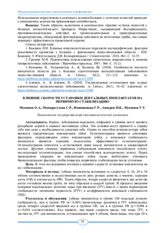 Влияние скорости установки дентальных имплантатов на первичную стабилизацию
Topical issues of surgical dentistry and dental implantologyКариес, заболевания пародонта, инфекции и травмы могут вызвать резорбцию корней и альвеол постоянных зубов. Эти условия могут привести к отрыву зуба или даже к необходимости удаления зуба. Одним из способов лечения потери зубов является хирургическая имплантация зубов. Остеоинтеграция является ключевым фактором, определяющим успех имплантации зубов, а начальная стабильность имплантата является хорошим показателем эффективности остеоинтеграции. Остеоинтеграция относится к естественной связи между имплантатом и альвеолярной костью. Другими словами, первичная стабилизация имплантата способствует успеху последующей остеоинтеграции, тем самым способствуя долгосрочному успеху. Перед проведением операции по имплантации зубов также необходимо учитывать биомеханические факторы, чтобы повысить первичную стабилизацию после установки.
-
 Economic and mathematical methods for assessing the economic sustainability of industrial enterprisesThis article describes how to ensure the economic sustainability of the industrial enterprises of Uzbekistan. Also, scientists analyzed the approaches of economists estimation of economic and mathematical methods of «sustainability» and «economic sustainability» and their studies. On the basis of these data, the author proposed a system of evaluation of the economic sustainability in industrial enterprises. Description of the comparative evolution of the industrial enterprises of statistical and dynamic methods in economic sustainability.
Economics and innovative technologies -
 Methodology for assessing the financial stability of industrial enterprisesIn the article have been discussed the theoretical aspects of the financial sustainability of enterprises and systematizes the assessment of financial sustainability based on the specifics of industrial enterprises. In addition, was assessed and analyzed financial sustainability of industrial enterprises. Based on the results of the analysis, were proposed scientific recommendations and practical recommendations aimed at improving the financial sustainability of industrial enterprises.
Economics and innovative technologies -
 Ensuring sustainable economic development of industrial enterprises in the world pandemic
Economics and innovative technologiesThe article examines the problems of sustainable development of industrial enterprises, analyzes the factors affecting the sustainability of enterprise development, gives a definition of various types of economic sustainability during a pandemic, and highlights the internal and external factors of the sustainability of enterprises. It is shown that of the most
important factors affecting the sustainability of the development of industrial enterprises, first of all, economic stability stands out. The factors in the conditions of a deteriorating economic situation are investigated and identified, which are advisable to take into account when solving the problem of sustainable development of industrial enterprises. When conducting the study, the main sources of data were the materials of state statistics committee of the Republic of Uzbekistan. The methodological developments are based on comparative methods of analysis. Proposals have been developed to ensure economic sustainable development of industrial enterprises during a pandemic. -
 Improving the treatment of methods odontogenic cysts of the jawsDuring the period 2010-2015 were observed 42 patients with odontogenic cysts of the jaws. From them, extended defects of the jaws have been observed in 32 patients. For suffusion of osseous cavity with platelets rich plasma of the blood were used to these patients. In the first 12 hours of postoperative period the infiltra-tion margins of the wound were expressed in 2 patients. Elimination of postoperative wealing were observed for 6-7 days. In a month and following periods of observation there wasn’t any complaints by patients, the mu-cous membrane of surgical operation was in pale pink color, without wealing. In the 6th month were observed full restoration of defects in rentgenological checkup. The usage of osteoplastic materials in right way will con-tributes for restoration of large bone defects with organotypical bone formation, corresponding the anatomy of the area, in optimal period, which makes short the period of post operational rehabilitation period of the pa-tients and will contributes early functional exertion of the organs
Journal problems of biology and medicine -
 КОМПЛЕКСНЫЙ ПОДХОД ПРИ ДЕНТАЛЬНОЙ ИМПЛАНТАЦИИ ПАЦИЕНТОВ С ХРОНИЧЕСКИМИ ЗАБОЛЕВАНИЯМИ, ПЕРЕНЕСШИХ КОВИД-19
Medicine and innovationsНесмотря на преимущества дентальной имплантации, имеются некоторые сложности в использовании этого метода у пациентов с заболеваниями, возникшими после перенесенной коронавирусной инфекции. Количество и качество костной ткани является важным фактором, который может отразиться на исходе лечения дефектов зубных рядов методом дентальной имплантации. Установлено, что с общим снижением костной массы в иериимплантационной зоне больных, страдающих хроническими заболеваниями и перенесенной инфекцией Кавид-19, уменьшаются темпы физиологической и репаративной регенерации, уменьшается удельный вес непосредственно костных структур и параллельно увеличивается доля губчатого компонента, а также отличается нарастанием примитивной грубоволокнистой кости и хондроидной зоны в имплантационной зоне.
-
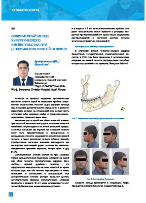 The experience of surgical treatment of maxillofacial deformation with Intraoral access is given. Preference Is given to the retromolar osteotomy, which Is based on the Dal Pont operation, the bone fragments are strengthened by titanium miniplates. Patlentstreatment Is conducted In conjunction with an orthodontist.