Library search
Search Results
Found 22 items.
-
 Defects of the cornea of the lower jaw of the metatarsal bone restoration using an autograftIn the process of reconstruction of the detects in the branch of lower jaw through metatarsal autogenous transplant method, studied of changes hi supportive system after the removal of metatarsal bone, the foot were based on the results of thorough examination of computer podometrics before the autogenous transplant was taken. This way was achieved to decrease the symptoms and development of flat-foot, the length of stay of patients in the hospital, the symptoms of quick tiredness, the pain in thigh, fibula and the pain in foot after a long walk after the surgery of autotransplant removal.
Doctor's Herald -
 The number of patients with tumors of big salivary glands is increasing. Most tumors are found in the parotid salivary gland. The most common benign tumors of the parotid salivary glands are pleomorphic adenomas. In the complex of medical and diagnostic measures, the surgical stage of treatment remains the main one.
 -
 Methodic of sonoelastography in the time of examination in patients with soft tissular new formations of jaw-fecial area is used m this work. Also, we can see high information of the method m time of differentiated detecting of benign, pre-cancer and malignant tumors, because of different staining of tissue depending on degree of its malignancy and deformation coefficient.
 -
 A brief literature review' was conducted, during which the current state of prevention of dental morbidity was identified and assessed. Hie reasons for the high dental morbidity among the population, including both poor sanitary culture and die level of public knowledge on die basics of hygiene and prevention, as well as poor quality of preventive work ar e indicated. The importance of organizing primordial prophylaxis was accented.
 -
 In the department of head and neck tumors of the Tashkent regional oncological dispensary complex treatment of locally spread laryngopharyngeal cancer is performed witli the use of the combined operation «Laryngopharyngoesophagectomy witli one-stage pharyngoesophagoplasty with the help of the gastric stem». Despite the traumatism and high risk of postoperative complications, laryngopharyngoezofagectomy with a one-stage gastric stem plasty and pharyngogastroanastomosis is an alternative treatment of locally spread forms of cancer of the laryngopharynx and cervical esophagus resistant to chemoradiotherapy.
 -
 A new approach to the surgical treatment of atrophy of the alveolar process of the jawAtrophy of alveolar bone often is the limiting factor for dental implantation. Decision of the issue of this problem is augmenting the alveolar process, uith different osteoinductive and osteoconductive materials. Comparative studies proved that use of umbilical membrane in combining with osteoplastic materials stimulates the regenerative processes, and thus facilitates the formation of mature bone in the defect area.
Stomatologiya -
 Statistics show that injuries that occur in the maxillofacial apparatus account for about 16% of the total number of injuries, while mandibular fractures are the most common among all fractures of the bones of the facial skeleton and, according to various authors, range from 75 up to 96.5%, and from the total number of inpatient dental profile patients 28-36% (Yu.I. Bernadsky, 1999; N.V. Novosyadlaya, 2003; R.S. Matveev, 2002; S.N. Fedotov, 2002 and etc.) [7]. In 67-82% of cases, mandibular fractures are localized within the dentition and, therefore, are open. Concerning some foreign authors call such infections of the bone wound pathogenic microflora (E. Kruger, 1986). In case of fractures of the lower jaw, due to the presence of fixing structures in the oral cavity, the process of self-cleaning in the oral cavity is sharply disrupted. AT In connection with this, the number of pathogenic microorganisms on the surface of the teeth and mucous membrane increases, the likelihood of infection of the wound substrate increases (A.I. Kaspina, 1981; Zh.B. Urazalin, 1985, Adzhiev K.S. 1991) [2].
 -
 Comparative characteristics of the methods of treatment of patients with posttraumatic defects and deformations of the bottom wall of orbitIncreasing the number of patients with injuries of the lower wall of orbit helps highlighting the development of new and improvement of existing treatments. That, in turn, leads to the need for thorough research posttraumatic de-formation of the bottom wall of the orbit in the diagnostic aspects, as well as for improving methods of medical reha-bilitation
Journal problems of biology and medicine -
 Dependence of the initial results of treatment for fractures of the face and jaw area on the duration of osteosynthesis4s a result of our study ustanovleno that more than half of the victims (55.6 per cent) were operated on during the first day after admission to the hospital, and 34.2% In the period from 2nd to 4th day and only 10.2% of the cases osteosynthesis was performed later than 4 days. We obtained data Indicate a high risk of development of complications and lethal outcomes when performing osteosynthesis Immediately upon receipt and In the period after 4 days. The next most favorable outcomes obtained from the victims, which the osteosynthesis of bones of the maxillofacial region was undertaken In the period up to 24 hours after Injury.
Stomatologiya -
 Methodic of sonoelastography in the time of examination in patients with soft tissular new formations of jawfacial area is used in this work. Also, we can see high information of the method in time of differentiated detecting of benign, pre-cancer and malignant tumors, because of different staining of tissue depending on degree of its malignancy and deformation coefficient.
 -
 A new approach to the surgical treatment of atrophy of the alveolar process of the jawAtrophy of alveolar bone often is the limiting factor for dental implantation. Decision of the issue of this problem is augmenting the alveolar process, with different osteoinductive and osteoconductive materials. Comparative studies proved that use of umbilical membrane in combining with osteoplastic materials stimulates the regenerative processes, and thus facilitates the formation of mature bone in the defect area.
Stomatologiya -
 Examination and treatment of 30 persons have been done In the age group of20-57years old Including 27 men and 3 women with complicated unilateral and bilateral lower jaw fractures. Traditional surgical and pharmacological treatment methods were applied for the Indications to all patients. Lymphotroplc regional administration of heparin anticoagulant and ceftriaxone antibiotic was additionally used In the group of patients with Inflammatory infiltrates In the fracture zone, and It promoted cure with complications reduction. The authors note an Improvement In the patient's condition and a reduction of treatment duration.
 -
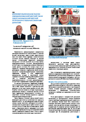
 Objective assessment of osseointegration of dental implants by resonance frequency analysis in patients with diabetes mellitusResults of intrabone dental implants osteointegration after restoration of an atrophy of an alveolar bone and dental implantation are shown on a material of 220 patients. Patients with the second type of diabetes at a stage of compensation are in comparative group. For the analysis of results of osteointegration and stability implants we were used the device «Osstell ISQ», it was manufactured by firm Integration Diagnostics (Sweden), it defining frequency-resonant analysis and stability factor of implants by method RFA (Resonance Frequency Analysis), which registries of resonant electromagnetic fluctuations of implants and a surrounding bone influences on them with electromagnetic field. ISQ (Implant stability Quotient) is expressed on a scale from one to hundred. The obtained data allows assuming influence of objective methods of research of osteointegration dental miplants with the subsequent reduction of terms of orthopedic treatment and choice of optimum term of prosthetics. The general average value of implants stability is about 70 units ISQ.
Stomatologiya -
 Methodic of sonoelastography in the time of examination in patients with soft tissular new formations of jaw-facial area is used in this work. Also, we can see high information of the method in time of differentiated detecting of benign, pre-cancer and malignant tumors, because of different staining of tissue depending on degree of its malignancy and deformation coefficient.
 -
 Comparative characteristics of the methods of treatment of patients with posttraumatic defects and deformations of the base of the orbitAmong the various deformities resulting from injuries of the middle zone of the face, one of the most severe in their consequences are defects and deformations of the edges and walls of the orbits, since in 90% of cases they cause functional disorders of the organs of vision and account for 6-28% of the total number of facial injurie skeleton (Ippolitov V.P. 1986, 1995; Belchenko V.A., 1988, 1996) [2, 3, 5]
Journal problems of biology and medicine -
 Morphofunctional assessment of remodeled bone tissue after guided bone regeneration in patients with diabetes mellitusMaterials of this study were the clinical and radiological survey of such as the cone-beam computed tomography, rodlovlsiography, frequency resonance analysis, which was carried out bone plastic Intervention on the upper and lower jaws. From all used In Implantology X- ray methods, cone beam computed tomography Is the most preferred for the analysis of morphofunctional of the state of remodeled bone tissue, as allows to visualize the bone regenerate In multi-plane and exchange of modes, estimate its topography, length, structure and plan the next stage of the Implant treatment.
Stomatologiya -
 Osteoplastic materials for replacement of defects and deformities of the maxillofacial regionThis article presents current information about the regeneration of bone tissue. The main directions of research and an analysis of the work to replace bone tissue defects in oral and maxillofacial surgery. The characteristics of the osteoplastic materials emphasize their positive and negative characteristics in different clinical situations, principles for the use of various forms of biomaterials. The mam provisions, which shall be guided by the doctor when choosing a replacement of the bone defect.
Stomatologiya -
 This article presents the results of treatment of patients with ankylosis of temporomandibular joint (TMJ) . The patients were divided into 2 groups. In the first group. Group 1 as a graft material used artificial Biositall, in group2 was used as a graft IV-metatarsal bone. Comparative evaluation between the first and the second group showed that the use of the metatarsal bone of the sculpture TMJ improves the outcome of surgery and provides new possibilities in the treatment of bone ankylosis of the temporomandibular joint.
 -
 Comparative analysis of tactics of treatment of patients with concomitant trauma of the maxillofacial regionTreatment of patients with associated oral and maxillofacial Injury with an objective assessment of the severity of Injury in the choice of surgical treatment tactic, rate of complications was 20.396 and the mortality rate - 4.296. Average time of death was 7,3 ±2,3 days.
Stomatologiya -
 Digital method of radiation diagnostics in the assessment of bone structure during dental implantation in patients with diabetes mellitusResults digital method of radiodiagnosis in assessment of bone structure at the dental implantation at patients with diabetes and of intrabone dental miplants osteointegration after restoration of an atrophy of an alveolar bone and dental implantation are shown on a material of 220 patients. Patients with the second type of diabetes at a stage of compensation are in comparative group. For the analysis of results of osteointegration and stability miplants we were used the device «Osstell ISQ». it was manufactured by firm Integration Diagnostics (Sweden), it defining frequency-resonant analysis and stability factor of implants by method RFA (Resonance Frequency Analysis), which registries of resonant electromagnetic fluctuations of implants and a surrounding bone influences on them with electromagnetic field. ISQ (Implant stability Quotient) is expressed on a scale from one to hundred. The obtained data allows assuming influence of objective methods of research of osteointegration dental implants with the subsequent reduction of terms of orthopedic treatment and choice of optimum term of prosthetics. The general average value of implants stability is about 70 units ISQ.
Stomatologiya -
 Оценка эффективности метода растягивания мягких тканей при восстановлении головы и шеи после склерозирования обширных гемангиом лицаРубповые деформации волосистой части головы, липа и шеи являются наиболее трагичными последствиями ожоговой травмы. [1.2,3.4]. Повреждение липа, кроме тяжелых функциональных нарушений, влечет за собой серьезные психические расстройства пациента [4.5,11.13]. Поэтом}7, основная задача пластических хирургов - решить не только функциональные. но и эстетические проблемы пациента [15]. Особую сложность представляет восстановление кожного покрова липа, поврежденного после склерозирования обширных гемангиом. В результате склерозирующей терапии образуются грубые рубцы. вызывающие функциональные и эстетические нарушения Последующая тактика заключается в иссечении рубцов на липе, что ставит перед хирургом сложную задачу - выбор метода пластического закрытия обширного дефекта ввиду дефицита прилегающих донорских участков кожи [3.4.6.10.15].
Stomatologiya -
 Review of technique of tissue expansion in maxilla-facial surgery Analysis of literary data shows, that the tissue expander is one of the leading methods, used practically at all anatomical areas for all increasing spectrum of operations. Nowadays this method is used in the plastic and reconstructive surgery. It gives possibility to create the skin with practically absolute accordance of color. texture and sensibility without creating of additional donor defects. However essential lacks of tissue expander are long duration and high frequency of complications. In the literatures methods of struggle with complications are not elucidated, concrete approbated recommendations on their prophylaxis and the methods of plasty by stretched tissue are not offered, that it is necessary for more wide and safe using of this method. Therefore overall analysis of large experience of using of the tissue expander with the use of various types of expanders and methods of stretching, working out of the methods of prophylaxis and adequate and opportune treatment of complications sen e for shortening of period of treatment, reduction of frequency of complications and achievement of the best cosmetic and functional results and it is actual problem of contemporary plastic and reconstructive surgery.
1 - 22 of 22 items