Library search
Search Results
-
 Biopotentials of masticatory muscles in musculoskeletal TMJ dysfunctions, before and after treatment.The article presents studies of the biopotentials of the masticatory muscles. In the course of their research, the authors recorded each indicator of the clcctromyograph before and after treatment for musculoskeletal dysfunctions of the temporomandibular joint. Electromyographic studies of the masticatory muscles before and after treatment reveal the completeness of the picture of its functional state, which affects the outcome of the correct treatment and normalization of the activity of the masticatory apparatus and TMJ.
Medicine and innovations -
 A COMPARATIVE APPROACH TO THE TREATMENT OF DISEASES OF THE TEMPERAMENTAL JOINTDiseases of the temporomandibular joint is the symptomatic complex characterized by dull pain, clicking sounds in the temporomandibular joint, ear pain, ear congestion, tinnitus, headache, dizziness, pain in the cervical spine, the back of the head and behind the auricle, heartburn in the throat and nose. Inflammatory disease of the temporomandibular joint is usually more common in old age with a functional structural change in the temporomandibular joint, at this time it is rejuvenating and the frequency of the formation of a permanent bite increases much more often. The problem is that in places where there is no specialist doctor, maxillofacial surgeon, patients are not diagnosed and appropriate treatment is not obtained, in turn, the acute process takes place in a chronic stage.
Medicine and innovations -
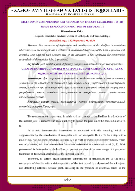 METHOD OF COMPRESSION ARTHRODESIS OF THE SUBTALAR JOINT WITH SIMULTANEOUS CORRECTION OF DEFORMITYFor correction of deformities and stabilization of the hindfoot in conditions where the latter is combined with a violation of the axis and shortening of the tibia, especially with extensive scar changes with concave side of the curvature, a new technique for compression arthrodesis of the subtalar joint is proposed.
Modern Science and Research -
 Possibilities of radiation research methods for damages of soft woven structures of the knee jointDamage to the knee joint is a serious problem in clinical medicine, as it is a fairly common cause of disability and disability [4, 8, 21]. The knee joint is involved in the pathological process in a variety of diseases - deforming osteoarthritis, rheumatoid arthritis, seronegative spondyloarthritis, microcrystalline arthropathies, chondromatosis, injuries. Injuries of the knee joint occupy one of the first places among all cases of pathology of the musculoskeletal system, and from 43% to 80% of cases are injuries of its ligamentous apparatus [4, 5, 8]. Due to the peculiarities of the anatomical structure of the knee joint, its soft tissue structures are damaged more often than the bone components. Ligamentous apparatus injuries occupy the first place and account for up to 50% of injuries of the knee joint, up to 24% of injuries of the lower limb. Chronic injuries of cartilage, menisci, and cruciate ligaments, which are recorded with a frequency of up to 79%, are the cause of the development of degenerative-dystrophic changes in the knee joint [2]. Degenerative-inflammatory diseases of the joints are a common cause of persistent disability in the population of various age groups. Osteoarthritis is the most common joint disease, occurring in 10-20% of the adult population.
Journal problems of biology and medicine -
 Артроскопическая характеристика синдрома хронического синовита коленного сустава различной этиологииАктуальность темы: При длительном течении синдрома хронического синовита (СХС) нарушается состояние костно-хрящевых структур, образующих коленный сустав. Поэтому рентгенологическое исследование проводится каждому пациенту с хронической патологией коленного сустава. Оно позволяет оценить форму и структуру костей, выявить некоторые косвенные признаки поражения суставного хряща [3,4].
Doctor's Herald -
Zamonaviy ortodontiyani barqaror tayanchdan foydalanmasdan tasavvur qilish qiyin. Distalizatsiya, retraktsiya, hatto mesializatsiyaga erishish qiyin, suyak darajasida kengayishning qiyin holatlar va boshqalar - bularning barchasi tayanch tufayli mumkin bo'ladi.
-
 Here are presented the results of conservative treatment of 199 patients wint congenital hip dislocation (hip dysplasia). After 30 years, the results of treatment has been studied in 102 patients. Good results were in 44 patients, satisfactory in 31 and unsatisfactory in 27 patients. The reasons for unsatisfactory results have been identified and methods of their prevention have been recommended.
 -
 Особенности медико-социальной реабилитации больных и инвалидов после эндопротезирования тазобедренного суставаАктуальность. Количество повреждений и заболеваний тазобедренного сустава составляет 8,1% среди всех патологии опорно-двигательной системы, поэтому проблема восстановления полноценной функции суставов является достаточно актуальной в современной ортопедии. В настоящее время наиболее эффективным среди оперативных методов лечения заболеваний тазобедренного сустава является эндопротезирование (6,7,9,11).
Journal problems of biology and medicine -
 Surgical treatment of dysplastic instability of hip joint in children and adolescents by creating the long canopy over head of the femurSurgical treatment of congenital dislocation of the hip in the late period, especially in older children and adolescents, with high standing of the femoral head and secondary changes of elements of the hip joint, and also the muscles surrounding it is a difficult and not solved the problem. Objective: Based on the study results exist, methods of surgical interventions aimed at restoring the supporting ability of the limb in congenital dis-location of the hip in older children and adolescents, the most effective options work out operational interven-tions in this group of patients depending on the severity of hip dislocation and implement them in practical medicine. Material and Methods: The study is based on a survey and study of the results of surgical treatment of 70 patients’ older children and adolescents with unilateral congenital hip dislocation. To study the results of treatment have been used clinical, radiological and statistical methods. Results: The results of treatment were followed in the period from 1 to 6 years. Positive results were re-ceived in 82% of patients
Journal problems of biology and medicine -
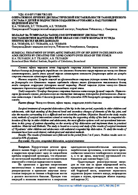
 Эндопротезирование коленного сустава при опухолях проксимального отдела большеберцовой косгиПроблема лечения опухолей костей, составляющих коленный сустав, в частности проксимального отдела большеберцовой кости, представляется чрезвычайно актуальной
Doctor's Herald -
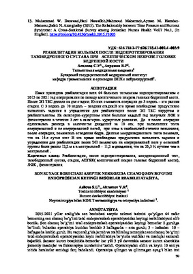 REHABILITATION OF PATIENTS AFTER ENDOPROSHETICS OF THE HIP JOINT IN ASEPTIC NECROSIS OF THE FEMORAL HEADWe have carried out the rehabilitation of all 64 patients with total arthroplasty from 2015 to 2021, surgery for aseptic necrosis of the femoral head. After EP, the hip joint was divided into two stages: Of these, from the moment of surgery to 3 weeks, this is the early stage. From 3 weeks to 10 weeks - late stage. It is necessary to efficiently perform tasks and exercises for rehabilitation after EPHT by a surgeon and a rehabilitation therapist. At the sanatorium-resort stage, every year patients get physical therapy and physiotherapy for 3 years in a sanatorium-resort environment. Before and after the operation, the difference in the number of movements in 10 seconds was assessed. When performing the test with the operated and unoperated leg, the index of hip abduction increased the most after the operation. The data of the coordination test showed that on the 14th day, it is necessary to productively perform tasks and exercises for rehabilitation after EP while the indicator on the operated leg in the main group was equal to 12.2 m, and in the control group - 11.2 m of movement, which is 25.3% better than the control.
Medicine and innovations -
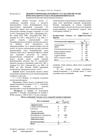
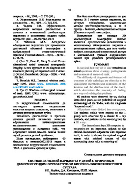 State of periodontal tissues in children with secondary deforming osteoarthrosis of the temporomandibular jointin article results or research oi influence oi anomalies and deformations maxilofacial systems (MFS) among children and teenagers with secondary deforming osteoarthrosis temporo-mandibular joint (SDO TMJ) on a periodontal fabric are stated. SDO TMJ promoted the expressed infringement of separate teeth, tooth alignments and a bite. Also at children with SDO TMJ at presence periodontitis in comparison with control group all nonspecific factors of protection of an oral cavity (p <0.005) have been lowered. Adequate treatment of the given pathology should be directed on normalisation microbiocinosis oral cavities and the prevention of development of a dysbacteriosis of an oral cavity and intestines that will promote increase of level of nonspecific factors of protection of an oral cavity and improvement of a condition of periodontal fabrics.
Stomatologiya -
 Ранняя диагностика и профилактика изменений внчс при наличии трем в зубных рядах
Actual problems of dentistry and maxillofacial surgery 4В настоящее время заболевания ВНЧС являются актуальной проблемой современной стоматологии. В результате расширения промежутка между зубами в ВНЧС начинают происходить различные изменения.
-
 Osteoarthritis of the knee joint: causal factors and methods of conservative treatmentSummarized scientific works devoted to the methods of conservative treatment of arthrosis of the knee joint describe generalized treatment of joint analgesics aimed at restoring the excretory and excretory function of the joint, the characteristic features of the action of homocysteine in the concentration of non-inflammatory prsparates against inflammation, and the periods of excitation of the knee joint. Cure mud-dat far from the effectiveness of kilib in kiska run and slow exposure to the landscape of the disease.attention was paid to the group of prsparats and their characteristics. Treatment is effective-maximizing the effectiveness of treatment with ointment Lori, creams, gels, which affect the gums, as well as physiotherapy-rapsvtik procedures, it is possible to obtain better results from the use of sleeping pills.
Doctor's Herald -
 Совершенствование ранней диагностики при остром гематогенном остеомиелите костей тазобедренного сустава у детейОстрая гнойная хирургическая инфекция занимает одно из ведущих мест в структуре заболеваемости детского населения. Среди нозологических форм острой хирургической инфекции по тяжести течения, трудностям диагностики и неблагоприятным исходам доминирующее место принадлежит острому гематогенному остеомиелиту, наблюдаемому преимущественно в детском возрасте [2,7,8].
Journal problems of biology and medicine -
Оценить эффективность реабилитационных мероприятии у детей после хирургического лечения патологии тазобедренного сустава.
-
 RECONSTRUCTION OF TEMPOROMANDIBULAR JOINT PATHOLOGY USING TITANIUM NICKELIDE MATERIALSSurgical treatment of patients with diseases and injuries of the temporomandibular joint (TMJ) is carried out in persons with traumatic injuries and their complications, secondary deforming osteoarthrosis, destructive changes resulting from purulent-inflammatory, tumor and tumor-like processes, congenital and acquired abnormalities, bone ankyloses, as well as destructive processes of bone-cartilage structures with their replacement to transplantation and implantation materials, and is one of the complex, important problems of modern maxillofacial surgery. Today in clinical practice from the traditional materials most widely used: auto-, allo-xenografts, complex autovascularized autografts, as well as various types of implant structures. However, as recent publications show, and clinical practice, the use of these materials for these purposes tends to decrease due to their shortcomings due to resorption or rejection of transplanted materials, death of osteocytes, osteoblasts and destruction of other living cellular structures, including difficulties in using microsurgical technology [1,4,6,8]. Endoprosthesis of the head of the temporomandibular joint, body and branch of the lower jaw is shown in cases where the use of bone grafts of allo- and autogenic origin is impossible.[ 1,4,7,8]. In this direction, studies related to the development of technology for the production of porous and nonporous materials based on titanium nickelide made at the Siberian Institute of Physics and Technology [2] made a huge contribution. Endoprostheses made from this alloy are well tolerated by body tissues, have high biological inertia, lack of toxicity and reply all the requirements for implant materials.
Medicine and innovations -
 Tactics of surgical treatment in acute hematogenous osteomyelitis of the hip bones in children86 children with acute hematogenic osteomyelitis of bones of coxofemoral joint have been studied. According to the form of performed surgical interferences three clinical groups have been selected/ In the follow-up period good results have been received in 15 (57.7%) patient of the 3d group that is considerably more in comparison with the 1st group 7(33.3%) and the 2nd group 9(45%). The method of draining osteoperforations of acetabular tegmen (DOAT) made it possible to improve the outcome of the disease to decrease the number of complications twice less and exclude transfer to a chronic form.
Journal problems of biology and medicine -
 Коррекция фармакотерапии аденоидов в комплексной реабилитации детей с вторичным деформирующим остеоартрозом височно-нижнечелюстного суставаУ детей с вторичным деформирующим остеоартрозом и анкилозом височно-нижнечелюстного сустава выявлены значительные нарушения во всех звеньях гемостаза. увеличение содержания гемолитических форм золотистых стафилококков, пиогенных стрептококков, грибов рода Кандида, лактозонегативных бактерий на фоне снижения количества лактозопозитивных форм. Предложена коррекция базисной фармакотерапииназначением антикоагулянтов, антнагрегантов. пробиотиков, антибиотиков и фунгицидов.
Stomatologiya -
 Мышечно-суставная дисфункция височнонижнечелюстного сустава, ассоциированная с дистопией третьих моляров у подростков и лиц молодого возраста
Actual problems of dentistry and maxillofacial surgery 4По данным современных авторов частота встречаемости дисфункции височно-нижнечелюстного сустава (ВНЧС) у подростков и лиц молодого возраста достигает 60%, а у взрослого населения - 89%. Как известно, дисфункция ВНЧС - патология, включающая комплекс анатомических и функциональных нарушений, состоящих из окклюзионного, суставного и мышечного компонентов, которая может сопровождаться болевым синдромом
-
 Сравнительная оценка возможностей визуализации травматических повреждений коленного сустава с использованием магнитно-резонансной томографии и артроскопииТравмы коленного сустава представляют собой актуальную проблему современной травматологии и ортопедии [1,10]. Они занимают ведущее место среди патологий опорно-двигательного аппарата (9,8%) и собственно суставов (50%). Нераспознанные или поздно диагностированные травмы с трудом поддаются лечению и приводят к длительной потере трудоспособности больных
Doctor's Herald -
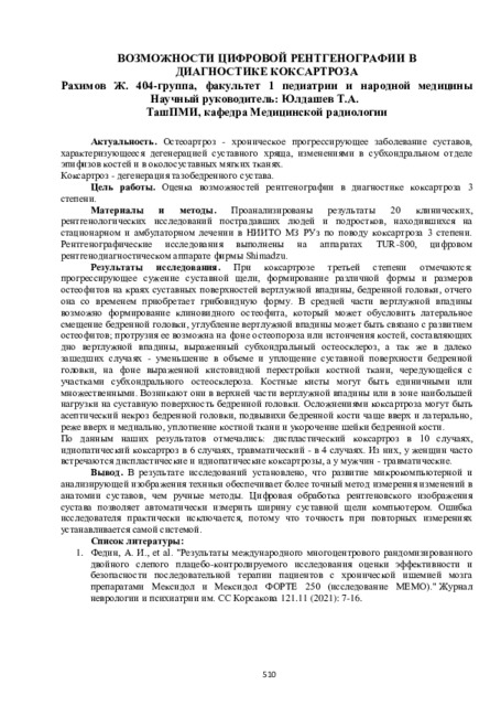 Возможности цифровой рентгенографии в диагностике коксартроза
Prospects for the development of medicineОстеоартроз - хроническое прогрессирующее заболевание суставов, характеризующееся дегенерацией суставного хряща, изменениями в субхондральном отделе эпифизов костей и в околосуставных мягких тканях. Коксартроз - дегенерация тазобедренного сустава.
-
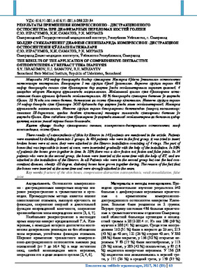 The results of the application of compressesive-distractive osteosynthesis at refract tibia diaphysisThere results of osteosynthesis of tibia by Ilizarov in 540 patients are mentioned in the article. Patients were examined by dividing them into 2 groups. In 486 patients who were in the first group, it was tried to insert broken bones were at once, they were attached to the Ilizarov installation consisting of 4 rings. The part of bones that was impossible to insert at once, were inserteded gradually with the help of the installation. In 80% of patients the bones grew together in time. In 20% there was a slow fusion not fusion and false joints. In 54 patients who were in the second group, the bones were inserted at the same time with the help of IIT, and are attached to the installation of the Ilizarov. In all Patients who were in the second group but one (he had con- comitant diseases, obesity -III degree, diabetes) bones have grown together during. The reason of the fact that the bones were inserted at the same time and were strongly attached to the seam.
Journal problems of biology and medicine -
 Анкилоз височно-нижнечелюстного сустава (ВНЧС) фиброзное или костное сращение суставных поверхностей обусловливающее частичное или полное исчезновение суставной щели. Врожденные анкилозы наблюдаются исключительно редко. Согласно имеющимся данным, до 80% анкилозов ВНЧС развиваются у детей в возрасте до 10-15 лет. Однако многие больные поступают в лечебные учреждение значительно позже.
 -
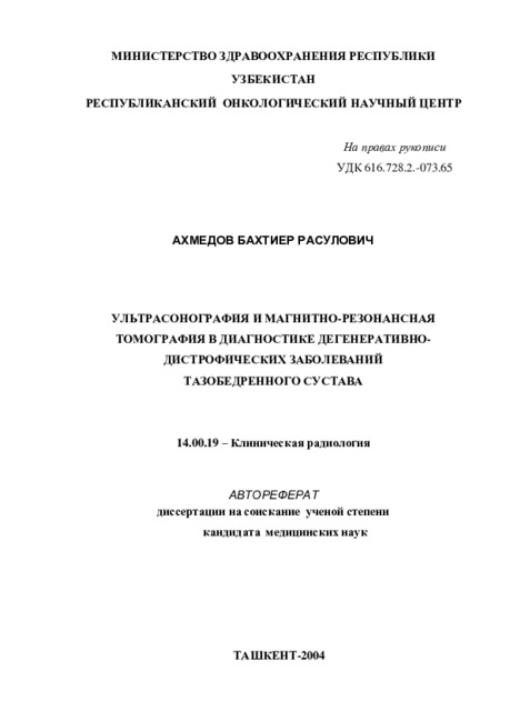 Ultrasonography and magnetic resonance imaging in the diagnosis of degenerative dystrophic diseases of the hip joint
Catalog of abstractsSubject of the inquiry: 148 patients with degenerative dystrophic diseases of the hip joint, of them 103 with osteoarthritis and 45 with avascular necrosis of the femoral head.
Aim of the inquiry: improvement of the diagnosis of degenerative-dystrophic diseases of the hip using possibilities of ultrasonography and magnetic resonance imaging.
Methods of research: X-ray, ultrasonography and magnetic resonance imaging.
The results achieved and their novelty: For the first time, the role of ultrasonog-aphy and magnetic resonance imaging in osteoarthritis and avascular necrosis of the hip was established in comparison with conventional X-ray. The result have shown that sensitivity of ultrasonography in osteoarthritis was low (57.7%) in the detection of femoral head deformation. In avascular necrosis of the hip detection of femoral head deformation was revealed better - 89.7%, which can be explained by differences in the mechanism and localization of deformation in these diseases. Ultrasonography was quite sensitive to changes of the joint capsule both in osteoarthritis and avascular necrosis, the sensitivity 86.0% and 84.6% respectively. MRI was superior to X-ray in the assessment of space orientation of the joint surfaces, necrosis zone, synovitis, subchondral cysts, structural changes and joint effusion. X-ray was more preferable for the detection of ostephytosis, subchondral sclerosis and changes of the joint space in osteoarthritis.
Practical value: of the work consisted in the concretization of sonographic and MRI signs of degenerative dystrophic diseases of the hip joint and in the suggested algorithm of using radiological methods for establishing the nature of hip joint pathology.
Degree of inculcate: the results of research were introduced in the Radiology Department of First Tashkent Medical Institute and the Department of Large Joint Pathology Department of the Traumatology and Orthopedy Research Institute.
Sphere of usage: radiology, traumatology and orthopedy, rheumatology.