Library search
Search Results
-
 ВЛИЯНИЕ СОДЕРЖАНИЯ ВИТАМИНА Д НА ПОКАЗАТЕЛИ МИНЕРАЛЬНОЙ ПЛОТНОСТИ КОСТНОЙ ТКАНИ У ЖЕНЩИН В МЕНОПАУЗЕАктуальность. В настоящее время несмотря на значительные успехи, достигнутые в области диагностики и лечения остеопороза (ОП), изучение факторов, влияющих на состояние минеральной плотности костной ткани (МПКТ), остается актуальной научной проблемой, все еще далекой от окончательного решения. Медико-социальная значимость её обусловлена неуклонным ростом распространенности заболевания, существенным снижением качества жизни пациентов и драматическими последствиями патологических переломов.
Modern Science and Research -
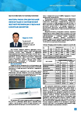
 STRUCTURAL AND FUNCTIONAL STATE OF BONE TISSUE WHEN PLANNING DENTAL IMPLANTATION IN PATIENTS WITH SOMATIC DISEASES.In everyday practice, at a doctor’s appointment with a dentist, about 7-10% of cases are patients with periodontal diseases on the back ground of somatic pathology, who turn for dental implants with the aim of prosthetics. In this study, the structural and functional state of bone tissue was determined in 72 patients who were planning this type of treatment, the following patients were determined by densitometric parameters: ultrasound propagation velocity - (SRS, m / s), broadband ultrasound attenuation - (SHO, dB / MHz), density index- (PI,%) In the course of work, only 25 (34.7%) patients had indicators within the normal range. The remaining 47 (65.3%) patients showed violations of the structural and functional properties of bone tissue in the form of osteopenia of varying severity in 36 (79.6%) and osteoporosis in 11 (23.4%) peoplе. The main parameters of the structural and functional state of bone tissue, osteogenesis markers, and levels of calcium-regulating hormones in patients with future dental implantation were studied. Violations of the strength characteristics of bone tissue, changes in the levels of calcium- regulating hormones and markers of osteogenesis have been identified, indicating the need for preventive and therapeutic measures in all patients at this stage of observation and treatment.
Journal of oral medicine and craniofacial research -
 FEATURES OF THE STATE OF BONE TISSUE DURING DENTAL IMPLANTATION IN PATIENTS WITH SOMATIC DISEASESIn everyday practice, at a doctor’s appointment with a dentist, about 7-10% of cases are patients with periodontal diseases on the back ground of somatic pathology, who turn for dental implants with the aim of prosthetics. In this study, the structural and functional state of bone tissue was determined in 72 patients who were planning this type of treatment, the followmg patients were determined by densitometric parameters: ultrasound propagation velocity - (SRS. m / s), broadband ultrasound attenuation - (SHO. dB / MHz), density index- (PI.0 o) In the course of work, only 25 (34.7%) patients had indicators within the normal range. The remaining 47 (65.3%) patients showed violations of the structural and functional properties of bone tissue in the form of osteopenia of varying severity in 36 (79.6%) and osteoporosis mil (23.4%) people. The main parameters of the structural and functional state of bone tissue, osteogenesis markers, and lex-els of calcium-regulating hormones in patients with future dental implantation were studied. Molations of the strength characteristics of bone tissue, changes in the levels of calcium-regulating hormones and markers of osteogenesis have been identified, indicating the need for preventive and therapeutic measures in all patients at this stage of observation and treatment.
Stomatologiya -
 Osteoporosis is a bone disorder in which bones become weak and prone to fracture. Osteoporotic bone has less density or mass, and the structure of bone tissue is far from normal. As bones become less dense, they become weaker and more likely to fracture. Women are four times more likely to have osteoporosis than men. Numerous studies show that weakness and atrophy can also affect the bone protrusions that hold dentures, resulting in improper fit. Regular dental visits, a healthy lifestyle, and a balanced diet high in vitamin D and calcium with regular physical activity are essential to strengthen and maintain good bone health.
 -
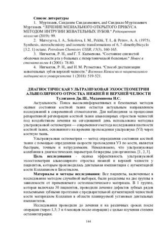 Диагностическая ультразвуковая эхоостеометрии альвеолярного отростка нижней и верхней челюсти
Actual problems of dentistry and maxillofacial surgery 4Поиск высокоинформативных и безопасных методов оценки состояния костной ткани остается актуальным направлением исследований в современной стоматологии. Для наблюдения за процессами репаративной регенерации костной ткани альвеолярных отростков челюстей под воздействием лечения на сегодняшний день использована методика ультразвуковой остеометрии — современного метода исследования плотности костной ткани, основанного на времени прохождения ультразвука (УЗ) через костную ткань.
-
 VIOLATION OF BONE MINERAL DENSITY IN DISEASES OF THE STOMACH AND DUODENUMSeveral scientists partially explained in the literature that the decrease in mineral density in patients with diseases of the stomach and duodenum occurs due to antisecretory and cytoprotective drugs. However, with acid-dependent gastrointestinal diseases, i.e., with chronic diseases of the stomach and duodenum, there are practically no signs of a violation of bone mineral density. Therefore, in this study, we attempted to determine that acid- and HP-associated diseases of the gastrointestinal tract, i.e., chronic gastritis and ulcers, are important risk factors for low bone mineral density. And we set ourselves the goal of developing methods for the prevention and treatment of complications that develop in the bone tissue in these diseases.
Journal of Cardiorespiratory Research -
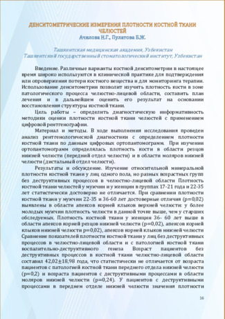 Денситометрические измерения плотности костной ткани челюстей
Актуальные вопросы профилактики стоматологических заболеваний и детской стоматологииРазличные варианты костной денситометрии в настоящее время широко используются в клинической практике для подтверждения или опровержения потери костного вещества и для мониторинга терапии. Использование денситометрии позволит изучить плотность кости в зоне патологического процесса челюстно-лицевой области, составить план лечения и в дальнейшем оценить его результат на основании восстановления структуры костной ткани.
-
 POSSIBILITIES OF DIGITAL RADIOGRAPHY AND DUAL-ENERGY X-RAY ABSORPTIOMETRY IN THE DIAGNOSIS OF OSTEPOROSIS IN THE AGE ASPECTOsteoporosis is an interdisciplinary problem, occupying one of the leading places among non-communicable diseases in terms of its medical and socio-economic consequences. Osteoporosis is also an age-related disease characterized by a decrease in bone mass and a violation of the microstructure of bone tissue, which leads to a decrease in bone strength and an increase in the risk of fractures. Currently, two-energy X-ray absorptiometry is used in practice to assess osteoporosis, which is the gold standard for diagnosing a decrease in bone mineral density.
Modern Science and Research -
 The effect of infusion of saline and succinasol on the functional and metabolic activity of neutrophils during reparative regeneration of a defect in bone tissue of the diaphysis was studied. The stereotype of mobilization of the functional and metabolic activity of phagocytic blood cells and connective tissue allows us to consider the dynamics of the activity of lysosomal and membrane enzymes, energy and oxidative metabolism, synthesis and secretion, migration, proliferation and differentiation.
 -
 Дентальной имплантации для сохранении зубо-альвеолярного сегмента верхней челюсти с помощью метода “root membrane”
in LibraryПротезирование вторичных адентий, которые длительное время присутствуют в верхней и нижней челюстях, является одной из актуальных проблем стоящих перед стоматологами. Одним из современных методов устранения этой проблемы, является дентальная имплантология. Одним из сдерживающих факторов для широкого распространения имплантации является недостаточный объем костной ткани для установки имплантата. Процесс дентальной имплантации во фронтальную часть верхней челюсти, которая на высоком уровне атрофирована или сопровождается вертикальным переломом, зубов, требует дополнительных костных изделий и длительного реабилитационного периода, причина этого в том, что вестибулярная пластинка фронтальной части верхней челюсти тонкая и он характеризуется переломом во время удаления зуба. Атрофия костной ткани после удаления зубов является одним из важнейших вопросов современной стоматологии, так как значительная атрофия костной ткани челюстей делает невозможным выполнение внутрикостной имплантации, а также создает серьезные трудности при ортопедическом лечении пациентов.
-
МORPHOLOGICAL CHARACTERISTICS OF REPLACING BONE TISSUE DEFECTS WITH THE SILICOPHOSPHATE GLASSThis article describes the characteristics of a new bioactive silicophosphate glass, created at the Turin Polytechnic University (Italy). The morphological analysis of newly formed bone tissue developed in the experimental animals' femur defect after implantation of silicophosphate glass 47.5B was carried out. It was noted that with an increase in the healing time, there is an increase in the reparative regenerative capacity of bone tissue in the area of the defect, which is expressed in an increase in bone density with the presence of mature osteocytes. The newly formed bone in the area of implantation was characterized by a high degree of maturity at the final stages of regeneration, which corresponds to the results of the study of domestic bioactive paste- like glass. The results of the experiment revealed both osteointegrative and osteoconductive features of the studied silicophosphate glass.
Medicine and innovations -
 Risk factors in dental implantation and guided bone regeneration in patients with diabetes mellitusGiven that diabetic patients have low regenerative potential of bone tissue, which Is associated with Infringement of microcirculation (angiopathy), atherosclerosis, tissue hypoxia, and increased levels of lipid peroxidation .That leads to the urgency of this problem which Iles in the optimization of reparative regeneration conditions, providing cellular material and macromolecules In dental Implantation. It is particularly important In case of simultaneous recovery of jaw bone tissue. The ability of diabetic patients, In the surgery with bone augmentation and dental Implantation, to differentiate osteogenic cells also depend on the preservation or restoration of blood vessels. In the case of poor circulation In the postsurgical field occurs oxygen deficiency, which In turn stimulates the proliferation of fibrous and chondroid tissue Instead of bone mineralization process.
Stomatologiya -
 Aim. To study the histological structure of regenerating bone tissue of rabbits jaw after the operation of dental implantation and magnetophoresis of 10% calcium gluconate solution and 5% rctabolil solution. Materials and methods. In Chinchilla rabbits under sodium thiopental anesthesia, a Verline screw dental implant, 3x5 mm with a passive thread and a smooth surface made of GRADE4 titanium, was installed on the side of the cuts of the lower jaw. The animals were observed for 30, 45,60 and 90 days. The animals of the experimental group underwent 10 magnetophoresis procedures with a 10% calcium gluconate solution and a 5% rctabolil solution in turn. For histological examination, a fragment of the lower jaw of experimental and control animals with a dental implant, external and internal compact plate and spongy substance was taken. The prepared sections were stained with hematoxylin and eosin. Results. In the control group, the histological structure of the bone tissue on the surface of the dental implant is normalized in 90 days after dental implantation operation. After magnctophorcsis of a 10% solution of calcium gluconate and a 5% solution of retabolil, the histological picture of bone tissue does not differ from normal in 60 days after the implantation operation.
 -
 Дентальной имплантации для сохранении зубо-альвеолярного сегмента верхней челюсти с помощью метода “root membrane”
in LibraryПротезирование вторичных адентий, которые длительное время присутствуют в верхней и нижней челюстях, является одной из актуальных проблем стоящих перед стоматологами. Одним из современных методов устранения этой проблемы, является дентальная имплантология. Одним из сдерживающих факторов для широкого распространения имплантации является недостаточный объем костной ткани для установки имплантата. Процесс дентальной имплантации во фронтальную часть верхней челюсти, которая на высоком уровне атрофирована или сопровождается вертикальным переломом, зубов, требует дополнительных костных изделий и длительного реабилитационного периода, причина этого в том, что вестибулярная пластинка фронтальной части верхней челюсти тонкая и он характеризуется переломом во время удаления зуба. Атрофия костной ткани после удаления зубов является одним из важнейших вопросов современной стоматологии, так как значительная атрофия костной ткани челюстей делает невозможным выполнение внутрикостной имплантации, а также создает серьезные трудности при ортопедическом лечении пациентов.
-
 Microbiological studies of osteoplasty of the alveolar process in patients with diabetes mellitusTake account that patient with diabetes mellitus have low regeneration potential bone tissue , connected with infringement microcirculation (angiopathy), atherosclerosis . hypoxia of tissue and rise level peroxide oxidation lipids flow a currency of this problem consist optimization condition reparative regeneration provision with cells material and macro molecule by dental miplantation especially important in simultaneous restoration bone tissue of jaws. Establishment feature of microflora in pathologic nidus may be begin base for new approach for planning dental miplantation with bone plastic and monitoring result by patient with inflammatory process in periodontal . and to the chose preventive measure selectively antibacterial action, including base on lytic bacteriophage to periodontalpatagen. What is given microbio logic research allow suppose positively influence ground use antimicrobial and antiseptics preparation on regeneration of wound after dental miplantation.
Stomatologiya -
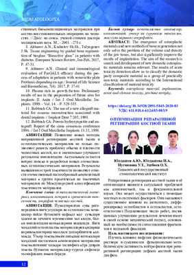
 Повышение эффективности дентальной имплантации при сохранении зубо-альвеолярного сегмента верхней челюсти с помощью метода “root membrane”
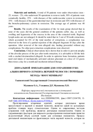
in LibraryПротезирование вторичных адентий, которые длительное время присутствуют в верхней и нижней челюстях, является одной из актуальных проблем, стоящих перед стоматологами. Одним из современных методов устранения этой проблемы, является дентальная имплантология. Одним из сдерживающих факторов для широкого распространения имплантации является недостаточный объем костной ткани для установки имплантата. Процесс дентальной имплантации во фронтальную часть верхней челюсти, которая на высоком уровне атрофирована или сопровождается вертикальным переломом, зубов, требует дополнительных костных изделий и длительного реабилитационного периода, причина этого в том, что вестибулярная пластинка фронтальной части верхней челюсти тонкая и он характеризуется переломом во время удаления зуба. Атрофия костной ткани после удаления зубов является одним из важнейших вопросов современной стоматологии, так как значительная атрофия костной ткани челюстей делает невозможным выполнение внутрикостной имплантации, а также создает серьезные трудности при ортопедическом лечении пациентов.
-
 A bone density scan, also called dual-energy X-ray absorptiometry (DEXA) or bone densitometry, is an advanced form of X-ray technology that is used to measure bone loss. DEXA is the current standard for measuring bone mineral density. X-rays help doctors diagnose and treat diseases. It exposes you to a small dose of ionizing radiation to produce images of the inside of your body.
 -
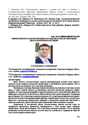
 REPLACEMENT OF THE WELL OF THE REMOVED TOOTH WITH AN AUTOGENOUS BONE GRAFT IN ORDER TO PREVENT THE OCCURRENCE OF DEFECTSIn many (63%) cases, after tooth extraction, there are bone defects and deformities in the alveolar processes of the jaws. Preservation and restoration of the volume of bone tissue of the alveolar processes of the jaws after tooth extraction, periodontal and tooth-preserving operations is an important problem of surgical dentistry and maxillofacial surgery. The difference between the bone tissue of the jaws and any other segment of the skeleton is that when the functional load is distributed or lost, the processes of resorption begin. In this case, bone loss occurs not only in the area of the removed tooth, but affects about 20% of the volume of the hole around it. 2-3 years after removal, there is usually a decrease in the anatomical size by 40-60% of the alveolar ridge, and this is typical for all population groups (A. Ashman). In the available literature, we have not found any works devoted to a detailed study of changes in bone density in the osteoplasty zone using modern methods of replacing jaw defects.
Journal of oral medicine and craniofacial research -
 Morphological features of the liver parenchyma in polypharmacy with anti-inflammatory drugs
Catalog of dissertations and abstractsThe purpose of the study was to determine and evaluate the features of morphological changes in the liver parenchyma of 5-month-old white outbred rats under the influence of anti inflammatory drugs under conditions of polypharmacy.
The object of study for experimental studies was taken 250 white male rats weighing 200-250 g.
The scientific novelty of the research is as follows: polypharmacy of anti-inflammatory drugs negatively affects all parameters of liver structures. Under the influence of polypharmacy, there is a decrease in the absolute mass of the liver, volume and morphological parameters of the liver parenchyma. The decrease in morphometric parameters depends on the number of drugs in polypharmacy; under conditions of polypharmacy, the state of the hepatic capillaries and internal bile ducts, as well as biological membranes, was studied, as a result of which the structural structure of the liver, the development of destruction of the hepatic tissue were studied, and the morphofunctional foundations of this condition were shown.
Implementation of the research results. Based on the obtained scientific results, the morphofunctional characteristics of the liver of rats in the norm and under the influence of polypharmacy were determined:
Approved methodological recommendations: "Methodology for determining the morphometric parameters of the liver during polypharmacy of anti-inflammatory drugs" (Conclusion No. 8n-r / 265 dated 14.03.2022 of the Ministry of Health of the Republic of Uzbekistan) and "Methodology for determining the morphometric parameters of the liver during polypharmacy of anti-inflammatory drugs" (Uzbekistan, Conclusion of the Ministry of Health of the Republic of Uzbekistan No. 8 n-z 180 of 2022), electronic program No. DGU 1038 "Program for studying the comparative characteristics of morphological changes caused by polypharmacy in the liver."
The scientific results obtained in the study of morphological and functional properties and morphometric changes in the structure of the liver under the influence of polypharmacy have been introduced into the practice of the Samarkand branch of the Republican Specialized Oncological and Radiological Scientific and Practical Medical Center of the Samarkand City Medical Association (Order of the Ministry of Health of the Republic of Uzbekistan dated March 14, 2022, 8n-r / 265-No. and conclusion No. 8 n-z 180 of 2022). The implementation of the obtained research data allows developing methods for early diagnosis, treatment and prediction of organopathology by morphological parameters, improving the quality of life and reducing the number of complications.
The structure and scope of the dissertation. The structure of the dissertation consists of an introduction, three chapters, a conclusion and a list of references. The volume of the dissertation was 103 pages. -
 Indicators of hydroxyprolin and mineral imbalance in children with clinical manifestations of connective tissue dysplasia
Journal of Biomedicine and PracticeConnective tissue dysplasia is a unique ontogenetic anomaly in the development of the body, which is one of the complex, far from studied questions of modern medicine, and is the morphological basis of functional changes in cardiac activity. We studied 115 children of preschool and school age with connective tissue dysplasia and small heart development abnormalities. A high frequency of occurrence of external phenotypic markers was revealed for the syndrome of dysplasia of the connective tissue of the heart and stigma of embryogenesis. An increase in the average level of hydroxyproline in blood serum in children with small abnormalities of the development of the heart in combination with cardiovascular pathology and patterns characterizing the relationship of the clinical manifestations of the disease and mineral imbalance has been established. Some pathogenetic lines of development of the pathological process in children with diselementosis have been determined. It has been proven that the state of the elemental status is an important informative criterion for assessing the severity of the underlying disease.
-
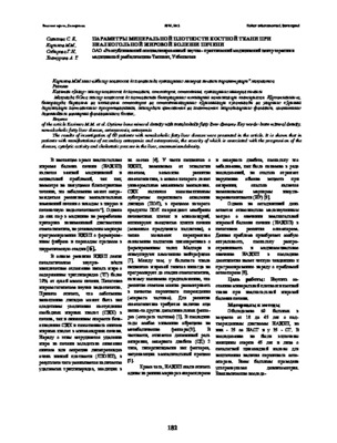
 Остеопороз — распространенное системное заболевание скелета, характеризующееся низкой костной массой и нарушениями микроархитектоники костной ткани, что приводит к увеличению ломкости костей и увеличивает риск переломов. Все большее внимание к проблеме остеопороза проявляют стоматологи. Для практических врачей достаточно остро стоит вопрос выбора адекватной комплексной терапии генерализированного пародонтита (ГП). тканей Альвеолярная кость является одной из составляющих пародонта. При ГП происходит резкая убыль костной ткани. Главная задача врача-стоматолога — замедление ее разрушения для предотвращения потери зубов[1.3]. Распространенность заболеваний пародонта среди населения Узбекистана составляет 80%, а изменения в пародонтальных тканях выявляются у 100% лиц старше 40 лет. (Юсупов С.Х. 1999)
 -
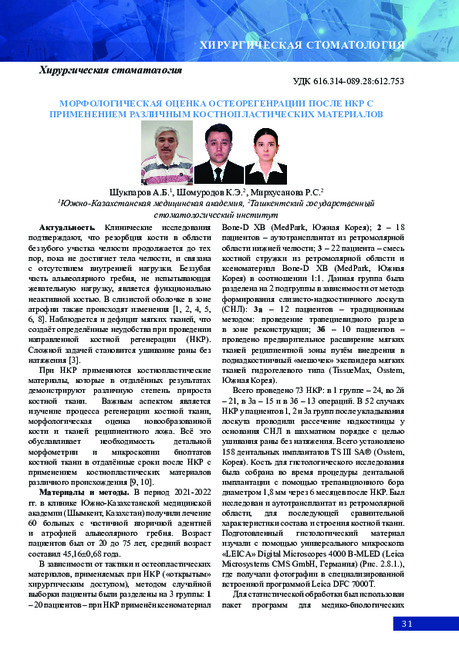 Morphological assessment of osteoregeneration after NCR using various osteoplastic materials
StomatologiyaSoft tissue deficiency in atrophy of the alveolar ridge creates difficulties for performing guided bone regeneration (GBR), the success of which to a certain extent depends on the closure of the wound without tension. The study examined the effect of the method of preliminary expansion of soft tissues on the subsequent processes of bone remodeling by histological examination of biopsies after NCR with the use of osteoplastic materials of various origins.
-
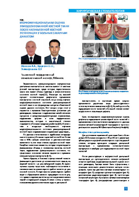 Morphofunctional assessment of remodeled bone tissue after guided bone regeneration in patients with diabetes mellitusMaterials of this study were the clinical and radiological survey of such as the cone-beam computed tomography, rodlovlsiography, frequency resonance analysis, which was carried out bone plastic Intervention on the upper and lower jaws. From all used In Implantology X- ray methods, cone beam computed tomography Is the most preferred for the analysis of morphofunctional of the state of remodeled bone tissue, as allows to visualize the bone regenerate In multi-plane and exchange of modes, estimate its topography, length, structure and plan the next stage of the Implant treatment.
Stomatologiya -
 The results of investigation of 60 patients with nonalcoholic fatty liver disease were presented in the article. It is shown that in patients with manifestations of secondary osteopenia and osteoporosis, the severity of which is associated with the progression of the disease, cytolytic activity and cholestatic process in the liver, anamnesisandobesity.
 -
 The article analyzes the diseases related to myositis, gives examples of diagnoses that may be in the practice of the maxillofacial surgeon, in particular ossifying the myositis of the masticatory muscles. The clinical and histological picture of ossifying myositis is analyzed, the clinical case is presented, with step-by-step analysis.