Library search
Search Results
-
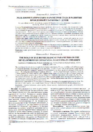
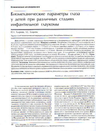 Purpose: to study the interrelation between the biomechanical and the biometric parameters of the eye in different stages of infantile glaucoma. Material and methods. A total of 19 patients (37 eyes) aged from 3 to 10 years with a non-operated primary infantile glaucoma were examined. Of these 6 patients (9 eyes) had initial stage, 7 patients (9 eyes) had advanced stage), 6patients (9 eyes) hadfar advanced stage, and 7patients (10 eyes) had terminal stage, respectively. A combination of different stages of the disease was found in 11 children, while 8 children had the same stage of the disease in both eyes. Examination methods included visometry, ophthalmoscopy, determining the excavation of the optic disc, A -scan recording the anterior-posterior axis (APA) of the eye, Filatov — Kalfa elastotonometry with weights of 5g, 10g, 15g. Results. The gradient of elastic curve rise was noted in all stages, but it was the highest in the terminal stage, where APA was greater according to the severity of the disease. APA and elastic curve raise was found to correlate in the far-advanced and the terminal stages of glaucoma. Conclusion. The changes in the biomechanical properties of the fibrous membrane of the eye in children with infantile glaucoma depend on the severity of the disease, which is manifested in changes in the biometric parameters. Sclera rigidity is reduced in the far-advanced and terminal stages of infantile glaucoma, which can lead to an underestimation of the true level oflOPand the risk of glaucoma development in children.
 -
 The role of eye biomechanical parameters in the development of congenital glaucoma in children
in LibraryTug'ma glaukoma bilan og'rigan bolalarda ko'zning biomexanik ko'rsatkichlari uning turli bosqichlari bilan o'rganildi. Oddiy birlamchi tug'ma operatsiyasiz glaukoma bilan og'rigan 1 oydan 3 yoshgacha bo'lgan 20 bemor (40 ko'z) tekshirildi. Ulardan 1-guruhga (ilg'or bosqich) mos ravishda 7 (14 ko'z), 2-chi (ilg'or bosqich) - 8 (16 ko'z), 3-chi (terminal bosqich) 5 bemor (10 ko'z) kiradi. Tadqiqot usullari orasida visometriya, refraktometriya, oftalmoskopiya, optik diskni qazib olishni aniqlash, ko'zning old-orqa o'lchamini ro'yxatga olish bilan A-skanerlash, 5 g og'irlikdagi elastotonometriya; janubiy; 15d, shuningdek, Filatov-Kalf usuli bilan. Olingan ma'lumotlarning tahlili shuni ko'rsatdiki, elastokrivning ko'tarilish gradienti barcha guruhlarda qayd etilgan, ammo eng yuqori ko'rsatkich terminal bosqichi bo'lgan bolalar guruhida bo'lgan, ko'zning anteroposterior hajmi esa mos ravishda kattalashgan. kasallikning og'irligi bilan. Ko'z ichi bosimining oshishi kasallikning og'irligiga mutanosib ravishda qayd etilgan. Konjenital glaukomaning oddiy shakli rivojlanishi bilan ko'zning biomexanik ko'rsatkichlari bosqichlarga ko'ra ortadi. Shu bilan birga, tolali membrananing qattiqligining pasayishi ko'zning to'qimalarida morfologik o'zgarishlarni va glaukoma jarayonining zo'ravonligini ko'rsatadi.
-
 The role of eye biomechanical parameters in the development of congenital glaucoma in children
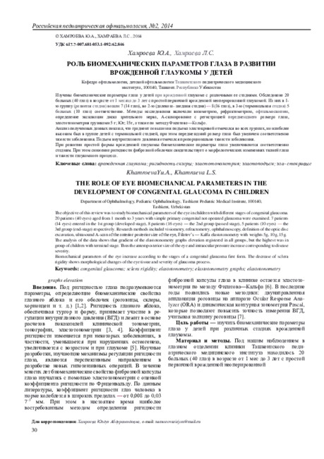
in LibraryThe objective of this review was to study biomechanical parameters of the eye in children with different stages of congenital glaucoma. 20 patients (40 eyes) aged from 1 month to 3 years with simple primary congenital not operated glaucoma were examined. 7 patients (14 eyes) entered in the 1st group (developed stage), 8 patients (16 eyes) — the 2nd group (passed stage), 5 patients (10 eyes) — the 3rd group (end stage) respectively. Research methods included visiometry, refractometry, ophthalmoscopy, definition of the optic disc excavation, ultrasound А-scan of the anterior posterior size of the eye, Filatov’s — Kalfa elastotonometry with weights 5g, 10g, 15g.
The analysis of the data shows that gradient of the elastotonometry graphs elevation registered in all groups, but the highest was in group of children with terminal stage. Thus the anteroposterior size of the eye and intraocular pressure increase corresponding to disease severity. Biomechanical parameters of the eye increase according to the stages of a congenital glaucoma first form. The decrease of sclera rigidity shows morphological changes of the eye tissue and severity of glaucoma process. -
 Age-related macular degeneration of the retina: the search for ways to prevent and early detection
in LibraryThe aim of the study was to study the possibility of using the electronic program for predicting the risk of occurrence and early detection of AMD developed by us. The program is used by 120 persons. 53 (44.2%) of them were diagnosed with "Macular Dystrophy", 27 (22.5%) - "Early stage of macular degeneration (AREDS category 2)", 22 (18.3%) - "Intermediate stage of macular degeneration (category 3) AREDS)", 18 (15%) - "Late stage of macular degeneration (category 4 AREDS) ".Thus, the electronic program we developed can be used as a method for predicting development, early detection of AMD and providing recommendations for further tactics.
-
 Evaluation of the effectiveness of the biopeptide preparation corticin in the non-proliferative and preproliferative stages of diabetic retinoblastoma
in LibraryPurpose of work w as the cliniko-functional assessment of efficiency of a preparation “CORTEXIN” in the complex treatment of a diabetic retinopathy at non proliferative and preproliferative stages. Under supervision there were 30 patients (60 eyes) aged from 40 till 81 years old, with a diabetic retinopathy at nonproliferative and preproliferative stages who were divided into 2 groups. The first group (main) - 15 people (30 eyes) in addition to traditional treatment took “CORTEXIN” preparation of 10 mg on 0,5ml parabulbarly 1 time within 10 days. The second group (control) — 15 people (30 eyes) underwent traditional therapy. There were 12 males (40%) and 18 females (60%). The efficacy of treatment was studied by methods of visiometry, ophthalmoscopy and perimetry. The examinations have been performed before and 1, 3 and 6 months after treatment. During the carried out treatment in the main group of patients the improvement of visual acuity, field of vision and condition of retina w ith keeping the stable effect till 6 months was fixed. And ali this allows authors to recommend “CORTEXIN” application in the complex treatment of diabetic retinopathy at nonproliferative and preproliferative stages.
-
 Performance evaluation of local therapy in the treatment of cervicalThe article is presented, evaluation result was local antimicrobic treatment in FarGALS domestic preparation in basic of express-test and cytological study in order to treat with the diagnosis of women with cervisitis. It is motivated that photological point of view was over-said choise in preparation. Overall, in ex-perimentation was detected by 60 women with the age of from 20 to 35 years old through the 5 days duration of disease during the 1,5 years period. In dependency from delivered statement, all patients were divided into 3 groups. The first group was 30 women with the diagnosis of cervisitis, in 9 of them were acute period , and 21 of them were chronic stage of disease. Complex treatment in these group was assigned by FarGALS in cervix in terms of treating in traditional forms with the treatment type of tampons 3,5ml course from 3 to 7 days. Evaluation of efficiency in antimicrob treatment was installed in base nitrite express-treatment, result of subjection to statistical analysis was used t-criterion of Styudenta and Vilkokson-Manna-Uitn, also in basic of analyzing bacterial data in cervix, cytological examination of cervical smear in Papanikalou. It was set up, after the treatment studying was showed by both groups in that age. In that case, 1gr efficient of anti-biotics by comparison of 2gr was felt in high by 45,8% and 40,7% factually. A period of time in preparations (drugs) were examined by FarGALS shortened in 3-5 days. During the treatment of women with the acute cervisitis were not become chronic stage during the examining. But women with chronic process of therapy was actually effective of situation 85%.
Journal problems of biology and medicine -
 Early stage of kidney damage in patients with hypertensive diseaseThe defeat of the kidney as the target organ of hypertensive disease in 40% of patients begins to form early - within 5 years from the debut of the disease. The early stage of hypertensive nephropathy is a dynamic process that has clinical and functional markers at each stage, so microalbuminuria, increased intrarenal vas-cular resistance, and changes in the total filtration function of the kidneys are the earliest
Journal problems of biology and medicine -
The aim of the study was to study the etiological and morphological features of large macular ruptures. The present study included 200 eyes (188 patients) who were diagnosed with a large macular rupture at initial examination. 96 eyes (48%) were diagnosed with stage 3, the remaining 104 eyes (52%) were diagnosed with stage 4 macular rupture. Large macular tears in 55.5% of cases are due to vitreomacular traction syndrome, in 24.5% of cases - myopia and in 20% of cases - eye trauma. Macular tears of various etiologies do not differ in visual acuity, intraocular pressure, diameter and stage of the rupture, duration of the disease. Macular rupture is associated with cataract in 51.5% of cases.
-
 Chronic endometritis is one of the most common inflammatory diseases of the female reproductive system. Timely treatment of the disease is insurance against serious complications, including oncology and infertility. The endometrium is the lining of the uterus. Acute endometritis develops when it becomes inflamed as a result of exposure to pathogenic bacteria. The chronic stage of the disease usually occurs due to lack of treatment.
 -
 REHABILITATION OF PATIENTS AFTER ENDOPROSHETICS OF THE HIP JOINT IN ASEPTIC NECROSIS OF THE FEMORAL HEADWe have carried out the rehabilitation of all 64 patients with total arthroplasty from 2015 to 2021, surgery for aseptic necrosis of the femoral head. After EP, the hip joint was divided into two stages: Of these, from the moment of surgery to 3 weeks, this is the early stage. From 3 weeks to 10 weeks - late stage. It is necessary to efficiently perform tasks and exercises for rehabilitation after EPHT by a surgeon and a rehabilitation therapist. At the sanatorium-resort stage, every year patients get physical therapy and physiotherapy for 3 years in a sanatorium-resort environment. Before and after the operation, the difference in the number of movements in 10 seconds was assessed. When performing the test with the operated and unoperated leg, the index of hip abduction increased the most after the operation. The data of the coordination test showed that on the 14th day, it is necessary to productively perform tasks and exercises for rehabilitation after EP while the indicator on the operated leg in the main group was equal to 12.2 m, and in the control group - 11.2 m of movement, which is 25.3% better than the control.
Medicine and innovations -
 Features morphogenesis lungs and diaphragm in the early period of ontogenesisWe elicited the features of the lung and the diaphragm growth and changes in its structure on 35 series of histological sections of human embryos and prefetuses by means of a complex of adequate anatomical and morpho-statistical study methods. Describe the structure, shape, topography lungs and diaphragm, revealed morphological conditions that cause the occurrence of certain congenital malformations of the respiratory system. At the end of the fourth - at the beginning of the fifth week of prenatal development of bronchopul-monary system is represented by an odd formation colbysimilar form, as the beginning of the formation of lung paired organs occurs in the middle of the fifth week. During prenatal development of the diaphragm through the following stages: Stage 1 - the tab aperture - the fourth to sixth week of fetal development; Stage 2 - the formation of the diaphragm as the phrenic obstacles - the seventh-eighth week of fetal development; Stage 3 - the formation of the definitive shape of the diaphragm - the ninth week of development
Journal problems of biology and medicine -
 Значение исследования содержания циркулирующих эндотелиоцитов в крови у больных диабетической ретинопатией
in LibraryВ настоящее время общепризнанно, что в патогенезе осложнений диабетической ретинопатии важную роль играют внутриглазные кровоизлияния различной локализации Частота осложнений многие авторы связывают с появлением новообразованных сосудов при препролиферативной и пролиферативной стадиях диабетической ретинопатии. |4| Пролиферативная стадия диабетической ретинопатии характеризуется ростом фиброзной, глиальной и неоваскулярной тканей в ответ на ишемию. Снижение перфузии капилляров сетчатки вызывает ангиогенез. Ведущим фактором в ангиогенезе являются фибробластические факторы роста (факторы ангиогенеза), количество которых при гипоксии и ишемии возрастает. [2]
-
 Ближайшие и отдаленные рентгенологические результаты лечения хронического генерализованного пародонтита в стадии обострения на фоне адъювантной терапии мелоксикамомВ наших предыдущих работах было показано, что включение мелоксикама в состав комплексного лечения обострения ХГП легкой и средней степеней тяжести позволяет оптимизировать традиционную пародонтальную терапию по клинико-лабораторным показателям [1,2,3]. Цель данного исследования заключалась в оценке влияния адъювантной терапии мелоксикамом обострения ХГП на ближайшие (1 мес) и отдаленные (1 год) рентгенологические результаты лечения.
Stomatologiya -
 Оценка эффективности различных методов лечения больных с местнораспространненным раком молочной железыВ статье анализируется оценка эффективности различных методов лечения больных с местно распространенным раком молочной железы (III стадия T3-4N1-2M0) с отсутствием отдаленных метастазов. Проанализировано 48 наблюдений с патологическими образованиями в молочной железе размерами более 5 см в диаметре с прорастанием и без прорастания в кожные покровы. Все пациентки были разделены на три группы. Первую группу составили пациентки, которым до радикальной операции была проведена лучевая терапия и пеоадь- ювантная химиотерапия. Вторую группу составили женщины, которым до радикальной операции была проведена только неоадь- ювантная химиотерапия. Третью группу составили женщины, которым была сразу проведена радикальная операция. Проведенный анализ показал, что наиболее благоприятный прогноз наблюдается у больных, которым до операции были проведены лучевая терапия и химиотерапия
Doctor's Herald -
 Morphological changes in the tissues of granulomas of teeth with chronic periodontitis in the acute stageFor a number of years scientists have been studying intensively the morphological changes taking place in periodontal tissues with variousforms of periodontitis. When studying teeth with various forms of apical destructive periodontitis, morph functional shifts in periodontal tissues were found [1]. Granulomatous periodontitis is a less active form of periodontitis, since inflammatory edema and hyperemia are less pronounced in this form of inflammation than proliferative processes. Morphologically characterized bytheformation ofa cellular and fibrous barrier around the focus of chronic inflammation [2].To understand the processes taking place in the tissues of periodontal, the researchers pay much attention to the study of granulomas [3,4,5,].
Stomatologiya -

In the databases of evidence-based medicine, studies that study the effectiveness of taking vitamin-mineral complexes indicate a high risk of age-related macular degeneration (AMD) as a prevention (primary prevention). The purpose of the study was to evaluate the effectiveness of the use of lutein-zeaxanthin vitamin-mineral complex containing preparations in individuals with a high risk of AMD for the prevention of the disease. The material of the study was 98 individuals (196 eyes) from the 1st (main) group with the highest risk of developing AMD, who agreed to participate in the prevention of AMD and 90 individuals (180 eyes) from the 2nd (control) group, for various reasons refused to take the drug, but agreed to participate in condition monitoring. The follow-up period was 3 years. The results of the observation showed that in persons of the 1st group, there was a stability in the indices of visual acuity and field of vision, ophthalmoscopic and tomographic picture of the macular zone during the entire period of observation. Whereas in persons of the 2nd group by the 3rd year of observation, visual acuity worsened by 2.5 times, the total boundaries of the peripheral visual field narrowed by 47.10, relative and absolute scotomas appeared (p<0.05). The appearance of drusen was observed and in 8 eyes (4.44%) a diagnosis of age-related macular degeneration of the retina, early stage, was made. Conclusions. The proposed scheme of drug prevention of persons with the highest risk of developing AMD (Group 1) showed a significantly positive effect on the functional state of the retina, leads to a stable preservation of visual functions during 3 years of observation and prevents the occurrence of AMD in 100% of individuals.
-
 Совершенствование диспансеризации и профилактики пациентов с первичной глаукомой
Topical issues of surgical dentistry and dental implantologyГлаукома – одно из самых значимых офтальмологических заболеваний, которое при позднем выявлении и отсутствии своевременного лечения, корректируемого в зависимости от состояния больного приводит к слепоте [1,2,3,4]. Это связано с тем, что наиболее распространенной является открытоугольная форма глаукомы, протекающая в большинстве случаев без каких-либо субъективных проявлений и незаметно для самого человека. По данным отечественных и зарубежных авторов, не смотря на национальные программы борьбы с данной патологией в мире на 80 - 105 миллионов учтенных больных глаукомой, насчитывается не менее 50-60 миллионов неучтенных [5,6,7,8].
-
 Мурожаатномада алоҳида урғу берилган муҳим масалалардан бири бу – жиноят ишлари юритувига дастлабки эшитув институтининг жорий этилишидир.Давлатимиз раҳбари таъкидлаганидек, “келгуси йилдан суд муҳокамасига қадар судда ишларни дастлабки эшитув амалиёти янги тартиб сифатида жорий этилади. Бунда ишни тўхтатиш ёки тугатишга асос етарли бўлса, суд ишни аввалгидай тергов ёки прокурорга қайтармасдан, ўзининг якуний қарорини қабул қилади”.
 -
 Epidemiology. Today, more than 230 million people worldwide have diabetes, which is about 6% of the adult population. Diabetes mellitus in Uzbekistan over the past 35 years the number of registered patients increased by 4 times. However, these data do not reflect the prevalence of diabetes in Uzbekistan. Epidemiological surveys were carried out in 3 cities of Uzbekistan showed that the prevalence of diabetes was 10 times higher than in registered patients. Over the past 35 years, 50% of diabetic patients are aged 40-59 years. He has diabetes the prevalence of cardiovascular diseases and stroke among patients by 2-3 times, the risk of vision loss by 10 times, the risk of developing nephropathy by 12-15 times,the development of gangrene of the foot is 20 times higher. Complications of various macro- and microvascular lesions are observed in 50% of patients with type 2 diabetes mellitus. The pre-diabetic state is characterized as a stage of changes in carbohydrate metabolism, leading to clinical manifestations of type 2 diabetes mellitus. This classification is purposefully a disease of the near future included to emphasize the high risk of development. At this stage of a person's carbohydrate metabolism, the disease either stops or diabetes mellitus develops can prevent dangerous complications. Type 2 diabetes develops in 15-30% of individuals who do not make lifestyle and nutritional changes in the pre-diabetic state. In Russia, according to the Nation Survey, 19.3% of the population, i.e. 20.7 million people suffer from this disease. Most of them are aged 45-54 and women (11.4 million).
 -
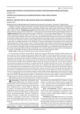
In the databases of evidence-based medicine, studies that study the effectiveness of taking vitamin-mineral complexes indicate a high risk of age-related macular degeneration (AMD) as a prevention (primary prevention). The purpose of the study was to evaluate the effectiveness of the use of lutein-zeaxanthin vitamin-mineral complex containing preparations in individuals with a high risk of AMD for the prevention of the disease. The material of the study was 98 individuals (196 eyes) from the 1st (main) group with the highest risk of developing AMD, who agreed to participate in the prevention of AMD and 90 individuals (180 eyes) from the 2nd (control) group, for various reasons refused to take the drug, but agreed to participate in condition monitoring. The follow-up period was 3 years. The results of the observation showed that in persons of the 1st group, there was a stability in the indices of visual acuity and field of vision, ophthalmoscopic and tomographic picture of the macular zone during the entire period of observation. Whereas in persons of the 2nd group by the 3rd year of observation, visual acuity worsened by 2.5 times, the total boundaries of the peripheral visual field narrowed by 47.10, relative and absolute scotomas appeared (p<0.05). The appearance of drusen was observed and in 8 eyes (4.44%) a diagnosis of age-related macular degeneration of the retina, early stage, was made. Conclusions. The proposed scheme of drug prevention of persons with the highest risk of developing AMD (Group 1) showed a significantly positive effect on the functional state of the retina, leads to a stable preservation of visual functions during 3 years of observation and prevents the occurrence of AMD in 100% of individuals.
-
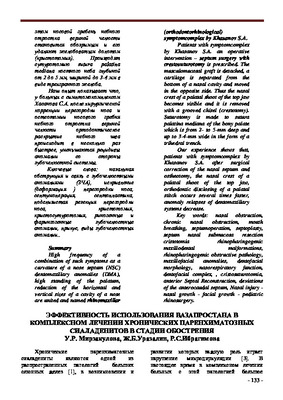 Efficiency of using vazaprostan in the complex treatment of chronic parenchymal sialadenitis in the acute stageUse of vazaprostan in complex treatment patients with parenchymatous sialadenitis in a stage of an aggravation in the field of amazed salivary gland renders good therapeutic effect, which is proved by clinical data and results of reological and biological researches (laktoferrin concentration, middlemolecularpeptids and sum of primary and secondary products of hyperxide acidification lipids).
Stomatologiya -
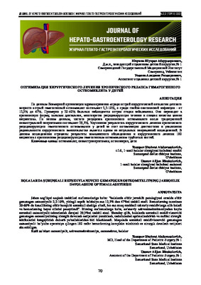 Optimization of surgical treatment of chronic relapse hematogenous osteomyelitis in childrenAccording to the World Health Organization: "among acute surgical pathology of childhood, acute hematogenous osteomyelitis is 2.2-10%, and among purulent septic infection - from 12.5% to 47%.In about 20-60% of patients, the acute stage of the disease turns into a chronic form, causing a long, repeatedly recurrent course and reducing the quality of life of patients." According to our data, the recurrence rate of chronic osteomyelitis after traditional sequestral necrectomy is 29.3%. Thus, improving the results of surgical treatment of chronic recurrent hematogenous osteomyelitis in children by optimizing the diagnosis and expanding the radicality of surgical intervention is one of the relevant areas for research. This study reflects the results of a comprehensive examination and surgical treatment of 180 patients with chronic recurrent hematogenous osteomyelitis of tubular bones.
Journal of hepato-gastroenterological research -
 The prevalence of diabetic retinopathy according to the optic center of "A.A.Yusupov" The city of samarkandSummary. In the ophthalmic center" A.A.Yusupov ” we examined 18880 patients, 384 (2%) of whom were diagnosed as diabetic retinopathy (DR).Proliferative stage of DR was found in 48.6 % of patients. On average, the rapid development of disease observed in age from 60-65 years. At this age the number of patients with proliferative stage prevailed. The findings suggest the need for and feasibility of population screening programs that may help identify the DR in the early stages of its development.
Doctor's Herald -
 The RSO and RSMC Samarkand included 68 patients with metacarpenic breast cancer from 2007 to 2017. More than half of the patients were women of reproductive age (26-49 years). The difference between the primary and metacarpal cancer in unilateral cancer was detected at a very early stage of the disease. This is because patients with cancer treatment are in deep dosing. Metaxron is one of the major factors affecting the survival of patients with ovarian cancer: the second stage of cancer, the size of the tumor in the lactation, the volume of treatment, the rate of damage to the regional lymph nodes and the patient's age
 -
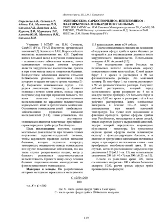 Учеными I МГМУ им. И.М. Сеченова, СамМИ (РУз), УРАН Института органической химии им.НД. Зелинского РАН, Всерос-сийского института гельминтологии им. К.И. Скрябина внесен большой вклад в изучении эхинококкоза - гельминтозного заболевания человека, почти единственным методом лечения которого является хирургическое вмешательство, причем и при этом методе высока смертность больных. Возбудителем заболевания является гельминт Echinococcus granulosus, личиночная стадия которого не щадит ни одного органа человека [1- 4]. Поражения эхинококкозом сердца - это редкая локализация. Например, у больного эхиноккоз печени и/или легких, однако сердце получило удар еще при заражении больного, что нами показано экспериментальными исследованиями по заражению гельминтозами каракульских ягнят в предлагаемом сообщении. Осложнение эхинококкоза детей грибковыми заболеваниями привлекло внимание исследователей [5-11]. Нами установлено, что спутниками эхинококков являются патогенные простейшие саркоспоридии и грибы рода Paecilomyces