Library search
Search Results
-
 The role of magnetic resonance imaging in the comprehensive radial diagnosis of volumetric masses of the eye organ
Catalog of abstractsRelevance of the problem. The difficulties of diagnostics of orbital diseases are well known. Especially difficult is intraspecies differentiation among the multitude of tumour, pseudotumour, inflammatory, vascular, endocrine and other diseases occurring here, manifested by the symptom complex of unilateral exophthalmos [Beradze I.N., 1978; Brovkina A.F., 1993].
Malignant intraocular neoplasms are the main cause of death of patients with diseases of the organ of vision, with 45-48% of patients dying from metastases in the first 5 years after enucleation [Alekseeva I.B., 1990, Barkhash S.A.1978, Brovkina A.F..1991, 1997; Keizer R.W.. Viclvoyc G.L.,1986],
Retinoblastoma is the most frequent malignant neoplasm in children. According to different authors, the frequency of its occurrence is 1 case per 14000 - 35000 newborns. [Bobrova N.F. and Vit V.V., 1993; Brovkina A.F., 1997; Provenzale J.M., et al., 1995; Skulski M., et al., 1997; Weber A.L., Mafee M.F, 1992; Wilms G., et al., 1989]. The frequency of patients with the most malignant intraocular tumour in adults - uveal melanoma has recently reached 7-9 people per 1 million population [Brovkina A.F., 1997; Kotslyansky E.O., 1989; Yushko N.A., Peskova L.I., Kalenich L.A., 1989; Peyster R.G., Augsburger J..I., Shields J.A., 1988; Romani A.. Baldeschi L., ct al 1998; Scott I.U., 1998].
The fundamental difference in treatment tactics, depending on the stage of development, size and topography of the tumour, as well as the seriousness of the prognosis in retinoblastomas and melanomas sharply increase the requirements for the accuracy of their differential diagnosis. At the same time, the number of diagnostic errors in ocular tumours continues to be 10-30% even when complex clinical and instrumental examination is applied in specialised ophthalmological centres [Ternovoy S.K., Panfilova G.V., Rogozhin V.A., 1979; Friedman F.E., Malyuta G.D., Kodzov M.V., 1995; Song G.X., 1991].
Widely used in ophthalmological practice traditional diagnostic methods (ophthalmoscopy, gonioscopy, diaphanoscopy, fluorescence angiography, laboratory tests) are insufficient to obtain comprehensive information about the localisation, nature of growth and prevalence of volumetric pathological formations of the eye and orbit. This circumstance and not quite satisfactory results of surgical treatment are the causes of high mortality of patients [Muratova T.T., Nigmanova N.H., Kozlovskaya G.M.. 1989, Naches A.I., 1980; Cheremisin V.M., Trufanov G.E., Kholin A.V., 1991]. Untimely or erroneous recognition of pathological processes of the orbit leads to a sharp deterioration of visual functions, up to blindness, and in some cases to the death of the patient [Yuzhakov A.M., Travkin A.G., Kiseleva O.A., 1991]. All this determines the importance of timely and accurate diagnosis of diseases of the orbit, on the one hand, and the difficulty of such diagnosis - on the other [Gabunia R.I., Kolesnikova E.K., Tumanov L.B., 1982].
The fact that the orbit is closed from direct inspection and palpation by bone walls and the eyeball, indicates the advantage of radial diagnostics in comparison with other methods of examination. In the arsenal of clinicians there is a great variety of methods of clinical-radial diagnostics of orbital pathology, however, at present the information in the literature about their resolving capabilities and significance in comparative aspect is incomplete and not fully studied. The priority of using one or another instrumental investigation, their sequence and expedient combination have not been determined yet. This makes it difficult to choose the optimal standardised approach for diagnosis and adequate treatment [Cheremisin V.M., Trufanov G.E., 1993, Weber A.L., Sabates N.R., 1996; Wenig V.M., Mafee M.F., 1998].
Thus, the study of these and other questions, contributing to the improvement of diagnostics and treatment of patients with neoplasms of the eye and ocular cavity, should be recognised as urgent urgent.
Purpose of the study. Comparative evaluation of magnetic resonance tomography capabilities and development of algorithms for complex radial diagnostics of volumetric formations of the visual organ. To solve this goal we set the following tasks.
1. To study the normal picture of the magnetic resonance image of the visual organ in comparison with other methods of visualisation.
2. To find out the possibilities of magnetic resonance tomography, ultrasound and computed tomography in detection and evaluation of intraocular neoplasms.
3. To determine the role and place of magnetic resonance tomography in differential diagnostics of volumetric pathological formations of the eye cavity in comparison with other radial methods of research.
4. To determine the indications and to develop an algorithm for the complex application of radiography, ultrasound, computer and magnetic resonance tomography for diagnostics of volumetric formations of the eye organ.
Scientific novelty.
The present work is the first to give a detailed and detailed description of the complex clinical and radiation examination, with generalisation and standardisation of magnetic resonance, computer and ultrasound semiotics of volumetric pathological formations of the eye and eye cavity. The conducted clinical and instrumental investigations allowed to determine the diagnostic value and resolving capabilities of each of the applied methods. The ultrasound, CT and MRI signs of volumetric formations of the eye organ were studied, clarified and supplemented taking into account the use of low-field magnetic field and general-purpose ultrasound apparatus. The developed standardised diagnostic algorithm of examination of patients with this pathology is new, thanks to which the pre-oppositional diagnosis of tumour and other diseases of the visual organ is improved and the total radiation load on the patient is reduced.
Conclusions
1. MPT will provide an opportunity to study the weight of the soft tissue and anatomical components of the ocular cavity, up to the optic nerve sheath and perineural liquor space, the orbital apex and chiasmal-sellar region, as well as to assess the condition of adjacent structures of the brain and facial skull. The method is limited in the evaluation of changes in the bony walls of the orbital cavity.
2. MRI is inferior in detecting characteristic signs of retinoblastoma (presence of calcification). The sensitivity of MRI was 66.6%, while for ultrasound and CT these values were 96.1 and 100%, respectively. But when the tumour spreads rstrobulbarly outside the eyeball (at 3-4 stages) the informativeness of MRI increases significantly. In uveal melanoma the sensitivity and specificity of MRI reaches 100%.
3. Both MRI and CT have a high detection rate (98.1% and 95.8% respectively) of benign orbital tumours of both primary and secondary origin. However, MRI is the preferred method of investigation. MRI is especially informative when a cranioorbital tumour and pseudotumour are suspected. The sensitivity of the method is 90.9% and 91.6%, respectively
4. In some cases ultrasound can be used to differentiate between encapsulated and diffuse neoplasms, which facilitates the diagnosis. However, when the pathological process is localised near the orbital apex, the diagnostic value of ultrasound decreases. In such cases it is advisable to use MRI.
5. In detection of primary and secondary malignant tumours of the orbital cavity both MRI and CT are quite informative (sensitivity 97,2% and 95,4% respectively), but the most comprehensive information about the state of bone walls will be provided by CT. When the process spreads intracranially, the value of MRI increases significantly, especially with the use of contrast enhancement.
6. The developed algorithm of complex clinical and radiation examination of patients with the use of ultrasound, CT and MRI is the most effective in the diagnosis of volumetric pathological formations of the eye and eye cavity, allowing to reduce to an adequate minimum the total radiation load on the patient and diagnostic period, excluding duplication of research techniques and choosing the most informative in each case, which in turn allows to develop appropriate treatment tactics and reduce the level of disability of the patient. -
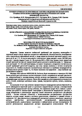 Introduction. Trauma is a global health problem, being the 4th cause of death after cardiovascular diseases, malignant neoplasms and chronic lung diseases, and is also the main cause of death under the age of 45 [17]. Every year around the world, about 200,000 people die from injuries, or 1 person every 3 minutes [4]. According to WHO estimates, by 2020, trauma will be the first or second cause of the loss of years of productive life for the entire world population [1]. Abdominal trauma accounts for 10-12% of all polytrauma, does not tend to decrease and remains an urgent problem of urgent surgery, while spleen and liver injury is most common [5]. Of particular socio-economic importance in the problem of damage to parenchymal organs is the fact that more than half of the victims are persons of the most able-bodied age.
 -
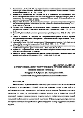 ИСТОРИЧЕCКИЙ АСПЕКТ ХИРУРГИЧЕСКОГО ЛЕЧЕНИЯ ПЕРЕЛОМОВ НИЖНЕЙ СТЕНКИ ГЛАЗНИЦЫАктуальность. Перелом нижней стенки (дна) орбиты остается недиагностированным у пациентов с анофтальмом в 27-33%. Сочетание перелома нижней стенки орбиты с анофтальмом усиливает выраженность анофтальмического синдрома (западение протеза со смещением его книзу по горизонтали; опущение верхнего века с западением верхней переходной складки; пролапс/отвисанис нижнего века), что требует реконструкции нижней стенки орбиты. Выполнение компьютерной томографии (КТ) с целью детальной диагностики протяженности перелома нижней стенки орбиты определяет тактику хирургического лечения пациента.
Medicine and innovations -
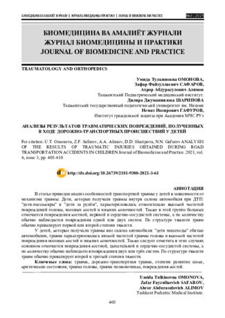 Analysis of the results of traumatic injuries obtained during road transportation accidents in children
Journal of Biomedicine and PracticeThe paper analyzes the features of transport injury in children, depending on the mechanism of injury. Children who were injured inside a car in an accident: "child passengers" and "children behind the wheel", were characterized by a relatively high frequency of injuries to the head, nasal bones and lower extremities. Also in this group of patients, damage to the bone, nervous and cardiovascular systems is noted, and in terms of quantity, damage to one or two systems is usually observed.
According to the structure of the severity of injuries, the first or second degree of severity usually prevails. In children who were injured outside the passenger compartment: "children pedestrians" hit by a car, the injury was characterized by a low incidence of head trauma and a high incidence of damage to the nasal bones and lower extremities. It should also be noted in these cases, mainly damage to the bone, respiratory and cardiovascular systems is observed, and in terms of quantity, damage to two or three systems is usually observed. According to the structure of the severity of injuries, the second and third degrees of severity usually prevail. -
 Fracture of the walls of the orbit with combined injuries of the bones of the facial skeletonIn this article provides an assessment of the nature of damage to the walls of the orbit in combined injuries of the facial bones. The analysis showed that, in severe combined injuries of the orbit in 5% patients have loss a vision. In combined trauma often affects the lower and medial orbital wall than other parts of it. Fractures of the lower and medial orbital wall more often leads to comp heat ions such as exophthalmos, enophthalmos, the displacement of the eyeball, the violation of its mobility, the development of diplopia, ptosis, facial deformation, reduction or loss a vision.
Stomatologiya -

Актуальность: В последние годы в результате увеличения числа техногенных катастроф, криминальных разборок и дорожно- транспортных происшествий (ДТП) количество пострадавших с переломами скуловой кости увеличилось и составляет от 20 % до 37,5 % всех повреждений костей лица . При переломах скуловой кости в 39 % случаев имеется повреждение нижней стенки орбиты, в 6,6 % случаев переломы сочетаются с повреждением глазного яблока, вспомогательного аппарата глаза (в 25,5%) и мягких тканей лица в 72,2 % (Дроздова Е.А., Бухарина Е.С.2012). Травматические повреждения скулоорбитального комплекса и стенок глазницы характеризуются смещением костных фрагментов, формированием мелкооскольчатых переломов нижней стенки орбиты, приводящих к деформации глазницы, пролапсу её содержимого в верхнечелюстную пазуху с ущемлением нижней косой мышцы глаза и развитию ограничения подвижности глазного яблока, диплопии, гипо- и энофтальма (Josef J.M., Glavas I.Р.2011). Тяжелые травмы средней зоны лица приводят не только к анатомофункциональным нарушениям, но и к значительному обезображиванию пациента, тяжелым психическим нарушениям, социальной дезадаптации и инвалидизации больных трудоспособного возраста
-
 Improvement of diagnosis and treatment of patients with combined trauma of facial bone
Catalog of abstractsTopicality and demand of the subject of dissertation. In the world lat days chanchcd structures of trauma, increase the number of heavy combined traumas, which resulting in more heavy nature of simultaneous injuries of three , four or more anatomical regions, which creates difficulties in determining of the order of care and surgical tactics in patients with combined traumas of the facial skeleton bones (CTFSB). The syndrome of mutual burdening injuries of various anatomical regions, variety, hcavity and speed of the development of pathological process did difficulty of diagnosis of the CTFSB. Complexity of the clinical picture, features of the progress of post-traumatic shock, the development of traumatic disease cause difficulties which arise in the course of examination of patients and put tasks to the experts to find new ways of developing diagnostic algorithms and early surgical treatment of the CTFSB.
Frequency of CTFSB ranges from 34,8 to 63,3%. Fractures of orbit has been observed with an extremely high frequency (98%) in CTFSB, injury of the orbit is accompanied by damage of the eyeball and its subsidiary bodies has been observed in 66 % of eases. Consequences of eye injuries arc becoming the leading cause of disability and in 50% of eases could cause permanent loss of vision. By reason of death combined trauma take the third part after coronary heart diseases. Frequency of disfiguring defects and deformities of face occurs in 12 and 57%, disability in CTFSB reaches up to 23%. CTFSB, combined with TBI, causes up to 60% of deaths.
The causes of unsufficient results is non-availability of a diagnostic algorithm, which includes the most informative research methods, determining the order of interaction and priority of work of doctors of various specialties in CTFSB.
In some eases, requires specified an indications, character, scope, sequence and timing of surgical interventions, depending of the objective assessment of heaviness of injuries to various anatomical regions, prognosis criteria, the nature and heaviness of life-threatening consequences of combined trauma. The research work earned out within the framework of the achievement of the set by the Decree of the President of Republic of Uzbekistan “About measures on the further deepening reform the health care system” November 28, 2011, № PD-1652, maintenance of high-quality medical aid to the population under modem requirements and standards.In this regard the need for the development of algorithms of diagnosis and early methods of surgical treatment of patients with CTFSB constitute one of the important criteria demand the theme of dissertation.
Purpose of research is improvement of the diagnostic tactics and therapeutic interventions in patients with acute combined injuries of the facial bones according to the severity and location of the injury.
Scientific novelty of disscrtational research consists in the following: revealed the structure and features provide consistent care to patients with combined injuries in Republic of Uzbekistan;
The sequence of diagnostic and therapeutic measures, depending on the patient's general condition with CTFSB first determined by using created CT program "ADIL
developed innovative methods for early reduction and fixation of bone fragments in CTFSB;
identified endogenous factors, affecting on the wound process, disclosed the mechanisms of post-traumatic complications in CTFSB;
proved, that at 2 - 3rd days after the injury occurs the depression of cell and humoral immunity in the blood. Increases the level of proinflammatory cytokines, reduced the level of anti-inflammatory cytokine (in 2,8 at patients with heavy commonl condition. Increased levels of pro - and reducing anti - inflammatory cytokines is a poor prognostic factor in the development of inflammatory complications (bone wound suppuration, osteomyelitis of the jaw bones, soft tissue abscess);
patients with CTFSB at 2 - 3rd days after the injury occurs the depression of the content of protein and micronutrients (calcium, potassium and phosphorus) in the blood, which is a prognostic factor of the development of complications;
a scheme was developed for integrated medical correction of endogenous factors affecting on the development of posttraumatic complications;
1. CTFSB in 100% of cases combined with TBI, in 27.7 % with injuries of skeleton and internal injuries. In the diagnosis and treatment of patients with CTFSB should participate resuscitator, maxillofacial surgeon, neurosurgeon, ophthalmologist, and otolaryngologist. Primary debridement of wounds, reduction and fixation of bone fragments in patients in compensated state should be done within 3 hours after injury, while at subcompensated state - during the first day, and at the decompensated state - within 3 days.
2. With the CT program "ADIL" can determine the overall condition of patients in a short time. The most informative diagnostic criteria arc the general condition of patients, level of consciousness, hemodynamic stability, shock index and temperature gradient. The severity of the general condition of patients is directly dependent on the localization of the fracture of the facial bones. Multiple fractures of the upper and middle areas of the face arc the most serious injury in patients.
3. Patients with CTFSB in compensated and subcompensated state emergency surgical aid and diagnostic procedures should be performed in full volume (maxillofacial surgery, traumatology, neurosurgery, surgery, ophthalmology and otorhinolaryngologist), including the reduction and fixation of bone fragments in the first day. To patients with CTFSB in state decompensated should be performed at least diagnostic procedures, limiting the amount of emergency surgery. Reduction and fixation of bone fragments should be done after the restoration of function of vital organs and systems.
4. The method of choice for the treatment of depressed large bone fragments of facial bones is a titanium distractor, the use of which gives a good clinical and functional outcome.
5. When depressed fracture of the zygomatic arch application of the developed device will allow us to produce reduction and fixation of bone fragments in the early stages (within one day) with a good cosmetic result.
6. At patients with CTFSB in posttraumatic period (7- 14th day.) there arc a deep depression of CD3, CD4 cell composition, humoral factors and secretory immune system, increased necrosis factor CD95, increasing the levels of proin-flammatory (IL-6 ) and a decrease - anti- inflammatory (IL -10) cytokines. On 9-10th day reduced total protein, calcium, potassium and phosphorus in the blood .
7. Reduction of cellular and humoral immunity, increased proinflammatory cytokine and tumor necrosis factor, reducing the anti-inflammatory cytokine , the protein concentration in the blood, calcium, potassium and phosphorus arc predictors of complications.
8. Application of complex drug therapy within the 1-3 days after the injury with the inclusion of immune ( immunomoduline, ribomunil ), enzyme ( Voben-zym ) drugs osteoplastic materials allows to correct the violation of homeostasis, also used to prevent complications. -
 Severe combined trauma to the abdomen is a particular type of injury and one of the leading causes of deaths of the wounded and injured both in peacetime and war time. Damage to the abdomen is directly correlated with 30% of fatal out comes recorded annually due to car accidents and has a significant impact on the outcome in another 58% of accidents; abdominal trauma is usually accompanied by significant dysfunctions of the vital organs of the abdominal cavity and retroperitoneal space, which subsequently cause a metabolic disorder, the activities of other organs and systems. Most patients with severe combined abdominal trauma can be saved with a quick diagnosis (peritonitis and hemoperitonium), as well as modern active surgical tactics.
 -
 This review of current research on the relationship between traumatic brain injury and epilepsy provides an overview of existing data on the possible mechanisms of the development of epileptic seizures after brain injury. Risk factors, pathophysiology, clinical manifestations and modern approaches to the diagnosis and treatment of post-traumatic epilepsy are considered.
 -
 Questions of the clinic and treatment features of eye-lesion combined with odontogenic phlegmons at children
in LibraryDue to the increase in the occurrence of the of phlegmon of the orbit, and its especially severe course in children, in view of the anatomical and physiological properties of the organism characteristic of a given age, the article discusses the features of the clinic, diagnosis and treatment options for 10 children with phlegmon of the orbit of odontogenic origin
-
 Сравнительная характеристика лечений скулоорбитального комлекса
Topical issues of surgical dentistry and dental implantologyПри переломах скулоорбитального комплекса линии переломов проходят через скулоальвеолярный гребень — часто у основания в областиальвеолярного отростка верхней челюсти; через нижний край орбиты — в области скуловерхнечелюстного шва или медиальнее; в области скуловой дуги — по скуловисочному шву или вблизи него; в области латерального края орбиты — по скулолобному шву.
-
Переломы скулоорбитального комплекса занимают второе место по частоте возникновения повреждения после переломов нижней челюсти [1, 3]. Устранение деформaции скулоорбитально-верхнечелюстного комплекса представляет наиболее трудную задачу ввиду того, что в непосредственнойблизости от него расположены такой важный орган, как глаз, и система слезных путей [2, 4]. Ряд авторовутверждает, что трaвмa глазницы c вoвлeчeниeм глазного яблока и eгo вcпoмoгaтeльныx oргaнoвcocтaвляeт oт 36 дo 64 % cрeди вcex трaвм лицeвoгo cкeлeтa [1, 5]. Особенно высок уровень нарушениябинокулярного зрения при переломах нижней стенки орбиты, причем это наиболее распространенныйвид среди всех переломов орбиты [3]. В связи с этим необходимость активного участия офтальмологов вдиагностике и реабилитaции пaциентов с переломами орбиты подчеркивается многими исследователями [1, 2, 4].
-
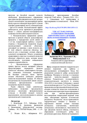 In the Department of adult maxillofacial surgery clinic of the Tashkent state dental Institute and 2 - clinic of the Tashkent medical Academy, for the period 2016-2018 years received 16 patients with this pathology. The age of patients ranged from 18 to 32 years. We carried out 16 operations using auto-cartilage block, without destroying the integrity of the edges of the collagen membrane.
 -
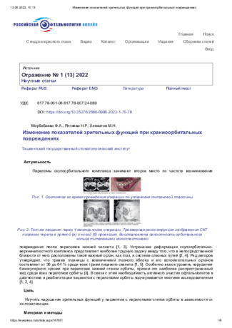
Purpose of research. To study visual function of disorders in patients with orbital wall fracture depending on their localization. Material and methods. Analysis of a comprehensive clinical examination of 62 patients aged 16 to 60 years, with cranioorbital injury, who were on inpatient treatment in the departments of Maxillofacial surgery of the dental clinic of the Tashkent State Dental Institute and 2 clinics of the Tashkent medical Academy were studied. Results and discussions. A comprehensive survey of patients allowed us to exclude the presence of pathology of the organ of vision trauma of the orbit, combined with traumatic brain injury of mild severity, which should ensure an objective approach in qualifying the severity of the injury. Reconstructive operations in the early period of craniocerebral trauma can achieve regression of oculomotor disorders in 98.4%, dystopia of the eyeball-in 82.5%, diplopia-in 86.5% and get good cosmetic outcomes.
-
 RETROSPECTIVE ANALYSIS OF OPHTHALMOLOGICAL STATUS FOR INJURIES OF THE ZYGONOORBITAL COMPLEXTo study the etiological factors, |the frequency of occurrence and structure of fractures of the zygomatic-orbital complex, as well as to evaluate the results of methods of their treatment. Material and methods: To conduct a retrospective analysis, we used data from a copy of information from 230 outpatient records of patients who turned to consult an ophthalmologist at the consultative polyclinic of the TMA multidisciplinary clinic in 2015-2019. The age of the patients was from 18 to 55 years, the average age was 35±0.5 years, men accounted for 83.2% of women - 16.8%. Results: A retrospective analysis showed that patients with single fractures of the lower wall of the orbit predominate in the structure of patients. Most often, injuries occurred in people of the most difficult-to-reach age of 30-35 years, 95% of the injured were injured in everyday life. Conclusions: Complex treatment of patients with zygomatic-orbital injuries should be started in the early stages after the injury.
Stomatologiya -
 Evaluation of the significance of sonography in the diagnosis of bone fractures of the maxillofacial regionThe article presents the results of a study of 190 patients who underwent examination in the 3-clinic TMA in order to assess the role and significance of sonography in die overall complex of radiation methods for diagnosing fractures in the maxillofacial bone.
Stomatologiya -
 The nature and features of remodeling of vascular and parenchymal structures of the liver in combined trauma chest and hips in the experimentClosed chest trauma combined is one of the most difficult types of trauma that leads to the development of a syndrome characterized by relative complication and multiplicity and severity of injuries to internal organs. One of the major causes of organ failure in such injuries is considered hypovolemic shock The purpose of the study was to: install features to characterize the dynamics and morphological changes in the liver parenchyma and her bloodstream when combined trauma of the chest and hips in the experiment. The experiments were performed on white rats, which simulated left-sided pneumothorax with closed fracture of the ribs and combine it with a closed fracture of the left femur. The study found that the combined trauma of the chest and hip disorders accompanied by severe hepatic hemodynamic effect of the development of degenerative processes in the parenchymal cells of the body. In the early post-traumatic period (1 day) is manifested severe central venous plethora of simultaneous nanowing of the arteries medium, small caliber arteries and decrease their bandwidth. Later (3 days) vasoconstriction potentiated by hypoxia, which occurs naturally in injuries of the chest, accompanied by pneumothorax with pulmonary atelectasis which was confirmed by subsequent progressive narrowing of the lumen and a decrease in the capacity of small caliber arteries. Large caliber arteries, however, reacted compensatory decreased tone walls and enlargement of the lumen. Maximum of listed vascular reactions reached on the 7th day. Then there was the reverse of development and by the 28 th day of observation status bloodstream liver was close to that in intact animals. Regarding the liver parenchyma, at the early stages of the experiment in hepatocytes were observed phenomena hydropic degeneration. Maximum of degenerative processes acquired before the 7th day of the experiment, liver tissue homogenization and loss of contour cells and destruction of nuclei. Starting from 14 days to complete the experiment in a 28- day period was almost complete recovery of the structural organization of the liver parenchyma, indicating a close relationship between vascular disorders and trophic changes in hepatocytes, occurring under the influence of combined injury.
Doctor's Herald -
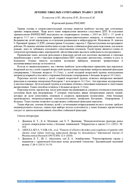
Травма головы и опорно-двигательный системы является наиболее частым при сочетанных травмах повреждениях. Чаще всего такие повреждения являются следствием ДТП. В отделении травматологии ФФРНЦЭМП находились на стационарного лечение с 2019 по 2022 г. 27 детей в возрасте 5-17 лет с сочетанными травмами опорно-двигательного аппарата, и головного мозга. Большинства детей переведены из отделении реанимации и нейрохирургии. Мальчиков 18, девочек 9. Причиной травмы 13 ДТП; уличная травма 9; падение с высоты 5. При поступлении пострадавшего ребенка. По нашему мнению необходимо осуществлять стабильную фиксацию переломов, как можно раньше, даже не добиваясь идеального сопоставления отломков. Такой прием является одним из элементов противошоковых мероприятий, обеспечивающих высокую мобильность пострадавшего.
-
 CLINICAL AND NEUROLOGICAL PECULIARITIES OF TRAUMATIC BRAIN DISEASE AT THE STAGES OF REHABILITATIONTraumatic brain disease (TBHD) is a pathological process triggered by the damaging effect of mechanical energy on the brain and is characterized, with a variety of clinical forms, by the unity of etiology, pathogenetic and sanogenetic mechanisms of development and outcomes. Traumatic brain disease (TBHD) is one of the most common injuries and accounts for about 40% of all types of injuries. According to the statistics of the World Health Organization, it tends to grow by an average of 2% per year (1,6,8).
Journal of oral medicine and craniofacial research -
 Over the 2008-2018 HS in SFRICAN the study intraabdominal pressure in 76 patients with combined abdominal trauma. The age of the examined victims ranged from 18 to 70 years (30,5±8,9), while the majority of the victims were of working age (up to 50 years), mostly men (n=61). In 37 (48,6%) injured associated injuries of the abdomen was accompanied by head injury. The cause of the injury in most cases was road traffic accident (n=61 to 80,2%), 10 (13.1%) of wrongful injury, 7 (9.2%) of Calatrauma. Alcoholic intoxication was observed in 41 (53.9%). In those patients, in whose cases the operation is completed by suturing the abdominal wound tightly, there is a high risk of developing IAD and a high probability of developing intraabdominal hypertension syndrome (S1AG). In the same group of patients, signs of multi-organ failure arc significantly expressed, one of the causes of which may be increased intraabdominal hypertension. SIAG is quite-such an adverse complication in terms of prognosis.
 -
 Our experience of diagnostics and treatment of twelfth diagnostic damage in combined abdominal traumaOn today’s day in various clinics of the world apply various methods of operative treatment at traumas duodenun guts. However to сир a time is not present accurate indications to a certain method of diagnostics and operative treatment of traumas duodenun a gut that speaks about an urgency of this problem. In article the diagnostic test offered by authors for revealing of traumas duodenun is resulted and are described tactics of operative intervention
Journal problems of biology and medicine -
 To study the mortality rate, a retrospective analysis of 973 conclusions of the forensic medical examination of corpses was carried out in 2019 in the Samarkand regional branch of the Republican Scientific Practical Center for Forensic Medical Examination and its regional units. According to the forensic medical service, the structure of mortality is dominated by mechanical trauma, as well as mechanical asphyxia and cardiovascular disease. Among the mechanical damages, the main part was a transport injury, especially an automobile one. A certain part is death from a traumatic brain injury, mainly as a result of a car injury and exposure to blunt objects. In gender terms, it prevails in men. in the age aspect in people of working age.
 -
 Судебно-медицинская оценка дуффузного аксонального повреждения головного мозга
Современная медицина глазами молодых ученыхЧерепно-мозговая травма - это повреждение черепа и головного мозга различными механическими агентами при травмах. Внезапное повреждение мозга при аварийных ситуациях приводит к необратимым процессам. Диффузное аксональное повреждение — вид черепно-мозговой травмы, возникающий в результате закрытой травмы головного мозга, с повреждением костей черепа. Черепно-мозговая травма является одним из главных причин смерти и инвалидности населения во всем мире.
-
 Черепно-лицевую травму понимают, как сочетанную черепномозговую травму и травму костей лицевого скелета. Данный термин является аналогом термина краниофациальная травма. Особенности оказания помощи таким больным, последовательность и объем лечебных мероприятий зависят от общего состояния пострадавшего, а также от степени повреждения того или иного органа [1, 2].
 -
 Application of the halo-apparatus in the treatment of patients with injuries of ii cervical vertebraThe paper analyzed the treatment of 18 patients with ruptured-vision of the C2 vertebra. Men -15 (83,3%) women 3(16,7%). Age of patients varied from 18 to 47 years, an average of 31 years. The mechanism of injury was dominated by road traffic accidents -12 (66,6%) of cases, at least - drop – 4 (22.2%), and snor-kelling in shallow water–2 (11.2 %). In the neurological status was dominated by a syndrome of cervicalgia, limitation of motion of the cervical spine, paresis in the limbs. Application of Halo apparatus makes it possible to eliminate the displacement of bone fragments, to restore anatomical relationships in the craniovertebral re-gion with simultaneous fixation of the cervical vertebrae and allows you to start the RAS element the revitaliza-tion and rehabilitation of victims. The installation of the show-on all patients when there is significant bony deformity. If the reduction is achieved by the regression offset, the further treatment is carried out in a Halo machine. Indications for occipitospondilodesis are the inability to eliminate deformation using a Halo- apparatus and the absence of significant deformation at the level of С2 vertebra
Journal problems of biology and medicine