Library search
Search Results
-
 The use of laser photocoagulation corneal herpetic keratitisOphthalmoherpes in the statistical analysis of ocular disease comes first corneal pathology in the world. We examined 18 (18 eyes) of patients with various forms of herpetic keratitis. Among the patients were 11 men and 7 women. All patients before and after treatment was performed standard ophthalmologic examina-tion. Laser coagulation of corneal infiltrates was performed by affecting the cornea by laser radiation with a wave length of 532 microns, the energy of 300-400 mJ / sm 2 , the laser spot diameter of 200 microns. Infiltra-tion of the cornea stained with Collargol. Visual function was assessed before treatment and after 1week. 1, 3 and 6 mth. after therapy. The use of laser photocoagulation in the treatment of herpetic keratitis leads to an acceleration of reparative processes, rapid relief of inflammatory processes and better healing of the corneal defect. Аfter laser coagulation of corneal visual acuity of patients with this pathology increased to 0.8-1.0. Average time epithelialization of corneal epithelium of the cornea in the application of LC was 6-7 days. Re-covery we noted in 82.6% cases. Relapses occurred 4.3% of cases. Conducting corneal infiltrates LC patients with herpetic keratitis in the early postoperative period leads to permanent removal of corneal syndrome, a certain increase in visual acuity development epithelialization of the cornea, reducing its thickness and re-duce swelling.
Journal problems of biology and medicine -
 Features of ultrastructural changes in the cornea of rabbits at its burns alkali and correction by the enterosorbent of polisorb30 rabbits took part at the experiment. Cornea of some of them was burned with alkali; conjunctival sac of the others was washed with the “Polisorb” sorbent solution after the combustion. Gross destructive chang-es in cornea arose already after the combustion and progressed till the end of the experiment. An hour after the combustion in the setting of homogenized tissue of the cornea frontal epithelium we also found cells with signs of gross destructive damages in its central parts. Cornea proper substance was destructed as well. De-fibring and fragmentation of collagen fibrillas was also discovered, their parallel orientation was getting lost. Changes in the group of animals, which were treated with “Polisorb”, were less significant than in the group of animals without correction. In an hour after the combustion the destructive changes in the frontal epitheli-um were almost the same as in the group of animals whose conjunctiva was not treated with “Polisorb” sorbent. Though, we discovered cells with partially residual structural elements more often. Destructive changes in the cornea proper substance were moderate. In the group of animals without correction destruc-tive changes in all layers of cornea progressed even later, their maximum development occurred in 24 hours after the combustion. In as little as 12 hours after the investigation start not only epithelial cells of the injured part of the cornea, but also its marginal area incurred obvious destruction. Intercellular spaces were exagger-ated and vacuolated. The proper substance was completely disorganized. In the contrast, in 12 hours after the combustion dehydration processes began to take off in the cornea of the animals whose conjunctive sac was washed with “Polisorb” sorbent. When morphological pattern at the combustion area was steady, we discov- ered residual and functionally active epithelial cells with moderately destructive changes in the marginal ar-ea. The eye structure was regenerating during 24 hours of the investigation
Journal problems of biology and medicine -
 Comparative analysis of the results of corneal diameters by different methods of measurement in children with primary congenital glaucom
Journal of Biomedicine and PracticeThe article provides a comparative analysis of the study of the diameter of the cornea in children with primary congenital glaucoma. Three methods, which are used in pediatric ophthalmology, are described. The first method is measuring the diameter of the cornea using a school ruler, the second method is using a surgical compass and the third is a new method developed with the use of a gauge in the form of glasses and a computer program. All the positive and negative aspects of methods for studying the diameter of the cornea are described in detail.
-
Purpose — to determine the values of central corneal thickness (CCT) in children depending on the level of intraocular pressure
(IOP) and the stage of congenital glaucoma (CG).
Material and methods. Clinical studies were carried out in the eye department of the clinic at the Tashkent Pediatric Medical In-
stitute. The study involved 18 patients (36 eyes) aged 9 to 11 years (mean age 9.3±1.6 years) with confirmed diagnosis of CG.
All patients underwent basic ophthalmologic examination prior to surgical and conservative treatment. In addition to basic meth-
ods, axial eye length and CCT were determined using an automatic non-contact tonometer/pachymeter manufactured by NIDEK
(USA).
Results. Analysis of the obtained data showed that in initial, moderate and advanced stages of glaucoma, the CCT values were sig-
nificantly lower than the age norm values. This indicates stretching of the fibrous capsule and thinning of the cornea in glaucoma.
In terminal stage CG, the CCT values practically did not differ from the age norm, but were higher than in initial, moderate and ad-
vanced stages of the disease. The noted thickening of the corneal membrane in terminal stage may be explained by edema of the cor-
neal tissue as a result of elevated IOP.
Conclusion. The age norm values of CCT should be taken into account when characterizing the severity of glaucomatous process
in children. Compared to the age norm, the cornea is significantly thinner in children aged 9 to 11 years with initial, moderate
and advanced stages of CG, and becomes significantly thicker in terminal stage, which is associated with edema caused by ele-
vated IOP. -
 EXPERIENCE IN THE TREATMENT OF PURULENT CORNEAL ULCERS WHILE WEARING SOFT CONTACT LENSESPurulent corneal ulcer – a serious eye disease, which is the outcome of gross corneal scarring with persistent depression of vision and, in extreme cases, loss of an eye as an organ. It proceeds most aggressively against the background of wearing soft contact lenses.
Science and scientific potential: the basis for sustainable innovative development of the society -
Inflammatory diseases of the cornea are one of the most common causes of blindness worldwide. In recent years, many modern methods of treating diseases of the cornea have appeared, including, in addition to traditional conservative methods, modern surgical methods, such as autoconjunctival plasty, amniotic membrane transplantation, therapeutic keratoplasty, corneal covering with a corneoscleral flap, microdiathermocoagulation, and corneal crosslinking. A promising direction in the treatment of corneal pathology is stem cell therapy and cell therapy.
-
 The role of central corneal thickness in the unterpretation of intraocular pressure readings in primary infantile glaucoma
in LibraryDetermine the indicators of intraocular pressure (IOP) tonometry, taking into account the parameters of the central thickness of the cornea (CTC) in children with congenital infantile glaucoma (CIG). Clinical studies were conducted in the eye department of the Tashkent Pediatric Medical Institute's clinic. A total of 14 patients (26 eyes) aged 3 to 10 years with a diagnosis of CG were examined. All patients underwent basic ophthalmologic examination prior to surgical and conservative treatment. In addition to basic methods, the axial length of the eyeball was determined, and the CTC was determined on an automatic non-contact tonometer - Reichert pachymeter (USA). An analysis of the data showed: IOP in 39% of cases was considered subnormal, after correction taking into account the CTC in 11% of cases, the IOP was high. This group (11% of cases) was composed of children with a “thin” cornea at the terminal stage of CIG and high myopia. The correct interpretation of IOP indicators will help in choosing an adequate treatment and monitoring the glaucoma process. The used indicator of the central nervous system has practical significance in the interpretation of tonometry according to Maklakov 5 gr.
-
 The role of central corneal thickness in the unterpretation of intraocular pressure readings in primary infantile glaucoma
Journal of Biomedicine and PracticeDetermine the indicators of intraocular pressure (IOP) tonometry, taking into account the parameters of the central thickness of the cornea (CTC) in children with congenital infantile glaucoma (CIG). Clinical studies were conducted in the eye department of the Tashkent Pediatric Medical Institute`s clinic. A total of 14 patients (26 eyes) aged 3 to 10 years with a diagnosis of CG were examined. All patients underwent basic ophthalmologic examination prior to surgical and conservative treatment. In addition to basic methods, the axial length of the eyeball was determined, and the CTC was determined on an automatic non-contact tonometer - Reichert pachymeter (USA). An analysis of the data showed: IOP in 39% of cases was considered subnormal, after correction taking into account the CTC in 11% of cases, the IOP was high. This group (11% of cases) was composed of children with a “thin” cornea at the terminal stage of CIG and high myopia. The correct interpretation of IOP indicators will help in choosing an adequate treatment and monitoring the glaucoma process. The used indicator of the central nervous system has practical significance in the interpretation of tonometry according to Maklakov 5 gr.
-
 Optical and biometric indicators of the eye in children with juvenile glaucoma combined with myopia
in LibraryPurpose. To analyze optical and biometric indicators of children's eyes with various stages of congenital juvenile glaucoma (CJG) combined with myopia. Material and methods. We examined 17 patients (31 eyes) aged 11 to 17 (averagely 14.0 ± 0.2 years) diagnosed with CYG who underwent, prior to surgical or conservative treatment, a regular ophthalmological examination supplemented with the measurement of the central corneal thickness (on an automatic contactless tonometer-pachymeter by NIDEK, USA), and the index of corneal deformation (ICD) by the Shkrebets technique. Results. The analysis showed a possible correlation between 1) tonometric intraocular pressure (P) and the axial length of the eye, 2) P t and the ratio of excavation to optic disk diameter (E/ON), 3) axial length of the eye and the central corneal thickness at the terminal CYG stage, and 4) the axial length and the refraction at the initial stage of CYG. Conclusion. As the glaucomatous process progresses, children with CYG combined with myopia show an increase of myopic refraction, a decrease in fibrous membrane rigidity, pretrabecular and trabecular changes, axial elongation, increased IOP due to an imbalance between the production of intraocularfluid and its outflow, an expansion of the excavation of the optic disk and a decrease in the central corneal thickness.
-
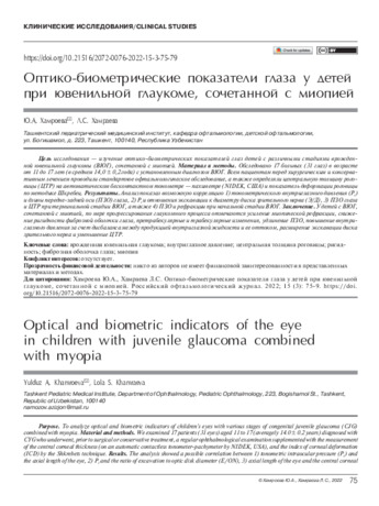 Optical and biometric indicators of the eye in children with juvenile glaucoma combined with myopiaPurpose. To analyze optical and biometric indicators of children's eyes with various stages of congenital juvenile glaucoma (CJG) combined with myopia. Material and methods. We examined 17 patients (31 eyes) aged 11 to 17 (averagely 14.0 ± 0.2 years) diagnosed with CYG who underwent, prior to surgical or conservative treatment, a regular ophthalmological examination supplemented with the measurement of the central corneal thickness (on an automatic contactless tonometer-pachymeter by NIDEK, USA), and the index of corneal deformation (ICD) by the Shkrebets technique. Results. The analysis showed a possible correlation between 1) tonometric intraocular pressure (Pt) and the axial length of the eye, 2) Pt and the ratio of excavation to optic disk diameter (E/ON), 3) axial length of the eye and the central corneal
in Library -
 Оценка эффективности применения препаратов кераторепарантого действия в терапии помутнений роговицы после перенесенного аденовирусного кератоконъюнктивит
in LibraryПо данным литературы, одним из самых распространенных вирусных заболеваний глаз является аденовирусный кератоконъюнктивит (АВКК). Сформировавшиеся в исходе АВКК помутнения (субэпителиальные инфильтраты) роговицы зачастую в значительной степени снижают остроту зрения и, соответственно, ухудшают качество жизни пациентов, в связи с чем важной проблемой является адекватное и эффективное лечение субэпителиальных инфильтратов роговицы после перенесенного АВКК.
-
 Results of surgical treatment of purulent corneal ulcers by autoconjunctive plastic method in combination with tarzoraphia
Journal of Biomedicine and PracticeThis article presents the results of surgical treatment of purulent corneal ulcers in 17 patients by the method of autoconjunctival plasty in combination with tarsorraphy. The article describes in detail the technique of the operation and its main advantages. The authors have shown the effectiveness of the proposed method, which is expressed in the fact that in ease of purulent corneal ulcers, the proposed method allows you to preserve the anatomical integrity of the eyeball and improve the patient's rehabilitation process after surgery.
-
To determine the hydrodynamic parameters of the uninjured fellow eye of children with combined injuries of the organ of vision. A prospective analysis of the hydrodynamic parameters of the fellow eye according to Friedenwald was carried out in 18 patients (18 eyes) aged 3 to 10 years 2–3 and 45–50 days after primary surgical treatment (PSD) of a penetrating wound of the cornea, who were hospitalized in the ophthalmological department of the clinic Tashkent Pediatric Medical Institute. Group I included 8 (44%) children with the following diagnosis: “Combined injury of the organ
of vision. Contusion of the eyeball severe. Complex penetrating wound of the cornea. Group II included 10 (56%) patients with complex penetrating wounds of the cornea. 2–3 days after PST of the wound, group I showed a statistically significant increase in Pt by 2.04±0.03 mm Hg. compared with the control group, while 1–2 days after the first measurement and 45–50 days after PST, the indicators decreased, on average, by 4.4±0.02 mm Hg. without the use of antihypertensive drugs. Changes in the hydrodynamics of the eye in children of group II were not statistically significant. The results of the examination of children revealed a transient increase in tonometric intraocular pressure in the paired uninjured eye 2–3 days after PST of a penetrating wound of the cornea with combined injuries of the organ of vision. -
 Effect of central corneal thickness on intraocular pressure in primary infantile glaucoma in children.
in LibraryThe importance of ophthalmotonometry in the diagnosis of ocular pathology cannot be overestimated. Not you- There are doubts about the need for this study in patients of all age groups [1]. For example tonometry data in eyes with a cornea having a thickness in the center of more than 580 microns need to be corrected lowering (real IOP is lower than the obtained data) [2].
-
 Results of treatment non-penetrating valved corneal injuries in children
Journal of Biomedicine and PracticeThe article provides an analysis of patients in terms of frequency, causes and epidemiological features of non penetrating valved injuries of the cornea according to the data of the clinic of the Tashkent Pediatric Medical Institute. Comparative analysis of the treatment of non-penetrating valved injuries of the cornea in 24% of children who did not undergo surgical treatment, give good functional results and contribute to a more rapid restoration of visual functions.
-

This article focuses on the interests of international system actors in Central Asia in modern global processes, such as the United States and China. In particular, Frederick Starr's concept of "Greater Central Asia", the US Strategy for Central Asia for 2019-2025 and China's "One Place - One Way" project were analyzed.
-
 Differential approach to the application of biomaterials in reconstructive surgery for thermochemical burns of the eyes in children
Journal of Biomedicine and PracticeSimblefaron is a scar fusion of the posterior conjunctival surface of the eyelid with the eyeball. It often develops as a complication of thermal burns of the eyes. Eye burns are severe damage to the organ of vision and occupy a significant share (6.9-30.5%) in the structure of eye injuries. The results of the study showed that the stage-by-stage surgical plastic surgery of the anterior segment of the eye performed by us, which consists in a combination of plastics of the fornix with flaps from the automucosal cavity of the mouth (lips) and transplantation of the amniotic membrane onto the surface of the cornea and conjunctiva with the capture of the limbal zone, allows you to eliminate the defect of the cornea and maintain its transparency and also contributes to the restoration of the conjunctival arches, which can significantly reduce the likelihood of developing complications of burn injury and shorten the rehabilitation period, improve the functional results of treatment.
-
 Early indicators of visiometry and refraction in children after intraocular lens implantation
in LibraryThe article presents the result of a survey of 35 patients after extracapsular extraction cataract with IOL implantation at the age of 8 months to 13 years.Patients underwent: viziometry, biomicroscopy, keratorefractometry, skiascopy, A, B-scan ultrasound, ophthalmoscopy, consultation of related specialists. The calculation of the optical power of IOL was performed using the SRK II formula. Refraction in the early postoperative period in children from 8 months to 6 years corresponded to the age range, from 6 to 13 years was presented in the form of ametropia.According to the authors , ametropia is a consequence of post-traumatic scarring of the cornea, tension of the stitches , swelling of the cornea and mistakes made at the calculation of the power of the IOL according to the formula SRK II.
-
 Clinical evaluation of biomechanical features of the fibrous membrane in children with congenital glaucoma
in LibraryThe clinical, functional and biomechanical properties of the fibrous membrane of the eye in children with
primary congenital glaucoma were studied using the method of elastotonometry. The results of a study of the
clinical, functional and biomechanical properties of the eyes in 57 children with primary congenital glaucoma and
in 11 healthy children aged 8 days to 7 years in the eye department of the clinic of the Tashkent Pediatric Medical
Institute are analyzed. Research methods included clinical and functional methods and special (to determine the
rigidity of the membranes of the eye). The results of the study showed that with an increase in elastopod values,
the indicators of corneal deformation decrease as a result of pronounced corneal edema; the level of IOP in the
advanced and terminal stages indicates the weak rigid properties of the fibrous membrane of the eye. Thus, the
elastotonometry method is an objective quantitative diagnostic criterion for assessing the biomechanical proper-
ties of the fibrous membrane of the eye in children with congenital glaucoma. -
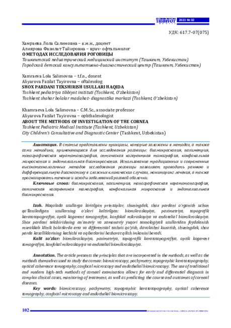
The article presents the principles that are incorporated in the methods, as well as the methods themselves used to study the cornea: biomicroscopy, pachymetry, topographic keratotopography, optical coherence tomography, confocal microscopy and endothelial biomicroscopy. The use of traditional and modern high-tech methods of corneal examination allows for early and differential diagnosis in complex clinical cases, monitoring of treatment, as well as predicting the course and outcomes of corneal diseases
-
 This article informs you about the influence of Russia on the countries of Central Asia. As we know, Central Asia is a post-Soviet space. And Russia is considered an equally important partner of the strategic, economic, and military plan. This article covers all the nuances of the international and domestic functioning of all five Central Asian states and the influence of Russia on these functions. You can also see the main changes in relations between the Central Asian countries and Russia in the period from the collapse of the Soviet Union to the present day.
 -

В данной научной работе изучались результаты больных с центральной серозной хориоретинопатией, которым было проведено лазерное микроимпульсное воздействие. В наше исследование было включено 39 человек, которых наблюдали в течение от 1 до 6 месяцев после проведенного лечения. Введение. По данным многих авторов и проведённых исследований, имеется множество заболеваний макулярной части сетчатки, в частности центральная серозная хориоретинопатия (ЦСХ), которые приводят к снижению остроты зрения, вплоть до слепоты. До настоящего времени причины возникновения ЦСХ окончательно не были установлены. При остром течении возникает идиопатическая отслойка нейроэпителия. Около в 80% случаев наблюдается самостоятельная резорбция субретинальной жидкости и прилегания нейроэпителия. В отличии от острой формы, хроническая форма встречается во взрослом возрасте, у лиц старше 45 лет и имеет двухсторонний процесс. Для этой формы характерно возникновение необратимых атрофических изменений в макуле и нарушения зрительных функций.
-
 This article mentions that the development of mutual cooperation is important for ensuring sustainable development in Central Asia. Today, the people of the region face the main goal of turning Central Asia into a stable economically developed and highly developed region with joint efforts. From this point of view, the mutual cooperation of the countries of the region is analyzed by many experts and shows its relevance.
 -
 Disadvantages and ways of improvement in diagnostics and surgical treatment of the diabetic foot syndromePatients with diabetes mellitus, often, there are changes in the vessels in the form of calcification of the middle shell of arteries and arterioles (arteriolosclerosis of Menkeberg, mediacalcinosis). The determination of the x-ray stage of calcification of the arteries, gives additional information on the state of the vessels of the foot. Destructive changes in blood vessels, soft tissues and bones, conducted surgical interventions are naturally related. The study of these patterns is possible with a timely, critical study of anamnestic, diagnostic data and therapeutic measures. In this regard, to date remain relevant issues related to the development of differentiated treatment, taking into account all possible diagnostic data and improving surgical methods for treating the diabetic foot syndrome.
Doctor's Herald -
В данной научной работе изучались результаты больных с центральной серозной хориоретинопатией, которым было проведено лазерное микроимпульсное воздействие. В наше исследование было включено 39 человек, которых наблюдали в течение от 1 до 6 месяцев после проведенного лечения. Введение. По данным многих авторов и проведённых исследований, имеется множество заболеваний макулярной части сетчатки, в частности центральная серозная хориоретинопатия (ЦСХ), которые приводят к снижению остроты зрения, вплоть до слепоты. До настоящего времени причины возникновения ЦСХ окончательно не были установлены. При остром течении возникает идиопатическая отслойка нейроэпителия. Около в 80% случаев наблюдается самостоятельная резорбция субретинальной жидкости и прилегания нейроэпителия. В отличии от острой формы, хроническая форма встречается во взрослом возрасте, у лиц старше 45 лет и имеет двухсторонний процесс. Для этой формы характерно возникновение необратимых атрофических изменений в макуле и нарушения зрительных функций.