Library search
Search Results
-
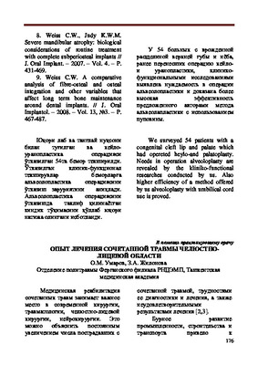
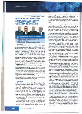 COMPOSITION AND MONITORING OF BUTTER DEFECTS OF YUTSORI LAB AND TANGLAI IN FERGANA REGIONThe article reports on the study of the structure of the congenital cleft lip and palate of sick children on the materials of the Fergana regional hospital specialized in maxillofacial surgery. Monitoring at the beginning of the year and compliance with the basic principles of rehabilitation of children with congenital cleft lip and palate requires long-term comprehensive treatment with the participation of many specialists. It is necessary to predict the expected results and a clear plan for the various stages. The list of activities carried out during the main stages includes early orthopedic treatment of the first days of the child's life, reasonable planning of surgical interventions. Orthodontic treatment, speech therapy training, rehabilitation measures together with the period of growth and development of the upper lip and palate, are pivotal in the main stages of surgical treatment, not only greatly improves the stages of surgical treatment, not only greatly improves the aesthetic and functional the results of treatment, but also minimizes the number of repeated corrective operations.
Stomatologiya -
 Using proverbs and sayings in Russian lessons
Traditions and innovations in language research and teachingСтатья затрагивает актуальную тему изучения языка русских пословиц и поговорок в контексте занятий по русскому языку, а также рассматривает использование этих языковых единиц при обучении студентов групп с нерусским языковым составом.
-
 The role of magnetic resonance imaging in the comprehensive radial diagnosis of volumetric masses of the eye organ
Catalog of abstractsRelevance of the problem. The difficulties of diagnostics of orbital diseases are well known. Especially difficult is intraspecies differentiation among the multitude of tumour, pseudotumour, inflammatory, vascular, endocrine and other diseases occurring here, manifested by the symptom complex of unilateral exophthalmos [Beradze I.N., 1978; Brovkina A.F., 1993].
Malignant intraocular neoplasms are the main cause of death of patients with diseases of the organ of vision, with 45-48% of patients dying from metastases in the first 5 years after enucleation [Alekseeva I.B., 1990, Barkhash S.A.1978, Brovkina A.F..1991, 1997; Keizer R.W.. Viclvoyc G.L.,1986],
Retinoblastoma is the most frequent malignant neoplasm in children. According to different authors, the frequency of its occurrence is 1 case per 14000 - 35000 newborns. [Bobrova N.F. and Vit V.V., 1993; Brovkina A.F., 1997; Provenzale J.M., et al., 1995; Skulski M., et al., 1997; Weber A.L., Mafee M.F, 1992; Wilms G., et al., 1989]. The frequency of patients with the most malignant intraocular tumour in adults - uveal melanoma has recently reached 7-9 people per 1 million population [Brovkina A.F., 1997; Kotslyansky E.O., 1989; Yushko N.A., Peskova L.I., Kalenich L.A., 1989; Peyster R.G., Augsburger J..I., Shields J.A., 1988; Romani A.. Baldeschi L., ct al 1998; Scott I.U., 1998].
The fundamental difference in treatment tactics, depending on the stage of development, size and topography of the tumour, as well as the seriousness of the prognosis in retinoblastomas and melanomas sharply increase the requirements for the accuracy of their differential diagnosis. At the same time, the number of diagnostic errors in ocular tumours continues to be 10-30% even when complex clinical and instrumental examination is applied in specialised ophthalmological centres [Ternovoy S.K., Panfilova G.V., Rogozhin V.A., 1979; Friedman F.E., Malyuta G.D., Kodzov M.V., 1995; Song G.X., 1991].
Widely used in ophthalmological practice traditional diagnostic methods (ophthalmoscopy, gonioscopy, diaphanoscopy, fluorescence angiography, laboratory tests) are insufficient to obtain comprehensive information about the localisation, nature of growth and prevalence of volumetric pathological formations of the eye and orbit. This circumstance and not quite satisfactory results of surgical treatment are the causes of high mortality of patients [Muratova T.T., Nigmanova N.H., Kozlovskaya G.M.. 1989, Naches A.I., 1980; Cheremisin V.M., Trufanov G.E., Kholin A.V., 1991]. Untimely or erroneous recognition of pathological processes of the orbit leads to a sharp deterioration of visual functions, up to blindness, and in some cases to the death of the patient [Yuzhakov A.M., Travkin A.G., Kiseleva O.A., 1991]. All this determines the importance of timely and accurate diagnosis of diseases of the orbit, on the one hand, and the difficulty of such diagnosis - on the other [Gabunia R.I., Kolesnikova E.K., Tumanov L.B., 1982].
The fact that the orbit is closed from direct inspection and palpation by bone walls and the eyeball, indicates the advantage of radial diagnostics in comparison with other methods of examination. In the arsenal of clinicians there is a great variety of methods of clinical-radial diagnostics of orbital pathology, however, at present the information in the literature about their resolving capabilities and significance in comparative aspect is incomplete and not fully studied. The priority of using one or another instrumental investigation, their sequence and expedient combination have not been determined yet. This makes it difficult to choose the optimal standardised approach for diagnosis and adequate treatment [Cheremisin V.M., Trufanov G.E., 1993, Weber A.L., Sabates N.R., 1996; Wenig V.M., Mafee M.F., 1998].
Thus, the study of these and other questions, contributing to the improvement of diagnostics and treatment of patients with neoplasms of the eye and ocular cavity, should be recognised as urgent urgent.
Purpose of the study. Comparative evaluation of magnetic resonance tomography capabilities and development of algorithms for complex radial diagnostics of volumetric formations of the visual organ. To solve this goal we set the following tasks.
1. To study the normal picture of the magnetic resonance image of the visual organ in comparison with other methods of visualisation.
2. To find out the possibilities of magnetic resonance tomography, ultrasound and computed tomography in detection and evaluation of intraocular neoplasms.
3. To determine the role and place of magnetic resonance tomography in differential diagnostics of volumetric pathological formations of the eye cavity in comparison with other radial methods of research.
4. To determine the indications and to develop an algorithm for the complex application of radiography, ultrasound, computer and magnetic resonance tomography for diagnostics of volumetric formations of the eye organ.
Scientific novelty.
The present work is the first to give a detailed and detailed description of the complex clinical and radiation examination, with generalisation and standardisation of magnetic resonance, computer and ultrasound semiotics of volumetric pathological formations of the eye and eye cavity. The conducted clinical and instrumental investigations allowed to determine the diagnostic value and resolving capabilities of each of the applied methods. The ultrasound, CT and MRI signs of volumetric formations of the eye organ were studied, clarified and supplemented taking into account the use of low-field magnetic field and general-purpose ultrasound apparatus. The developed standardised diagnostic algorithm of examination of patients with this pathology is new, thanks to which the pre-oppositional diagnosis of tumour and other diseases of the visual organ is improved and the total radiation load on the patient is reduced.
Conclusions
1. MPT will provide an opportunity to study the weight of the soft tissue and anatomical components of the ocular cavity, up to the optic nerve sheath and perineural liquor space, the orbital apex and chiasmal-sellar region, as well as to assess the condition of adjacent structures of the brain and facial skull. The method is limited in the evaluation of changes in the bony walls of the orbital cavity.
2. MRI is inferior in detecting characteristic signs of retinoblastoma (presence of calcification). The sensitivity of MRI was 66.6%, while for ultrasound and CT these values were 96.1 and 100%, respectively. But when the tumour spreads rstrobulbarly outside the eyeball (at 3-4 stages) the informativeness of MRI increases significantly. In uveal melanoma the sensitivity and specificity of MRI reaches 100%.
3. Both MRI and CT have a high detection rate (98.1% and 95.8% respectively) of benign orbital tumours of both primary and secondary origin. However, MRI is the preferred method of investigation. MRI is especially informative when a cranioorbital tumour and pseudotumour are suspected. The sensitivity of the method is 90.9% and 91.6%, respectively
4. In some cases ultrasound can be used to differentiate between encapsulated and diffuse neoplasms, which facilitates the diagnosis. However, when the pathological process is localised near the orbital apex, the diagnostic value of ultrasound decreases. In such cases it is advisable to use MRI.
5. In detection of primary and secondary malignant tumours of the orbital cavity both MRI and CT are quite informative (sensitivity 97,2% and 95,4% respectively), but the most comprehensive information about the state of bone walls will be provided by CT. When the process spreads intracranially, the value of MRI increases significantly, especially with the use of contrast enhancement.
6. The developed algorithm of complex clinical and radiation examination of patients with the use of ultrasound, CT and MRI is the most effective in the diagnosis of volumetric pathological formations of the eye and eye cavity, allowing to reduce to an adequate minimum the total radiation load on the patient and diagnostic period, excluding duplication of research techniques and choosing the most informative in each case, which in turn allows to develop appropriate treatment tactics and reduce the level of disability of the patient. -
 Бухоро областида янги ерларни ўзлаштириш (1950-1964 йй)
Sustainable directions of land management in Uzbekistan: problems and solutionsУшбу мақола иккинчи жаҳон урушидан сўнг ЎзбекистонССРда қишлоқ хўжалигини ривожлантириш программаси фонида Бухоро областида амалга оширилган янги ва қўриқ ерларни ўзлаштириш тарихига бағишланади. Хусусан, янги ерларни ўзлаштириш босқичлари, область ҳудудидаги массивлар ва уларни ўзлаштириш плани ҳамда режалар бажарилиш ҳолати вилоят архивларида сақланаётган материаллари асосида таҳлил этилган.
-
 Features and development trends of seasonal supply of tourist products in Urkhandarya regionThis article discusses the features of seasonality and trends in the development of tourism products in the field of improving regional tourism using the example of the Surkhandarya region. Surkhandarya region is not only the southern region of Uzbekistan, but also one of the regions rich in natural tourism resources. The rapid growth of tourism potential contributes to the awakening of positive experiences for visiting tourists. The rapid development of tourism in the region creates a number of problems, which can be called "problems of growth." Based on the results of the analysis conducted by the author, the development trends of regional tourism are forecasted.
Economics And Education -
 The anthropomorphic conceptual metaphors on the basis of natural phenomena in Spanish poetic textThe anthropomorphic conceptual metaphors based on the natural phenomena in the presence of certain features peculiar to the human are examined in this article. The examples from Spanish poetic discourse are given
Foreign philology: language, literature, education -
 Clinic, diagnosis and treatment of malignant non-epithelial tumors of the head and neckIn the article were analyzed data from the literature: the clinical currents and diagnostics and treatment of malignant of non-epithelial tumors of the head and neck. At intra arterial and systemic chemotherapy immediate effect depends on the method and long time of introduction, as well as therapeutic dose chemotherapy. The immediate effect of chemotherapy at intra arterial marked more than systemic chemotherapy.
Stomatologiya -
 This article examines the epizootiology of the origin of trichophytosis in cattle, depending on the season. In the research process, the distribution of the trichophytia fungus in cattle was highlighted in the cross section of the seasons. This is due to the wider development of international relations and the increase in animal exports. According to Rasulov's research in Uzbekistan in the 1970s, trichophytosis in cattle was exacerbated during the winter months, mainly in calves under one year of age. During the life of the animal, the diagnosis is made by laboratory examination of the fungus itself or its hairs under a microscope, taking into account the clinical signs of the disease, epizootiological data, age, type, and what type of fungus it has. Due to high humidity in the barns, the bottom of the cattle is filled with manure, the barns are not ventilated in time, pits and dust appear between the hairs of the cattle, the cattle are strongly disturbed and lick their outer bodies.
 -
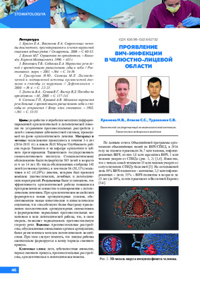 The current issue of clinical manifestations of HIV infection in the maxillofacial area has been discussed in the article. The clinical picture of HIV infection is described in detail, depending on the clinical stage of the disease. Clinical cases of HIV infection in the maxillofacial area are presented. Observance of HIV-alertness in the practice of a dentist will help reduce the risk of spreading this deadly infection to date.
 -
 Одонтогенные инфекции головы и шеи в хирургической стоматологии
Topical issues of surgical dentistry and dental implantologyЧастыми и потенциально опасными осложнениями в нелеченых стоматологических патологий или неправильных вмешательств, в особенности хирургических и терапевтических, являются одонтогенные инфекции (ОИ).
-
 The use of bacteriophages and ointment "gipofur" in the complex treatment of boils and carbuncles of the maxillofacial regionThis article presents the results of complex treatment of patients with boils and carbuncles maxillofacial region using bacteriophage Fagio and ointment of "Hypofur".
Stomatologiya -
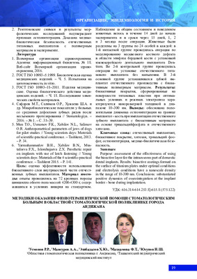 METHODS OF PROVIDING PHYSIOTHERAPEUTIC CARE TO DENTAL PATIENTS IN THE REGIONAL DENTAL POLYCLINIC OF THE CITY OF ANDIJANThis article shows the results of the physiotherapeutic assistance provided in outpatient and inpatient surgical conditions for dental patients, designed for 75 beds. We examined 391 patients (16.25%), with pulpitis - 317 (13.17%)), with acute and exacerbated chronic periodontitis - 416 (17.27%), with periodontitis - 312 (12.96%), with caries - 509 (21.15%), with glossalgia - 54 (2.2%). with alveolitis - 147 (6.4%). with trigeminal neuralgia - 21 (0.88%), with epulis - 12 (0, 49%), with sinusitis - 34 (1.4%), with other diseases - 193 patients. Physical methods of treating dental patients gave a positive effect in a short period of time and achieved a reduction m pam.
Stomatologiya -
Актуальность проблемы диагностики и лечения гнойно-воспалительных заболеваний ЧЛО определяется необходимостью дальнейшего изучения и разработки принципиально новых способов прогнозирования характера течения и повышения эффективности лечения. Существующие методы диагностики, включая клинические, не всегда позволяют адекватно отслеживать патологический процесс, что не дает врачу своевременную, эффективную коррекцию лечения больного. В обзоре описаны современные основы комплексного лечения гнойно-воспалительных заболеваний челюстно-лицевой области. Поднимается вопрос о необходимости разработки новых лечебных мероприятий по борьбе с этим заболеванием.
-
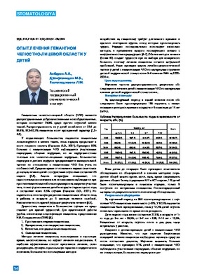 The analysis of department frequency and medical - diagnostic tactics at children with hemangioma maxillofacial area on materials of branch of children's surgical stomatology of the Tashkent medical academy with 2002 for 2006 is lead . The analysis of our researches has revealed necessity of perfection of diagnostics and tactics of treatment children with the hemangioma maxillofacial.
 -
 Despite the considerable difficulties in the diagnosis and examination of patients with combined trauma of the maxillofacial region, the success of the treatment depends on the developed tactics of specialized medical assistance. In this research we have studied therapeutic tactics of combined traumas of the maxillofacial region depending on the term of rendering specialized medical assistance. The algorithm of diagnosis and treatment methods are developed.
 -
 Osteoplastic materials for replacement of defects and deformities of the maxillofacial regionThis article presents current information about the regeneration of bone tissue. The main directions of research and an analysis of the work to replace bone tissue defects in oral and maxillofacial surgery. The characteristics of the osteoplastic materials emphasize their positive and negative characteristics in different clinical situations, principles for the use of various forms of biomaterials. The mam provisions, which shall be guided by the doctor when choosing a replacement of the bone defect.
Stomatologiya -
 Assessment of the severity of the inflammatory process in the maxillofacial region according to laboratory studiesCurrently, according to the literature, there is an aggravation of the severity of the course of phlegmonous processes in the maxillofacial region, accompanied by severe endogenous intoxication and characterized by a fulminant course. In addition, atypical forms of the clinical course of these processes are increasingly encountered, the number of which has doubled over the past ten years. Such life-threatening complications as mediastinitis, sepsis, thrombophlebitis of the veins of the face and sinuses of the brain, meningitis, etc. can develop. The number of deaths in patients with odontogenic phlegmon has increased.
Journal problems of biology and medicine -
 Подходы комплексного лечения гнойно-воспалительных заболеваний челюстно-лицевой области
Topical issues of surgical dentistry and dental implantologyВ настоящее время разработаны и внедрены в практику стандарты для лечения больных с гнойно-воспалительными заболеваниями лицевой области, которые включают в себя проведение адекватной хирургической санации и дренирование гнойного очага, антибактериальную, дезинтоксикационную, противовоспалительную терапию, коррекцию систем гомеостаза.
-
 Despite the considerable difficulties in the diagnosis and examination of patients with combined trauma of the maxillofacial region, the success of the treatment depends on the developed tactics of specialized medical assistance. In this research we have studied therapeutic tactics of combined traumas of the maxillofacial region depending on the term of rendering specialized medical assistance. The algorithm of diagnosis and treatment methods are developed.
 -
 The Article presents a brief re-view of the literature devoted to the actual problem of pediatric dentistry - acute odontogenic osteomyelitis. The article presents an analysis of the works of foreign and domestic authors on the etiology of purulent inflammatory diseases, abscesses and phlegmon of the maxillofacial region, risk factors contributing to them, the types and methods of antibacterial therapy The main modem methods of diagnostics and treatment, determination of antibiotics concentration in biological media and inflamed tissues are presented.
 -
 Pre-hospital factors affecting the severity of the course of odontogenic purulent-inflammatory diseases and their outcome
in LibraryThe analysis of prehospital factors influencing the severity of the course of odontogenic purulent- inflammatory diseases and its outcome was carried out. The total number of patients during the study period was 1305 patients (2018-675, 2019-630 patients). The most common cause of the development of pyoinflammatory diseases is the lower molars. A detailed study showed that in 5 5 % of cases the tooth was removed during surgery, which indicates a low level of therapeutic and surgical debridement among the population. In patients with acute toothache, from the first day of illness, pathological changes are observed in the tissues surrounding the tooth, underestimation of this fact can lead to the development of severe purulent-inflammatory complications.
-
 Comparative analysis of tactics of treatment of patients with concomitant trauma of the maxillofacial regionTreatment of patients with associated oral and maxillofacial Injury with an objective assessment of the severity of Injury in the choice of surgical treatment tactic, rate of complications was 20.396 and the mortality rate - 4.296. Average time of death was 7,3 ±2,3 days.
Stomatologiya -
 Influence of sorbents on the clinical course of periostitis of the maxillofacial region
StomatologiyaTo evaluate the effectiveness of a fibrous polypropylene sorbent in the complex treatment of patients with periostitis of the maxillofacial region.
-
 Methodic of sonoelastography in the time of examination in patients with soft tissular new formations of jaw-facial area is used in this work. Also, we can see high information of the method in time of differentiated detecting of benign, pre-cancer and malignant tumors, because of different staining of tissue depending on degree of its malignancy and deformation coefficient.
 -
 The article presents an analysis of modem methods for eliminating defects in facial soft tissues, classifies defects and deformations of the maxillofacial region, including the use of revascularized autografts, discusses the positive and negative aspects of surgical methods for eliminating defects m soft tissues of the face. Including analysis of the use of Pedicale Perforator Propeller Flap (PPPF) perforating propeller flaps. The use of this plastic technology made it possible to strategically simplify reconstructions, reduce the number of plastic steps and traumatize the flap formation zone, shorten the operation time, and keep the mam vessels intact in both the donor and recipient zones.