Library search
Search Results
-
 Evaluating the effectiveness of treatment of purulent-inflammatory diseases in the experiment by using laser (co2 laser and photodynamic therapy) treatment methodsObjective: Improve the treatment of purulent destructive wounds by topical application of CO2 laser and photodynamic therapy in the experiment. Materials and methods: purulent wound model reproduced in 80 male rats by MP Thick (2002) with some modification. Animals from the third day were divided into 4 groups: 1) 20 rats with physiological regeneration, 2) 20 rats with standard therapy, and 3) 20 rats PDT 4) rats 20 inclusion complex treatment with conventional methods, the CO2 laser (JZ- 3A) 3 times daily until the wound cleansing from necrotic raids and PDT. Studied plane geometry, healing time, peripheral blood counts and morphology of biopsies from the wound bed and the wall on the 1st, 3rd, 7th and 10th day of the experiment. Results. Photodynamic therapy in a CO2 laser complex was not sufficiently effective invasive treatment of purulent wounds and allows to recommend its use in clinical practice for the treatment of local purulent-destructive diseases of soft tissues. Complex application of PDT and CO2 laser leads to an earlier cleansing from the pus, the active formation of granulation tissue and shortening deadlines complete epithelialization of the wound surface. Conclusions: Complex treatment with CO2 laser and photodynamic therapy is the most effective, as compared to other methods studied separately
Journal problems of biology and medicine -
 MODERN APPROACHES TO TREATMENT OF PATIENTS AFTER EMERGENCY CORONARY INTERVENTIONSIn the article modern questions of treatment after percutaneous coronary intervention at ischemic heart disease and conducting these patients from the point of view of modern requirements and clinical recommendations are considered. Efficiency of antiaggregant and hypolipidemic therapy in patients with stable CHD who underwent planned DES-stenting of coronary arteries is estimated. Patients with stable angina pectoris (SS) of III-IV functional classes (FC) of the Uzbek population (average age 57.3±6.5 years, duration of the disease on the average 5.4±1.2 years) were under observation. According to the indications, a planned coronaroangiography was performed with subsequent implantation of DES-stents on GE OPTIMA angiography unit (USA). As the results of the study showed, the timely use of modern means to control platelet aggregation, lipid metabolism in the use of basic drugs, which include aspirin, clopidogger, statins, individualized approach, taking into account pharmacogenetic studies to treat patients with CHD undergoing stenting, increases the safety and efficiency of treatment.
Journal of Cardiorespiratory Research -
 Posstbitities of minilaparotamy choleclntectomy in patients with chronic calculous cholecystitis wift increased surgical risk
Catalog of dissertations and abstractsIn modern abdominal surgery, one of the current areas for research continues to be the improvement of various options for plastic surgery of postoperative hernias. More than 2,100,000 operations for ventral hernia are performed annually in the world, and 42% of them are postoperative hernias. In recent years, there has been a clear trend toward the expanded use of various types of biological meshes in hernioplasty. “A pooled analysis of seven PCSTAR studies for incisional hernias using retromuscular mesh showed a hernia recurrence rate of 5.7%”[1]. The use of standard surgical interventions such as alloplasty in the onlay position does not solve the problem of early postoperative complications: seroma discharge, mesh migration, adhesive disease, high frequency of hernia recurrence, etc. At the same time, hernia repair with local tissue creates the problem of increased intra-abdominal pressure and late complications in the form of recurrent hernias. “An increase in postoperative intra-abdominal pressure leads to multiple organ failure, then abdominal compartment syndrome, and even death.” [2]. Currently, there is no consensus on the surgical approach for giant postoperative abdominal wall hernias, and therefore the need to continue to develop new technologies and improve There is no doubt about the tactics. In world practice, at present, the most relevant studies continue to be studies aimed at studying the morphological and functional aspects of recurrent postoperative ventral hernias; electron microscopy reveals ultrastructural destructive changes in skin cells, aponeurosis and muscles, which indicates morphofunctional insufficiency of abdominal wall tissues; issues of cell engineering are discussed new innovative materials, experimental studies are being carried out on animals testing the biotechnical properties, texture and elasticity of new polymers, research is being conducted on open approaches with division of the posterior component with the release of the transverse abdominis muscle and a retrograde mesh, robotic operations have begun. Modern aspects of the development of domestic healthcare include many measures aimed at improving the results of treatment of patients with postoperative ventral hernias and associated pathological conditions through the introduction of modern principles of intensive care and surgical tactics. The development strategy of New Uzbekistan for 2022-2026 in seven priority areas includes tasks to improve the quality of provision of qualified medical services to the population[3]. The implementation of these tasks, including by optimizing tactical and technical approaches to the choice of hernioplasty method, as well as the development of methods for the prevention of purulent-inflammatory complications in the field of alloplastic material, is one of the current areas of abdominal surgery and medicine in general, due to the high medical and social the significance of this pathology.
This dissertation research to a certain extent serves to fulfill the tasks approved by the Decree of the President of the Republic of Uzbekistan “On comprehensive measures to radically improve the healthcare system of the Republic of Uzbekistan” No. UP-5590 dated December 17, 2018, the Resolutions of the President of the Republic of Uzbekistan “On measures to transform the surgical service, improving the quality and expanding the scale of surgical operations in the regions" for No. PP-5254 dated October 4, 2021 and "On additional measures to ensure public health by further increasing the efficiency of medical prevention work" for No. PP-4891 dated November 12, 2020, and as well as other regulatory documents adopted in this area. Compliance of the research with the priority directions of development of science and technology of the republic. The dissertation research was carried out in accordance with the priority direction of development of science and technology of the VI Republic “Medicine and Pharmacology”. Review of foreign scientific research on the topic of the dissertation.[4] Research work aimed at improving the quality of therapeutic and preventive care for patients with ventral hernias, carried out by many leading scientific centers and higher educational institutions in the world, including the Department of Surgical and Perioperative Sciences, Umeå University, Umeå (Sweden), Department of Surgery, Kingston General Hospital, 76 Stuart Street, Kingston (Sweden). nada), Department of Surgery, Helsinki University Hospital, Helsinki ( Finland), Service de chirurgie digestive et oncologique, CHU d'Amiens (France), Department of Biostatistics and Epidemiology, University of Oklahoma Health Sciences Center, Tulsa (USA), Department of Surgery, Howard University College of Medicine, Washington (USA) , Yong Loo Lin School of Medicine, National University of Singapore (Singapore), Department of Surgery, Michigan Medicine, Ann Arbor, MI, USA; University of Calgary, Calgary (Canada), Division of Plastic Surgery, Perelman School of Medicine at the University of Pennsylvania, University of Pennsylvania Health System, Philadelphia (USA), Brigham and Women's/Faulkner Hospital, Harvard Medical School, Boston (USA) , Department of Surgical Sciences, Uppsala University Hospital, Uppsala (Sweden), Department of Surgery, University of Texas Health Sciences Center at Houston, Houston (USA), Department of Surgery, Erasmus University Medical Center Rotterdam, Rotterdam (Netherlands), Department of Preventive Medicine and Public Health, Faculty of Medicine, Fukuoka University, Fukuoka (Japan), National Medical Research Center for Surgery named after A.V. Vishnevsky" (Russia), Republican Scientific Center for Emergency Medical Care (Uzbekistan), Tashkent Medical Academy (Uzbekistan), Republican Specialized Scientific and Practical Medical Center for Surgery named after Academician V. Vakhidov (Uzbekistan).
As a result of studies conducted around the world to increase the effectiveness of alloplasty for postoperative ventral hernias and reduce the risk of complications in the postoperative period, a number of scientific results were obtained, including: it was determined that patients who underwent reconstruction of the abdominal wall have an increased risk of postoperative respiratory failure, understanding the epidemiology of this complication can improve prevention (the Division of Plastic and Reconstructive Surgery, Department of Surgery, Oregon Health & Science University, USA); It has been proven that the larger the hernia, the higher the risk of early surgical complications, including such as respiratory decompensation, since hernias often increase in size over time, delaying surgery can lead to an increase in the size of the hernia and, therefore, a greater risk of complications (CentreforDigestiveDiseases, KarolinskaUniversityHospital, Stockholm, Sweden); it has been shown that the ratio of the hernia volume to the volume of the abdominal cavity <20% is an independent factor in tension-free closure, which justifies the interest in preoperative volumetry to adapt the tactics of surgical care (Servicedechirurgiegénérale, digestiveetendocrinienne, CHU LyonSud, HospicescivilsdeLyon, France); It has been determined that in patients undergoing elective laparoscopic hernia repair, predictors of mortality are older age and certain concomitant diseases: congestive heart failure, pulmonary circulatory disorders, coagulopathy, liver disease, metastatic cancer, neurological disorders and paralysis (Department of Surgery, College of Medicine, University of Oklahoma, Tulsa , USA); Older age, ascites, preoperative renal and pulmonary insufficiency have been found to be independent predictors of 30-day mortality, and in the presence of these risk factors, conservative treatment should be seriously considered (Department of Surgery, University of Kentucky College of Medicine, Lexington, USA); The American College of Surgeons (ACS) Universal Surgical Risk Calculator has been shown to accurately predict thirty-day outcomes, including major complications: venous thromboembolism, medical morbidity, surgical site infection, unplanned reoperation, mortality, and length of hospital stay (Department of Plastic Surgery, Brown University and Rhode Island Hospital, Providence, USA). At the present time in the world, the most relevant research in surgery continues to be the development of new methods of hernioplasty for large and giant ventral hernias, each of which has its own pros and cons depending on the complexity of implementation, the risk of postoperative complications and relapse, large randomized clinical trials are being conducted, comparing existing methods of traditional hernioplasty with laparoscopic access and robotic surgery, which has become increasingly widespread in the last 10 years, a search is being made for new synthetic and biological materials developed for the production and use of composite meshes that have the necessary strength and the ability to prevent fatal local complications in a contaminated environment. However, despite technical advances in this field, no modern hernia repair method or prosthesis meets all the requirements. One of the key problems is that existing synthetic endoprostheses do not have sufficient elasticity, resistance to infection, high mechanical strength and integrity over a long period of time. Further research into these clinical aspects will undoubtedly improve the current understanding of the capabilities of biocompatible endoprostheses and will make it possible to develop an optimal method for their placement during allohernioplasty. The degree of knowledge of the problem. The current period of development of abdominal surgery is characterized by an emphasis on the problems of the effectiveness of introducing new installation methods and techniques for attaching bioprostheses, options for various suture materials to determine the most promising directions for the development of these technologies [5]. Researchers led by BittnerR.[6] (2019) state that a giant postoperative abdominal wall hernia, the maximum diameter of which exceeds 12 cm or the ratio of the volume of the hernial sac to the abdominal cavity more than 20%, is difficult to treat, with a high recurrence rate and a large number of complications. One of the most challenging problems is that after the hernia contents return to the abdominal cavity, postoperative intra-abdominal pressure will increase, leading to multiple organ failure, then abdominal compartment syndrome (ACS), and even death. There is currently no agreement on the surgical approach for these giant incisional abdominal wall hernias. To prevent recurrences, some articles recommend placing the hernia mesh in the sublayer position and or linings (KirkpatrickAW.)[7]. According to CornetteB.[8], to prevent recurrence, it is recommended to place the hernia mesh in a sublayer or underlay position, and to achieve better mesh expansion, a component separation technique (CST) may be a suitable solution, but with a significant risk of complications and recurrence. JensenKK, et al. believe that truly successful giant hernia repair requires effective bridging or augmentation that will prevent recurrence with an acceptable risk of complications[9]. Another pressing issue in abdominal surgery is that patients with incisional hernias are extremely difficult to treat due to a number of factors including obesity, previous hernia repair, previous mesh placement, domain loss, and other variables.
The approach to patients with incisional hernias has changed significantly over the past 20 years due to both advances in mesh technology and surgical approaches. Key factors for successful outcome include modification of risk factors preoperatively, such as smoking cessation and weight loss, selection of mesh appropriate for the type of hernia and planned mesh location, and wide mesh coverage beyond the hernia defect. New techniques such as transabdominal muscle release and component separation with retrograde mesh placement and robotic approaches to abdominal wall hernia are increasingly being used in these patients[10]. Recent years have seen an increase in the number of biological meshes available for abdominal wall hernia repair. Biological meshes typically consist of materials obtained from humans, pigs, or cattle. The rationale for using biological meshes is that they can act as a scaffold for the growth of natural tissues. In addition, there are absorbable synthetic meshes that have properties similar to those of biological meshes, but with theoretically less risk because they are not derived from animal or human material. The choice of mesh for a ventral hernia depends on many factors, which include both the properties of the mesh and its location, for example, whether it should be placed intraperitoneally, preperitoneally, or retrorectus. BaierKF[11](2021) believes that the guiding principle should be to avoid placing uncoated polypropylene mesh in an intraperitoneal location where it may be in direct contact with internal organs. In addition, the type of hernia defect is another risk factor, such as whether the wound is clean or dirty, and whether the repair is performed with a bridge or abutment. Lightweight or biologic meshes to bridge the defect should be avoided due to increased recurrence rates. Holihan JL [12] (2016), Hodgkinson JD [13] (2018) believe that the principle of anatomical restoration to achieve a reliable, tension-free repair with reinforced mesh reduces the incidence of early postoperative complications and late recurrence of hernia compared with bridging mesh. The analysis of the literature concerning the theoretical aspects and clinical experience of using technologies for improving modern synthetic and biological prostheses that can provide a better plastic effect, as well as methods for preventing recurrence of ventral hernias, indicates that this is one of the priority areas in modern abdominal surgery. An unresolved issue remains the choice of the optimal endoprosthesis, which is highly effective and meets international standards for such properties as biological inertness and mechanical strength, as well as the method of positioning the mesh in relation to the layers of the abdominal wall. Considering that many of the allohernioplasty methods used today are not without drawbacks, the current direction is the development of new methods of repair for giant ventral hernias and methods for the prevention of postoperative complications in conditions of a contaminated wound, with justification of their effectiveness in a clinical experimental study. The connection between the dissertation research and the research plans of the research institution where the dissertation was completed. The dissertation research was carried out within the framework of the research work plan of the State Institution “RSNPMCH named after. acad. V. Vakhidov" under the project AL-422105574 "Development of new biocompatible mesh implants made of composite materials for reconstructive surgery of abdominal and diaphragmatic hernias" (2022-2024).
The purpose of the study is to improve the results of surgical treatment of large and giant postoperative ventral hernias by introducing new laser technologies and improving the tactical and technical aspects of surgical treatment. Objectives of the study: to study the structure of immediate complications after various types of prosthetic plastic surgery; to clarify the influence of obesity factors, primary or repeat hernioplasty on the incidence of immediate and long-term complications; to evaluate the role of the immediate complicated course of the postoperative period in the incidence of long-term complications of hernioplasty; to improve the technical aspects of alloplasty for large and giant postoperative ventral hernias (POVH); to improve the technique of photodynamic therapy (PDT) of the wound surface after prosthetic plastic surgery; to study in an experiment the effectiveness of using the proposed technique of alloplasty and PDT; evaluate the morphological features of the condition of tissues during prosthetic plastic surgery using the proposed method; in a comparative aspect, evaluate the clinical effectiveness of the proposed alloplasty options in the immediate and long-term periods. The object of the study was the results of allohernioplasty in 448 patients with extensive (large) and giant POVH, who were operated on at the surgical department of the 1st clinic of the Samarkand State Medical Institute in the period from 2012 to 2021, as well as experimental animals on which the effectiveness was assessed developed a technique for prosthetic repair of postoperative ventral hernias and applied the technique of photodynamic therapy.
The subject of the study is to analyze the effectiveness of the developed alloplasty of postoperative ventral hernias and intraoperative photodynamic therapy in abdominal surgery in experiments and in the clinic. Research methods. To achieve the goal of the study and solve the assigned problems, general clinical, instrumental, biotechnological, experimental, histomorphological, microbiological and statistical research methods were used. The scientific novelty of the study is as follows: it was established that the need for extensive tissue mobilization and, as a consequence, the intersection of lymphatic capillaries during implantation of the prosthesis in the onlay position, as well as the lack of sufficient resorption function of the hernial sac in the inlay position causes a high risk of the formation of clinically significant seromas; It was determined that with prosthetic hernioplasty, along with the volume of the defect, the type of plastic surgery and the degree of obesity, the most significant predictor of the risk of developing immediate complications is the factor of re-intervention in case of recurrent hernia with the presence of a “dormant infection” hidden in the remaining ligature granulomas or scar tissue; the structure and clinical features of the course of long-term complications of hernioplasty for giant and extensive hernias were clarified, taking into account the results of the immediate postoperative period, as well as the option of fixing the prosthesis, primary or repeated hernioplasty and the degree of obesity; the method of surgery for large hernias of the anterior abdominal wall has been improved, characterized by a combination of factors such as the formation of tension-free prosthetic repair, preservation of local resorptive function to prevent the development of fluid accumulations and reducing the risk of infection; the method of preventing the development or progression of wound infection during alloplasty of ventral hernias has been improved, aimed at enhancing the antibacterial effect and stimulating reparative activity through the photosensitizing and photodynamic effect of low-energy laser radiation; It was determined in an experimental model of prosthetic plastic surgery that the proposed method of fixing the prosthesis in combination with the use of the effect of photodynamic therapy through laser radiation helps to enhance reparative processes with a reduced risk of wound complications; It has been proven that all methods of antiseptic exposure and laser stimulation of the wound surface after prosthetic plastic surgery enhance preventive measures against the development of local infection, but are ineffective in the case of an already developed purulent-inflammatory process against the background of the use of alloplastic material. The practical results of the study are as follows: it has been determined that the implantation of synthetic materials for giant and extensive hernias is accompanied by a significant number of wound complications caused by both the surgical technique itself and the reaction of surrounding tissues to a foreign body, requiring improvement of tactical and technical approaches when performing hernioplasty; it was clarified that scar-degenerative changes in the tissues of the aponeurosis in giant and extensive hernias are a predisposing factor to the occurrence of post-prosthetic hernias, especially when implanting the prosthesis in the “inlay” position and, accordingly, require increasing the efficiency of their fixation and engraftment, as well as reducing the risk of developing local complications; it was determined based on the data of an experimental study that the proposed method of alloplasty for large ventral hernias makes it possible to achieve adequate reconstruction of the anterior abdominal wall, reduce the incidence of infection in the wound, and also use a smaller size of prosthetic material; it was determined that the proposed method of tension-free repair of large hernias of the anterior abdominal wall with strengthening of the aponeurosis with a mesh implant allows maintaining physiology, reducing the number of complications, shortening treatment time and reducing the risk of hernia recurrence; It has been determined that the proposed method for preventing the development or progression of wound infection during alloplasty of ventral hernias can reduce the frequency of suppuration, shorten the treatment time and the likelihood of relapse; It has been proven that the use of the proposed tactical and technical aspects of prosthetic repair for postoperative ventral hernias can reduce the incidence of specific complications, reduce rehabilitation time and the risk of hernia recurrence. Reliability of the research results. The reliability of the results is justified by the use of objective criteria for assessing the condition of patients, modern methods of diagnosis and treatment, the correct application of methodological approaches and sets of statistical analysis, methods for solving the problems discussed in the dissertation are based on modern scientific and practical concepts and approaches to the diagnosis and surgical treatment of patients with giant postoperative hernias. Scientific and practical significance of the research results. The results obtained make a significant contribution to the expansion of irradiation of existing ideas about the structure and clinical features of complications of hernioplasty for giant and extensive ventral hernias by identifying the morphological features of the development of a purulent-inflammatory process against the background of the use of alloplastic material, studying predictors of the risk of developing immediate complications, mechanisms for enhancing reparative processes through the use of an improved method of fixing the prosthesis in together using the effect of photodynamic therapy through laser radiation, which made it possible to enhance the antibacterial effect and improve the wound healing process. The practical significance of the study is that, based on the results obtained, the tactical and technical aspects of prosthetic hernioplasty have been optimized, the features of methods of antiseptic exposure and laser stimulation of the wound surface after prosthetic repair have been revealed, enhancing preventive measures for the development of local infection, and the method of surgery for large anterior abdominal hernias has been improved walls, characterized by a combination of factors such as the formation of tension-free prosthetic plasty, preservation of local resorptive function to prevent the development of fluid accumulations, thereby reducing the risk of developing postoperative complications, reducing the frequency of unsatisfactory results, length of hospitalization and the likelihood of relapse. Implementation of research results. According to the results of a scientific study to optimize the tactical and technical aspects of surgical treatment of large and giant postoperative ventral hernias: the “method for plastic surgery of giant hernias of the anterior abdominal wall” has been improved (invention patent No. IAP 2022 0148 dated April 18, 2022). The proposed method of tension-free repair of large hernias of the anterior abdominal wall with strengthening of the aponeurosis with a mesh implant made it possible to reduce the number of complications, shorten the treatment time and reduce the risk of hernia recurrence; the “method for preventing the progression of infection during alloplasty of infected hernias” has been improved (certificate of the Ministry of Health No. 08-32071 dated October 17, 2022). The proposed method made it possible to reduce the frequency of wound purulent-inflammatory complications and shorten the period of rehabilitation of patients after allohernioplasty; methodological recommendations “Tactical and technical aspects of prosthetic repair for large and giant postoperative ventral hernias” have been developed (certificate of the Ministry of Health No. 08-32071 dated October 17, 2022) . The developed recommendations made it possible to optimize the tactical and technical aspects of allohernioplasty in patients with large and giant postoperative ventral hernias; The scientific results obtained were introduced into the practical activities of healthcare, in particular, in the departments of surgery of the Khorezm and Andijan regional multidisciplinary medical centers, the clinic of the Samarkand State Medical University (certificate of the Ministry of Health No. 08-32071 dated October 17, 2022). Improving the tactical and technical aspects of prosthetic repair for postoperative ventral hernias has made it possible to reduce the incidence of specific immediate complications from 40.9% to 15.6%, to reduce rehabilitation time from 8.6±2.7 to 7.1±1.5 days, and also reduce the likelihood of long-term complications from 11.7% to 3.1%. Approbation of research results. The results of this study were discussed at 8 scientific and practical conferences, including 5 international and 3 republican ones. Publication of research results. 26 scientific works have been published on the topic of the dissertation, including 9 journal articles, 4 of which in republican and 5 in foreign journals recommended by the Higher Attestation Commission of the Republic of Uzbekistan for publication of the main scientific results of doctoral dissertations. Structure and scope of the dissertation. The dissertation consists of an introduction, seven chapters, a conclusion, conclusions, practical recommendations and a list of cited literature. The volume of work is 200 pages.
-
 The mechanism of therapeutical effect of laser therapy and cream chistotel at the patients with the skin angeitis and preparation lefno at the patients with the arthropatic form of psoriasis to the IgE.
Journal of Biomedicine and PracticeThe aim of work was to study the effectiveness of laser therapy in patients with arthropatic form of psoriasis and angiitis of the skin based on changes of immunological parameters. Forty-five patients were observed. There 25 patients with angiitis (10 males and 15 females), 25 patients with arthropathic form of psoriasis (12 males and 13 females). In combined use of laser therapy positive changes were revealed in all the patients. During the treatment concentration of IgE was reduced to normal limits
-
 The role of magnetic resonance imaging in the comprehensive radial diagnosis of volumetric masses of the eye organ
Catalog of abstractsRelevance of the problem. The difficulties of diagnostics of orbital diseases are well known. Especially difficult is intraspecies differentiation among the multitude of tumour, pseudotumour, inflammatory, vascular, endocrine and other diseases occurring here, manifested by the symptom complex of unilateral exophthalmos [Beradze I.N., 1978; Brovkina A.F., 1993].
Malignant intraocular neoplasms are the main cause of death of patients with diseases of the organ of vision, with 45-48% of patients dying from metastases in the first 5 years after enucleation [Alekseeva I.B., 1990, Barkhash S.A.1978, Brovkina A.F..1991, 1997; Keizer R.W.. Viclvoyc G.L.,1986],
Retinoblastoma is the most frequent malignant neoplasm in children. According to different authors, the frequency of its occurrence is 1 case per 14000 - 35000 newborns. [Bobrova N.F. and Vit V.V., 1993; Brovkina A.F., 1997; Provenzale J.M., et al., 1995; Skulski M., et al., 1997; Weber A.L., Mafee M.F, 1992; Wilms G., et al., 1989]. The frequency of patients with the most malignant intraocular tumour in adults - uveal melanoma has recently reached 7-9 people per 1 million population [Brovkina A.F., 1997; Kotslyansky E.O., 1989; Yushko N.A., Peskova L.I., Kalenich L.A., 1989; Peyster R.G., Augsburger J..I., Shields J.A., 1988; Romani A.. Baldeschi L., ct al 1998; Scott I.U., 1998].
The fundamental difference in treatment tactics, depending on the stage of development, size and topography of the tumour, as well as the seriousness of the prognosis in retinoblastomas and melanomas sharply increase the requirements for the accuracy of their differential diagnosis. At the same time, the number of diagnostic errors in ocular tumours continues to be 10-30% even when complex clinical and instrumental examination is applied in specialised ophthalmological centres [Ternovoy S.K., Panfilova G.V., Rogozhin V.A., 1979; Friedman F.E., Malyuta G.D., Kodzov M.V., 1995; Song G.X., 1991].
Widely used in ophthalmological practice traditional diagnostic methods (ophthalmoscopy, gonioscopy, diaphanoscopy, fluorescence angiography, laboratory tests) are insufficient to obtain comprehensive information about the localisation, nature of growth and prevalence of volumetric pathological formations of the eye and orbit. This circumstance and not quite satisfactory results of surgical treatment are the causes of high mortality of patients [Muratova T.T., Nigmanova N.H., Kozlovskaya G.M.. 1989, Naches A.I., 1980; Cheremisin V.M., Trufanov G.E., Kholin A.V., 1991]. Untimely or erroneous recognition of pathological processes of the orbit leads to a sharp deterioration of visual functions, up to blindness, and in some cases to the death of the patient [Yuzhakov A.M., Travkin A.G., Kiseleva O.A., 1991]. All this determines the importance of timely and accurate diagnosis of diseases of the orbit, on the one hand, and the difficulty of such diagnosis - on the other [Gabunia R.I., Kolesnikova E.K., Tumanov L.B., 1982].
The fact that the orbit is closed from direct inspection and palpation by bone walls and the eyeball, indicates the advantage of radial diagnostics in comparison with other methods of examination. In the arsenal of clinicians there is a great variety of methods of clinical-radial diagnostics of orbital pathology, however, at present the information in the literature about their resolving capabilities and significance in comparative aspect is incomplete and not fully studied. The priority of using one or another instrumental investigation, their sequence and expedient combination have not been determined yet. This makes it difficult to choose the optimal standardised approach for diagnosis and adequate treatment [Cheremisin V.M., Trufanov G.E., 1993, Weber A.L., Sabates N.R., 1996; Wenig V.M., Mafee M.F., 1998].
Thus, the study of these and other questions, contributing to the improvement of diagnostics and treatment of patients with neoplasms of the eye and ocular cavity, should be recognised as urgent urgent.
Purpose of the study. Comparative evaluation of magnetic resonance tomography capabilities and development of algorithms for complex radial diagnostics of volumetric formations of the visual organ. To solve this goal we set the following tasks.
1. To study the normal picture of the magnetic resonance image of the visual organ in comparison with other methods of visualisation.
2. To find out the possibilities of magnetic resonance tomography, ultrasound and computed tomography in detection and evaluation of intraocular neoplasms.
3. To determine the role and place of magnetic resonance tomography in differential diagnostics of volumetric pathological formations of the eye cavity in comparison with other radial methods of research.
4. To determine the indications and to develop an algorithm for the complex application of radiography, ultrasound, computer and magnetic resonance tomography for diagnostics of volumetric formations of the eye organ.
Scientific novelty.
The present work is the first to give a detailed and detailed description of the complex clinical and radiation examination, with generalisation and standardisation of magnetic resonance, computer and ultrasound semiotics of volumetric pathological formations of the eye and eye cavity. The conducted clinical and instrumental investigations allowed to determine the diagnostic value and resolving capabilities of each of the applied methods. The ultrasound, CT and MRI signs of volumetric formations of the eye organ were studied, clarified and supplemented taking into account the use of low-field magnetic field and general-purpose ultrasound apparatus. The developed standardised diagnostic algorithm of examination of patients with this pathology is new, thanks to which the pre-oppositional diagnosis of tumour and other diseases of the visual organ is improved and the total radiation load on the patient is reduced.
Conclusions
1. MPT will provide an opportunity to study the weight of the soft tissue and anatomical components of the ocular cavity, up to the optic nerve sheath and perineural liquor space, the orbital apex and chiasmal-sellar region, as well as to assess the condition of adjacent structures of the brain and facial skull. The method is limited in the evaluation of changes in the bony walls of the orbital cavity.
2. MRI is inferior in detecting characteristic signs of retinoblastoma (presence of calcification). The sensitivity of MRI was 66.6%, while for ultrasound and CT these values were 96.1 and 100%, respectively. But when the tumour spreads rstrobulbarly outside the eyeball (at 3-4 stages) the informativeness of MRI increases significantly. In uveal melanoma the sensitivity and specificity of MRI reaches 100%.
3. Both MRI and CT have a high detection rate (98.1% and 95.8% respectively) of benign orbital tumours of both primary and secondary origin. However, MRI is the preferred method of investigation. MRI is especially informative when a cranioorbital tumour and pseudotumour are suspected. The sensitivity of the method is 90.9% and 91.6%, respectively
4. In some cases ultrasound can be used to differentiate between encapsulated and diffuse neoplasms, which facilitates the diagnosis. However, when the pathological process is localised near the orbital apex, the diagnostic value of ultrasound decreases. In such cases it is advisable to use MRI.
5. In detection of primary and secondary malignant tumours of the orbital cavity both MRI and CT are quite informative (sensitivity 97,2% and 95,4% respectively), but the most comprehensive information about the state of bone walls will be provided by CT. When the process spreads intracranially, the value of MRI increases significantly, especially with the use of contrast enhancement.
6. The developed algorithm of complex clinical and radiation examination of patients with the use of ultrasound, CT and MRI is the most effective in the diagnosis of volumetric pathological formations of the eye and eye cavity, allowing to reduce to an adequate minimum the total radiation load on the patient and diagnostic period, excluding duplication of research techniques and choosing the most informative in each case, which in turn allows to develop appropriate treatment tactics and reduce the level of disability of the patient. -
 Sonography and magnetic resonance imaging of the hand joints in rheumatoid arthritis
Catalog of abstractsSubject of the inquiry: 116 patients with rheumatoid arthritis of the hand joints and 25 healthy subjects.
Aim of the inquiry: improvement of the diagnosis of rheumatoid arthritis of the hand joints using sonography and magnetic resonance imaging.
Methods of inquiry: X-ray, sonography, magnetic resonance imaging and magnetic resonance imaging with contrast enhancement.
The results achieved and their novelty: For the first time data were presented on the role of sonography, magnetic resonance imaging and magnetic resonance imaging with contrast enhancement in the diagnosis of rheumatoid arthritis of the hand joints. The findings have shown that sonography in hand joint rheumatoid arthritis allowed detection of changes in soft tissues, synovial capsule, joint surfaces and ligaments. Diagnostic value was given of sonography in revealing characteristic sonographic signs of rheumatoid arthritis. Magnetic resonance imaging was highly informative radiological method to detect synovitis, changes of synovial capsule and subchondral cysts. Magnetic resonance imaging with contrast enhancement reliably detected the degree of activity and severity of the rheumatoid process.
Practical value: consisted in revealing and describing characteristic sonographic and MRI signs of hand joint rheumatoid arthritis and in the developed radiological algorithm of the disease.
Degree of embed: the results of the investigation were introduced in the practice of rheumatology and radiology departments of the First Tashkent Medical Institute, in teaching process of the Radiology Chair of the First Tashkent Medical Institute.
Sphere of usage: radiology, rheumatology, traumatology and orthopedics. -
 According to the modem concept, pathological processes are realized on cell membranes, causing a violation of the structural and functional organization, up to complete destruction. The use of glucocorticoid and cytotoxic therapy in combination with heparin does not guarantee success and lead to complications, especially in children. Consequently, the search for drugs that remove the toxic effects of immunosuppressive therapy continues to be an urgent medical and social problem. What was the reason to study the features of antioxidant therapy in order to optimize the pathogenetic treatment of chronic glomerulonephritis in children. The advantage of therapy with the inclusion of actovegin antioxidant was that parallel to the normalization of malondialdehyde and lysophosphatidylcholine, the other cell membrane fractions increased, apparently due to the ability of aktovegin to reduce the activity of phospholipases. The conducted research will allow to review the tactics of treatment of nephropathy in accordance with the obtained research results, to include in the complex therapy a step-by-step correction of membrane destabilization with Actovegin. In addition, pathogenetic therapy for glomerulonephritis is fraught with complications, ultimately associated with a violation of cellular stability and requires corrective antioxidant therapy.
 -
 SOME ASPECTS OF MEDICAL REHABILITATION OF PATIENTS WITH CHRONIC CORONARY SYNDROME AFTER STENTINGModern medical advances in the treatment of chronic coronary syndrome (CCS), in particular, the use of high medical technologies have necessitated a more thorough study and implementation of complex medical rehabilitation of this category of patients into clinical practice. Purpose of the study: to study the effectiveness of complex medical rehabilitation with the inclusion of the herbal preparation Myocard in patients with CCS who underwent coronary artery (CA) stenting. Materials and methods: patients with CCS with stable exertional angina who underwent CA stenting (40) and received standard therapy were examined. The patients were divided into 2 groups: group 1 (20 patients) received additional herbal preparation Myocardin (APOLLO PHARM MED, Uzbekistan), group 2 received only standard treatment. Conclusions: Cardiac rehabilitation is an important method of prevention and treatment of CCS, a factor in improving health and improving the quality of life. Complex cardiac rehabilitation of patients with CCS who underwent stenting with the inclusion of Myocardin increases the efficiency and safety of treatment and medical rehabilitation of this category of patients.
Journal of Cardiorespiratory Research -
 Improvement of results of furnye gangrene complex treatment with co 2 laser and photodynamic therapy usingThe use of CO 2 laser has allowed for the possibility of early and bloodless nekroektomy, improve wound repair, and reduces microbial contamination of purulent wounds. Method has photo coagulating and sterilizing properties effects on the tissue. Photodynamic therapy is a very effective non-invasive and gentle treatment of purulent wounds and serve as justification for the use of the method of photodynamic therapy in clinical prac-tice for the treatment of acute local inflammatory processes combined with CO 2 laser and traditional treat-ments.
Journal problems of biology and medicine -
 The use of laser photocoagulation corneal herpetic keratitisOphthalmoherpes in the statistical analysis of ocular disease comes first corneal pathology in the world. We examined 18 (18 eyes) of patients with various forms of herpetic keratitis. Among the patients were 11 men and 7 women. All patients before and after treatment was performed standard ophthalmologic examina-tion. Laser coagulation of corneal infiltrates was performed by affecting the cornea by laser radiation with a wave length of 532 microns, the energy of 300-400 mJ / sm 2 , the laser spot diameter of 200 microns. Infiltra-tion of the cornea stained with Collargol. Visual function was assessed before treatment and after 1week. 1, 3 and 6 mth. after therapy. The use of laser photocoagulation in the treatment of herpetic keratitis leads to an acceleration of reparative processes, rapid relief of inflammatory processes and better healing of the corneal defect. Аfter laser coagulation of corneal visual acuity of patients with this pathology increased to 0.8-1.0. Average time epithelialization of corneal epithelium of the cornea in the application of LC was 6-7 days. Re-covery we noted in 82.6% cases. Relapses occurred 4.3% of cases. Conducting corneal infiltrates LC patients with herpetic keratitis in the early postoperative period leads to permanent removal of corneal syndrome, a certain increase in visual acuity development epithelialization of the cornea, reducing its thickness and re-duce swelling.
Journal problems of biology and medicine -
 Metabolic activity of erythrocytes and peculiarities of ferrokinetics in patients with ankylosing spondyloarthritis and ways of its correction
Catalog of abstractsSubject of the inquiry: 97 patients with ankylosing spondylitis, 20 healthy volunteers.
Aim of the inquiry: to assess metabolic activity of erythrocytes and peculiarities of fcrrokinctics in using Fhlogcnzymc in the complex therapy of patients with AS. Methods of research: functional-metabolic process erythrocytes and ferrokinctics (scrum iron, ferritin, saturation of transferrin with iron) lipid peroxidation and antioxidant system.
The results achieved and their novelty: For the first time purposeful complex study was conducted of metabolic process parameters in erythrocytes and fcrrokinctics, lipid peroxidation and antioxidant system processes, Willebrand factor, atherogenicity in relation to the clinical course of the disease. Functional-metabolic disorders in erythrocytes in fcrrokinctics determined systemic changes in blood, with their extent associating with severity of the clinical course of AS. For the first time it was found that the use of Fhlogcnzymc in the complex treatment improved clinical course of the disease. The shortening of clinical recovery period was associated with normalization of parameters characterizing functional metabolic activity in crythrocytea and ferrokinctics. Systemic enzyme therapy was proved to increase the effect of conventional therapy by a directed improvement of functional metabolic processes in erythrocytes, energetic activity and ferrokinctics which allowed inclusion of systemic enzyme therapy preparations for hemic hypoxia in patients with AS.
Practical value: Use of Fhlogcnzymc in the complex treatment of AS increased the efficacy of therapy, decreased the rate of drug side-effects, allowed us to decrease the doses of basic preparations and non-stcroid anti-inflammatory. Changes of laboratory parameters in Fhlogcnzymc using included more significant decrease of acute phase indices, increase of hemoglobin and erythrocyte concentration in the peripheral blood.
Degree of inculcate and economic efficiency: conclusions and practical recommendations were introduced into the practice of departments of rheumatology, nephrology of the third clinic of the Tashkent Medical Academy and into teaching process of departments of faculty and hospital therapy, folk medicine, internal diseases of the stomatological faculty and clinical pharmacology of the Tashkent Medical Academy.
Sphere of usage: rheumatology, therapy. -
 Features of coronary heart disease treatment in patients with iron deficiency anemia against the different severity levelsCurrently comorbidity is relevant in the outcome of many diseases [1]. In particular, we arc talking about 1HD, unstable angina and anemia of varying severity. In 30 patients with coronary artery disease in combination with anemia, 3-valence iron was changed, the drug Sufer (Yuria-Pharm Ukraine) at a dose of 5.0 ml iv for 5 days against the background of standard therapy. In examined patients, that an increase in hemoglobin levels contributes to the normalization of the vibros fraction reduce or disappear angina attacks, increased exercise tolerance [2,3].
Doctor's Herald -
 Evaluation of the effectiveness of laser radiation on the microcirculation of the eye in patients with diabetic retinopathy
in LibraryFor all successes in the treatment of diabetic retinopathy (DR), this disease remains one of the major problems of today’s ophthalmology. Examined 40 patients with diabetes mcllitus with difference studies of DR. the purpose of this study is assessment of the efficacy laser radiation in treatment DR. After laser therapy were noted to have stable and improved visual i acuity and microcirculation of the retina. Our studies showed improved intravascular and 1 perivascular conjunctival index’s after laser therapy.
-
 Usefullness of laser in oral and maxillofacial surgery
Actual problems of dentistry and maxillofacial surgery 4Lasers have revolutionized dental treatment since three and a half decades of the twentieth century. Theodore Maiman in 1960 invented the ruby laser, since then laser is one of the most captivating technologies in dental practice. Lasers have been used in initial periodontal therapy, oral surgical procedures, and also in implant treatment. Further research is necessary so that laser can become a part of the dental armamentarium. This paper gives an insight towards the uses of laser in Oral and Maxillofacial Surgery.
-
 The use of trt-therapy in the treatment of tinnitus caused by sensorineural hearing lossData of the results of research which are carried out at 40 patients with tinnitus in the ears caused by bilateral sensonevralny relative deafness are provided in article. The comparative characteristic drugs therapy and combinations of drugs therapy with TRT - therapy at patients with tinnitus is given. The received results showed efficiency of a combination drugs therapy with TRT - therapy.
Stomatologiya -
 EVOLUTION OF APPROACHES TO THE THERAPY OF REFRACTED CELLIACIA IN CHILDREN OF THE UZBEK POPULATIONExamined 200 children with celiac disease aged 1-14 years. The diagnosis of the disease was verified based on the criteria of the European Association of Pediatric Gastroenterologists (1999). Celiac disease was revealed as a typical form in 81%, an atypical form was found in 19%, of which 21.5% had a refractory course of the disease. Depending on the drug therapy, 4 groups were identified: -1st-30 children who, in addition to traditional therapy, received ecdisten 0.01 g at 1 mg / kg / day; -2nd- 37 children who additionally received antioxidant zinc with 20 mg / day; - 3rd - 28 children who additionally received ecdisten 0.01 g at 1 mg / kg / day in combination with antioxicaps with zinc at 20 mg / day; -4-I- 47 children who received the generally accepted basic therapy. The course of treatment is 1 month. The studies were carried out after 3, 6 and 12 months. The results obtained indicate the restoration of the intestinal absorption function against the background of the modified treatment, more significant changes were obtained in patients who received the combined use of ecdisten with antioxicaps with zinc. This combination, against the background of basic therapy in the refractory course of the disease, has a beneficial effect on the clinical course, reduces the degree of structural and functional disorders, improves the outcome of the disease. To obtain a stable clinical remission, it is necessary to carry out repeated courses of modified therapy in a differentiated manner.
Journal of hepato-gastroenterological research -

В статье отражены современные взгляды на патогенез остеоартрита. Показана роль хондропротекторов в комплексной терапии заболевания, приведены данные собственных исследований. Цель – изучить влияние комбинированного хондропротектора ИБРА на течение ОА различной локализации I–II стадии. Полученные результаты показали эффективность ИБРЫ в терапии и реабилитации ОА и целесообразность ее применения в общеврачебной практике. Патогенетически оправдано назначение препарата в качестве как старттерапии, так и для длительного лечения ОА и на этапе реабилитации.
-
Subjects of research: 815 patients with the various forms acute pancreatitis, 235 patients with primary erronous diagnosis of acute pancreatitis.
The purpose of research: improve the treatment results of the patients with acute pancreatitis by upgrading existing and developing new diagnostic methods in the treatment complex.
Methods of research: clinical, biochemical investigation.
The results achieved and their novelty. On the basis of the analysis of occurance frequency of various clinical symptoms, diagnostics system of the acute pancreatitis is offered, the criteria of the laboratory and ultrasonic diagnostics for given disease arc advanced, for a quantitative estimation of severity of a condition of the patients the ball scale is offered; the complex conservative therapy is advanced, the technique of realization of long intraarterial catheter therapy is optimized, the optimum indications to realization of retrograd and pcrcutanc endobiliar interventions in treatment of acute biliar pancreatitis arc determined, drainage and sanation of omental bag for open operative interventons is offered, the technique of treatment of the patients in postoperative period is advanced and the algorithm of medical measures is offered at acute pancreatitis.
The developed diagnostic and treatment complex has allowed to reduce frequency of diagnostic errors from 28,4% to 16,8%, to increase efficiency of conservative therapy from 89,8% up to 95,3%, long intraarterial catheter therapy -from 72,0% up to 82,8%, to reduce mortality from 7,4% to 3,4%.
The practical importance. The offered clinical system for diagnostics and the advanced criteria of laboratory diagnostics promote to increase reliability of diagnostics, the systematized ultrasonic criteria allows verify the form of acute pancreatitis. The offered scale of definition of severity of the acute pancreatitis allows quantitatively characterize a condition of the patients, dynamical changes of pathological process and efficiency of used of a complex of medical measures. The advanced complex conservative therapy, technique of realization long intraarterial catheter therapy, definition of the indications to performance retrograd and pcrcutanc endobiliar interventions, advanced draining method and sanation of omentum bag at pancrcanccrosis, combined treatment in postoperative period allow to improve results of treatment of the given category of the patients.
Degree of introduction and economic efficiency: the received results arc introduced into practical activity of surgical branches of second clinics TMA.
Area of application: emergency and abdominal surgery. -
 RADIATION THERAPY AS A METHOD IN THE COMBINED TREATMENT OF RECTAL CANCERThis article provides information on radiation therapy as a method of complex treatment of rectal cancer.
Modern Science and Research -
 This review summarizes the current evidence on the role of cytokines in the pathogenesis of ischemic myocardial damage. It described the important role of inflammation in the development of coronary heart disease. The role of individual cytokines in the pathogenesis of coronary artery disease and the most frequent forms of it - angina. It is shown that in patients with coronary heart disease progression of the disease is due to an imbalance in cytokine system, elevated pro-inflammatory cytokines (TNF, IL-1β, IL-6). Anti-inflammatory cytokines (IL-4, IL-10) inhibit the secretion of proinflammatory cytokines by limiting excessive intensity of the immune response. Revealed increasing levels of TGF-β1 as a key cytokine that promotes the development of fibrosis in the wall of the heart and blood vessels. The interrelation between improving markers of inflammation and the development of coronary heart disease, the predictive value of these markers of inflammation in patients with stable coronary heart disease: angina of effort of different functional classes.
 -
 Correction of disturbans of the liver at the patients with diabetic purulent – septic compliations of lower extremities
Catalog of abstractsPurpose of reseach: to optimize outcomes of therapy of the patients SD, purulent - septic complicated of lower extremities (PSCLE), by monitoring, duly revealing of character disturbans of function of the liver and purposeful correction them in complex therapy of the specified pathological condition.
Methods of research: clinical-biochemical researches, ECG, ultrasonic research of the liver, ultrasonic dopplerography, percutaneus definition a pressure (voltage) of oxygen, angiography, radionuclide researches, psichometric tests.
The results achieved and their novelty: the carried researches have shown, that the liver is not one of the basic organs - targets at SD (at the expense of features it blood circulation), at decompenciation SD, complicated PSCLE, there is an essential infringement liver blood circulation in system lever artery, to the subsequent disturbance of its numerous functions. The inclusion in complex therapy hepatoprotectory, antioxygents and preparations blocking ALT activity is pathogenic proved and promotes optimization of outcomes such of complication SD, as PSCLE.
Practical significance: the elaborated and offered complex therapy of the patients provides of optimization of outcomes, is an effective method of treatment and preventive maintenance of complications.
Degree of introduction and economic efficiency: the results of research have been introduced into practice of branches intensive therapy and reanimation 2-clinic TMA and Republican Centre of Purulent Surgery and Surgical Complication of Sugar Diabetes
Area of application: reanimation and intensive therapy, surgery. -
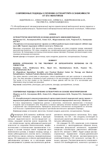
The article presents current data on phenotypes, clinical manifestations and pain syndrome in osteoarthritis (OA). The main principles of treatment of pain syndrome in OA and dorsalgia in spondylarthrosis are outlined. The results of our own studies on the treatment of these patients with the use of a non-steroidal anti-inflammatory drug (Lornado) are presented.
-
 The role of inflammatory fat tissue major markers in the development of arterial hypertensionArterial hypertension in patients with metabolic syndrome is an actual problem of modern medicine due to the significant influence of its components on the risk of cardiovascular complications. Visceral adipose tissue is an endocrine organ secreting a wide range of biologically active substances - adipokines, which influence the progression of arterial hypertension, atherosclerosis, thrombus formation, insulin resistance, etc.
Doctor's Herald -
 The effect of trimetazidine in complex treatment of chronic heart failure in Patients with myocardial infarctionChanges in hemodynamics in patients with CHF on the background of long-term use of Trimetazidine.
Doctor's Herald -
 STRATEGY FOR THE RATIONAL MANAGEMENT OF PATIENTS WITH GOUT, TAKING INTO ACCOUNT OPTIMAL REDUCING THERAPY ACCORDING TO MATERIALS OF THE AMERICAN COLLEAGUE OF RHEUMATOLOGISTS 2020 (ACR)The review is devoted to the management of patients with gout, taking into account the recommendations of the American College of Rheumatologists (ACR), updated in 2020, according to which it is recommended to treat exacerbations of the articular syndrome with colchicine, non-steroidal anti-inflammatory drugs (NSAIDs) or glucocorticoids. Uratus-lowering therapy (ULT) should be given to all patients with tofuses, radiological signs of joint damage, or frequent exacerbations of gout. Allopurinol is the first-line drug, including for patients with chronic kidney disease (CKD), at the initial dose (≤100 mg / day and lower with CKD), followed by titration of the dose under the control of serum uric acid (UA) and its reduction <6 mg / dl (<360 μmol / L), i.e. using the strategy "treatment to achieve the goal." Reducing therapy is strongly recommended against the background of prophylactic anti-inflammatory therapy lasting at least 3-6 months. The purpose of the review is to convey to doctors of various specialties who make decisions about the treatment of gout with reducing drugs, the necessity of compulsory achievement of the target level of UA, the prevention of new exacerbations of the joint syndrome and complications of comorbid diseases.
Journal of Cardiorespiratory Research