Library search
Search Results
-
 Brain tumors make up to 20% of malignant tumor diseases in children. These tumors differ from each other in their structure and characteristics. In children, medullablastomas (tumors that develop independently), as well as astrocytomas (malignant tumors of the central nervous system), glioblastomas that arise in the spinal cord, and ependymoma tumors that develop in the internal spaces of the brain are found in children.
 -
 Diagnosis and treatment of the retroperitoneal extra-organic tumorsThe article provides a data of immediate and long term results of surgical treatment in 208 patients with locally spread nonorganic retroperitoneal tumors. Malignant tumors constituted 152 (71,3%) while be-nign tumors 56 (26.9%). Radical operationswere performed to 64.8% of patients, palliative-26.7%and in8.3% of cases operations were cytoreductive. From these, in 52.8% of cases operations had combined character. General, Intra- and postoperative complications constituted 16,4%, meanwhile postoperative lethal outcomes – 2,4%. 5-year survival rate of patients with benign tumorsreached83.3%, and malignant-12.6%. The same index after radical operation constituted 41,3%, and after non radical -9,6%.Relapses within 5 years after surgery for a malignant tumor occurred in 73.7%, and after a benign tumor - 26.3% of patients
Journal problems of biology and medicine -
 In this article were controled 89 patients and analyzed treatment methods of giant cell tumors in pipe bones. Different size of excochleation and cement plastic surgical operation results were described to patients. There were 3 types of surgical operation depending its size. In the first group were 49 (55%) patients with excochleation and cement plastic operations. Second were 31 (34,9%) patients with excochleation, cryo and cement plastic operations and third group were 9 (10,1%) patients with excochleation, auto and cement plastic operations. In ail groups De-Rie medical cement (Johnson- Johnson Company, USA) which keeps antibiotics was used for cement plastic operations. Assisting results of surgical operations by recidivation of diseases, in the first group 5 (10,2%) patient out of 49. in the second group 1 (3,2%) patient out of 31, and in the last group 2 (22,2%) patient out of 9 recidivations were defined. In this article shows preference of excochleation, cryo and cement plastic perations then other methods in the pipe bones
 -
 GENERAL CHARACTERISTICS OF PSYCHIATRIC DISORDERS IN OLDER PEOPLE WITH SPINAL CORD TUMORS (LITERATURE REVIEW)primary spinal cord tumors are combined with many independent tumors that develop from the spinal parenchyma, its roots, membranes, vertebrae and other structures, which are involved in the formation of the spinal canal, differing in localization, histological structure, clinical development and course prognosis. According to the International Classification of diseases, 10 revisions (ICD 10) distinguish primary spinal cord tumors as benign, malignant histological structure, and cranial nerve spinal tumors.
Modern Science and Research -
Malignant tumors firmly occupy a leading place in the structure of morbidity and mortality in many leading countries of the world. As the authors of literary sources note, research in the field of certain areas of combined therapy of malignant neoplasms is currently being intensively developed. The development of drugs to combat neoplasms is developing especially intensively, since drug therapy is the leading component of the combined method of treating malignant tumors.
-
 Radiation exposure on a bed of tumor removal in patients with breast cancer who received complex treatment with breast-conserving surgeryIn the present work, we have considered the results of organ-preserving treatment of patients with breast cancer (BC) by the additional radiation exposure in the bed of the removed tumor. Patients underwent ablative surgery with / without subsequent systemic therapy - group 1 (11 patients). In group 2 (64 patients) treatment is supplemented by postoperative radiotherapy for the remainder of the breast at a dose of 50 Gy with / without lymph irradiation zone. The third group (15 patients) in addition to the tumor bed after exposure to radiation to the entire breast was applied dose of 10-16 Gy. Conducting additional radiation exposure on the bed of the removed tumor after radiation therapy to the entire breast in patients with breast cancer who received comprehensive treatment to include conserving surgery reduces the incidence of local recurrence.
Journal problems of biology and medicine -
 Siiionasal tumors (tumors of the nasal cavity and paranasal sinuses) are rare and make up 3% (according to some reports up to 6%) of all tumors of the head and neck. Malignant tumors in this anatomical region are more common than benign. Complex anatomy makes it difficult to diagnose tumors of a given location.
 -
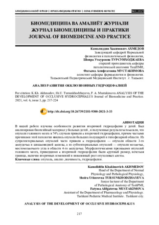
In our work, we studied the features of the development of secondary hydrocephalus in children . Biopsy material was analyzed in sick children, and the results showed that brain tumors in 74% of cases led to secondary hydrocephalus, and the most common causes of this pathology were tumors of the large hemispheres and the pituitary region. Supratentorial tumors often led to hydrocephalustumors of the 3rd ventricle and pineal gland, and from subtentorial tumors-tumors of the cerebellum, the cerebellar angle and the 4th ventricle. The morphological features of brain tumors leading to secondary hydrocephalus were large size, indistinct border, presence of secondary changes, and invasive growth of tumor cells.
-
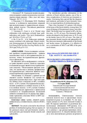
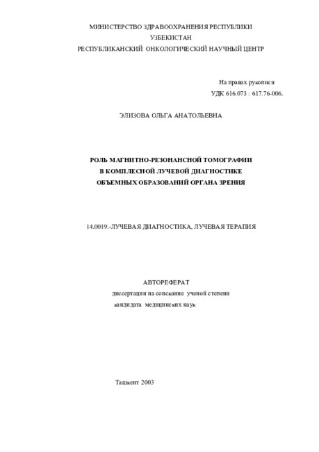 The role of magnetic resonance imaging in the comprehensive radial diagnosis of volumetric masses of the eye organ
Catalog of abstractsRelevance of the problem. The difficulties of diagnostics of orbital diseases are well known. Especially difficult is intraspecies differentiation among the multitude of tumour, pseudotumour, inflammatory, vascular, endocrine and other diseases occurring here, manifested by the symptom complex of unilateral exophthalmos [Beradze I.N., 1978; Brovkina A.F., 1993].
Malignant intraocular neoplasms are the main cause of death of patients with diseases of the organ of vision, with 45-48% of patients dying from metastases in the first 5 years after enucleation [Alekseeva I.B., 1990, Barkhash S.A.1978, Brovkina A.F..1991, 1997; Keizer R.W.. Viclvoyc G.L.,1986],
Retinoblastoma is the most frequent malignant neoplasm in children. According to different authors, the frequency of its occurrence is 1 case per 14000 - 35000 newborns. [Bobrova N.F. and Vit V.V., 1993; Brovkina A.F., 1997; Provenzale J.M., et al., 1995; Skulski M., et al., 1997; Weber A.L., Mafee M.F, 1992; Wilms G., et al., 1989]. The frequency of patients with the most malignant intraocular tumour in adults - uveal melanoma has recently reached 7-9 people per 1 million population [Brovkina A.F., 1997; Kotslyansky E.O., 1989; Yushko N.A., Peskova L.I., Kalenich L.A., 1989; Peyster R.G., Augsburger J..I., Shields J.A., 1988; Romani A.. Baldeschi L., ct al 1998; Scott I.U., 1998].
The fundamental difference in treatment tactics, depending on the stage of development, size and topography of the tumour, as well as the seriousness of the prognosis in retinoblastomas and melanomas sharply increase the requirements for the accuracy of their differential diagnosis. At the same time, the number of diagnostic errors in ocular tumours continues to be 10-30% even when complex clinical and instrumental examination is applied in specialised ophthalmological centres [Ternovoy S.K., Panfilova G.V., Rogozhin V.A., 1979; Friedman F.E., Malyuta G.D., Kodzov M.V., 1995; Song G.X., 1991].
Widely used in ophthalmological practice traditional diagnostic methods (ophthalmoscopy, gonioscopy, diaphanoscopy, fluorescence angiography, laboratory tests) are insufficient to obtain comprehensive information about the localisation, nature of growth and prevalence of volumetric pathological formations of the eye and orbit. This circumstance and not quite satisfactory results of surgical treatment are the causes of high mortality of patients [Muratova T.T., Nigmanova N.H., Kozlovskaya G.M.. 1989, Naches A.I., 1980; Cheremisin V.M., Trufanov G.E., Kholin A.V., 1991]. Untimely or erroneous recognition of pathological processes of the orbit leads to a sharp deterioration of visual functions, up to blindness, and in some cases to the death of the patient [Yuzhakov A.M., Travkin A.G., Kiseleva O.A., 1991]. All this determines the importance of timely and accurate diagnosis of diseases of the orbit, on the one hand, and the difficulty of such diagnosis - on the other [Gabunia R.I., Kolesnikova E.K., Tumanov L.B., 1982].
The fact that the orbit is closed from direct inspection and palpation by bone walls and the eyeball, indicates the advantage of radial diagnostics in comparison with other methods of examination. In the arsenal of clinicians there is a great variety of methods of clinical-radial diagnostics of orbital pathology, however, at present the information in the literature about their resolving capabilities and significance in comparative aspect is incomplete and not fully studied. The priority of using one or another instrumental investigation, their sequence and expedient combination have not been determined yet. This makes it difficult to choose the optimal standardised approach for diagnosis and adequate treatment [Cheremisin V.M., Trufanov G.E., 1993, Weber A.L., Sabates N.R., 1996; Wenig V.M., Mafee M.F., 1998].
Thus, the study of these and other questions, contributing to the improvement of diagnostics and treatment of patients with neoplasms of the eye and ocular cavity, should be recognised as urgent urgent.
Purpose of the study. Comparative evaluation of magnetic resonance tomography capabilities and development of algorithms for complex radial diagnostics of volumetric formations of the visual organ. To solve this goal we set the following tasks.
1. To study the normal picture of the magnetic resonance image of the visual organ in comparison with other methods of visualisation.
2. To find out the possibilities of magnetic resonance tomography, ultrasound and computed tomography in detection and evaluation of intraocular neoplasms.
3. To determine the role and place of magnetic resonance tomography in differential diagnostics of volumetric pathological formations of the eye cavity in comparison with other radial methods of research.
4. To determine the indications and to develop an algorithm for the complex application of radiography, ultrasound, computer and magnetic resonance tomography for diagnostics of volumetric formations of the eye organ.
Scientific novelty.
The present work is the first to give a detailed and detailed description of the complex clinical and radiation examination, with generalisation and standardisation of magnetic resonance, computer and ultrasound semiotics of volumetric pathological formations of the eye and eye cavity. The conducted clinical and instrumental investigations allowed to determine the diagnostic value and resolving capabilities of each of the applied methods. The ultrasound, CT and MRI signs of volumetric formations of the eye organ were studied, clarified and supplemented taking into account the use of low-field magnetic field and general-purpose ultrasound apparatus. The developed standardised diagnostic algorithm of examination of patients with this pathology is new, thanks to which the pre-oppositional diagnosis of tumour and other diseases of the visual organ is improved and the total radiation load on the patient is reduced.
Conclusions
1. MPT will provide an opportunity to study the weight of the soft tissue and anatomical components of the ocular cavity, up to the optic nerve sheath and perineural liquor space, the orbital apex and chiasmal-sellar region, as well as to assess the condition of adjacent structures of the brain and facial skull. The method is limited in the evaluation of changes in the bony walls of the orbital cavity.
2. MRI is inferior in detecting characteristic signs of retinoblastoma (presence of calcification). The sensitivity of MRI was 66.6%, while for ultrasound and CT these values were 96.1 and 100%, respectively. But when the tumour spreads rstrobulbarly outside the eyeball (at 3-4 stages) the informativeness of MRI increases significantly. In uveal melanoma the sensitivity and specificity of MRI reaches 100%.
3. Both MRI and CT have a high detection rate (98.1% and 95.8% respectively) of benign orbital tumours of both primary and secondary origin. However, MRI is the preferred method of investigation. MRI is especially informative when a cranioorbital tumour and pseudotumour are suspected. The sensitivity of the method is 90.9% and 91.6%, respectively
4. In some cases ultrasound can be used to differentiate between encapsulated and diffuse neoplasms, which facilitates the diagnosis. However, when the pathological process is localised near the orbital apex, the diagnostic value of ultrasound decreases. In such cases it is advisable to use MRI.
5. In detection of primary and secondary malignant tumours of the orbital cavity both MRI and CT are quite informative (sensitivity 97,2% and 95,4% respectively), but the most comprehensive information about the state of bone walls will be provided by CT. When the process spreads intracranially, the value of MRI increases significantly, especially with the use of contrast enhancement.
6. The developed algorithm of complex clinical and radiation examination of patients with the use of ultrasound, CT and MRI is the most effective in the diagnosis of volumetric pathological formations of the eye and eye cavity, allowing to reduce to an adequate minimum the total radiation load on the patient and diagnostic period, excluding duplication of research techniques and choosing the most informative in each case, which in turn allows to develop appropriate treatment tactics and reduce the level of disability of the patient. -
 The head and neck region is one of the most complex parts of the body not only because of its anatomical and functional features, but also because of the wide variety of lesions that occur, which can cause difficulties in verification even for the most experienced pathologists. Worldwide, in the general structure of oncological morbidity, head and neck tumors occupy (excluding the brain and spinal cord) approximately 20-22%. According to the world statistics among malignant tumors of other localizations, malignant tumors of the head and neck are about 10-15%. In 90% of cases of head and neck tumors this squamous cell carcinoma originating from the epithelium of the oral, oropharyngeal region, 7% is lymphoepithelioma
 -
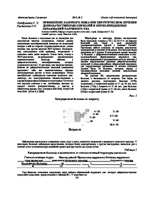 Применение лазерного ножа при хирургическом лечении доброкачественных опухолей и опухолеподобных образований наружного ухаЧисло больных с опухолями уха за последние два десятилетия заметно увеличилось. Однако данная локализация новообразований, несмотря на возросший интерес к ней со стороны оториноларингологов , менее изучена, чем другие опухоли ЛОР-органов. Исследования, посвященные этой проблеме, затрагивают в основном лишь узкие вопросы. Очень часто, говоря о ЛОР-онкологии, подразумевают исключительно злокачественные опухоли, забывая о доброкачественных, новообразованиях, которые встречаются почти в 10 раз чаще, чем злокачественные
Doctor's Herald -
 Clinical features of hepatoblastoma in young childrenHepatoblastoma, a liver tumor that develops in early childhood, is a common primary malignant tumor in children aged 0 to 4 years. In the structure of liver neoplasms, pathology is 72% and has been steadily growing over the past 30 years with an annual increase of 4-5%. All children were admitted to the oncohematological department with the presence of metastases, high values of alpha-fetoprotein - the main marker of the tumor process, thrombocytosis. Immunohistochemically, the fetal-epithelial tumor subtype was established in most children. All children received a full course of polychemotherapy (PCT) and tumor resection after completing the course of PCT. In 85.7% of cases, after surgical removal of the tumor and subsequent PCT, the outcome of the disease was favorable. The stages of diagnosis and the effectiveness of modern methods of therapy for malignant neoplasms of the liver in young children are shown.
Journal of hepato-gastroenterological research -
 Interventional performance of en-bloc transurethral resection of muscular non-invasive bladder cancer
Journal of Biomedicine and PracticeBladder cancer (BC) is a rather common disease. The incidence of bladder cancer in the population is gradually increasing[3,7] In non-invasive bladder carcinoma (NICM), transurethral resection of bladder tumour (TUR) is the cornerstone of treatment. Successful treatment of these tumours depends on adequate initial resection and an accurate histological diagnosis. After TUR alone, around 50 70% of patients develop recurrence. Reasons for this high rate of recurrence of NICMP have been cited for incomplete resection during the initial TUR, aggressive tumour biology, and implantation of tumour cells[6,8,9].
-
 To the features of diagnostics and treatment of cerebellar tumoursIn the present research are included the data of complex investigation and treatment of 35 (21 women, 14 men) patients, the average age of the patients consisted 30 years (from 3 till 69 years). To all patients was spent complex investigation, including brain CT, MRI and contrast CT, MRI. Postoperative mortality was observed in 6 (17.1%) patients, and it is dominated after subtotal removal of the tumor (11.4%), and in pa-tients after total removal of the tumor and biopsy death was registered only on one occasion.Results of treatment of cerebellar tumors depends on application of modern surgical methods and their efficacy.
Journal problems of biology and medicine -
 Clinic-diagnostics factors of the forecast at the choice of immunotherapy methods at patients with ovarian and cervical cancerThe purpose of the work to study of the molecular and biological markers in the ovarian and cervix can-cer as choice criteria methods such a extracorporal immunofarmakotherapy (EIFT) among the complex treat-ment. To study level of tumor markers was shown that the greatest predictive importance for treatment efficien-cy such markers as p53, VEGF and Ki-67, and level of tumor proliferative activity. The greatest effect obtained when carrying out an accompanying immunotherapy with EIFT and plasma exchange
Journal problems of biology and medicine -
 Comparative estimation of methods of treatment of a cancer of a bladder with metastasises in regional lympho noduses
Doctor's HeraldThe Tashkent Institute of improvement of doctors Results of treatment of 78 bladders sick by a cancer (BCa) with a lesion regional lymph nodes in stages T3-4 N1-2 MO are presented. The recurrent tumor is taped 19 (82,6 %) from 23 patients by whom the radical cystectomy (RCE) with standard lymph node dissection and the subsequent polychemotherapy (PCHT ) (I group was spent). On the average in 5,2 months after the treatment termination; in 11 group of 25 patients after RCE with dilated lymph node
dissection and the subsequent PCHT at 18 (72.0 %) the recurrent tumor, on the average in 4,8 months is taped. In 111 group after carrying out PCHT (without surgical treatment) advance of growth of a tumor after its regress and stabilization, was diagnosed at 27 (90,0 %) from 30
patients, on the average in 3,5 months. The survival rate median in 1 group of patients has made 9,4± 3,6 months, in II group - 12,5±4,2 months and in 111 group - 7,4 ± 2.9 months (p <0,05) -
 in this work, we have studied the macroscopic and microscopic features of clear cell ovarian adenoma. The results showed that clear cell ovarian adenoma is macroscopic, usually unicameral, up to 30 cm in diameter, often associated with endometriosis. The microscopic characteristic of clear cell ovarian adenoma is that the epithelium of the tumor has a diffuse, tubulocystic, tubular arrangement, sometimes with papillary growth. Sometimes the epithelial cells of the tumor with abundant light cytoplasm, rich in glycogen, are flattened, form nests, fields and contain a hyaline-like mass in the lumen of the cysts.
 -
 The analysis of observations of 68 patients with relapses of soft tissue sarcoma of extremities and trunk is assumed as a basis of this work. The relapses of soft tissue sarcoma of the head and neck arc not included into the work. There were 38 women and 30 men among the observed. The localization of the relapses was various: 22 patients had them on the upper extremity, 13- on the trunk, 33- on the lower extremity. There were 6 patients at the age of before 19, 14 - from 20 tO 29, 19- from 30 to 39, 12- from 40 to 49, 11- from 50 to 59, and 6 patients at the age of 60 and older. There were the most patients with relapses at the age of 30 to 39, as sarcomata occur more often at this age group. Two patients had relapsing tumours with the structure of rhabdomyosarcoma; 4 patients- with angiosarcoma, 20- with fibrosarcoma, 13- with synovial sarcoma, 3- with malignant schivanoma, 4- with liposarcoma, 2- with leiomyosarcoma. Twenty patients had unclassified tumours.
 -
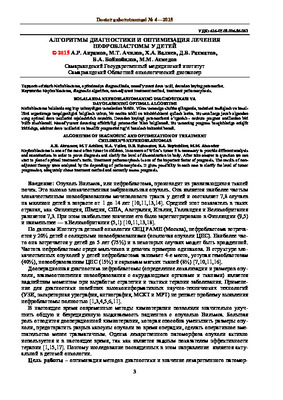
 «TUMOR AND TUMORLIKE FORMATIONS OF THE MAXILLOFACIAL REGION AMONG CHILDREN STRUCTURE MODERN METHOODS OF DIAGNOSIS AND TREATMENT»Annotation.Tumor and tumorlike formations of the maxillofacial region among children are very diverse. Most of them are of disontogenetic origin, combined with malformations of other organs and manifest in newborns or in the first years of life (up to 5 years). This article provides data on planned surgical interventions for tumors and tumor- lice formations of the maxillofacial region in children based on data from the departments of maxillofacial surgery in the CCH ()City Clinical Hospital) and the department of the ARCC (Almaty Regional Childrens Clinical Ho spital) for 5 years. The authors conducted an analysis of nosological units, diagnostic methods multidisciplinary approaches to the treatment of children.
Medicine and innovations -
 PECULIARITIES OF SURGICAL TREATMENT FOR MALIGNANT NON-ORGAN TUMORS OF THE RETROPERITONEAL SPACE AND THEIR RECURRENCEThe article describes a clinical case of a tumor of the retroperitoneal space with a recurrent course and the tactics of managing a patient with this pathology using the example of a specific clinical situation. The studies arc based on the analysis of 58 cases when, due to the local spread of the tumor, its removal was carried out simultaneously with the removal or resection of adjacent organs, for the period from 2018-2021 in the department of thoracoabdominal surgery of the Samarkand branch of the republican specialized scientific and practical center of oncology and radiology. In all patients, the surgical intervention was of a combined nature. In 58 patients, additional organs were removed and/or resected (72): of which 1 organ - in 18 (51.3%) cases. 2 organs - in 9 (27.6%), 3 organs - 5 (13.1%), 4 or more organs - 3 (7.8%). Combined and multivisccral resections for non-organ tumors of the retroperitoneal space arc traumatic interventions and are associated with various complications. The most frequent complications were massive bleeding from the tumor bed and as a result of damage to large great vessels.
Doctor's Herald -
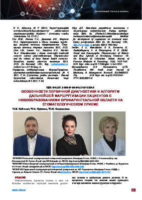 Features of the primary diagnosis and the algorithm for further routing of patients with oropharyngeal neoplasms at a dental appointmentOncological processes of the head and neck are a large number of tumors with a different course and requiring different treatment. In advanced stages,this treatment can lead to loss of vital functions and disrupt the appearance of a person with the appearance of bone and soft tissue defects [3]. But if this process is detected at the very beginning with an asymptomatic course or at the fi rst stage of the disease, then in 95-97% of cases the neoplasm can be removed without disability [5]. At this stage in the development of modern medicine with the help of radiation therapy and endoscopic surgery, it is possible to effectively treat oncological processes of the head and neck at stages I and II [2, 6]. With regard to the use of endoscopic surgery, the risk of developing psychological trauma in patients is reduced due to the absence of large incisions [8]. In this article, 217 patients, aged 30 to 89 years old, residents of the city of Chelyabinsk and the region with a confi rmed diagnosis of malignant neoplasm of the oropharyngeal zone were analyzed, examined and questioned, of which 127 were men and 90 were women. Of the total number of patients, 30% fi rst consulted a dentist. The largest number of cases of malignant neoplasms is determined at the age of 50 to 69 years in both sexes. Patients living in Chelyabinsk were statistically signifi cantly (p<0.05) less likely to meet, in aggregate, in terms of accessibility than patients living in the Chelyabinsk region. Primary T1 tumors were statistically signifi cant (p≤0.05) more often. The absence of regional metastases (N0) was statistically signifi cant (p≤0.05) more often. Also, neoplasms with a high degree of cell differentiation (G1) were also more often statistically signifi cant. Most often (p≤0.05),malignant neoplasms were localized on the tongue and lips. The keratinizing form of squamous cell carcinoma occurred statistically signifi cantly (p<0.001) more often.
Medicine and innovations -
 FEATURES OF MECHANISMS OF DEVELOPMENT OF CHRONIC OBSTRUCTIVE PULMONARY DISEASE AND CORONARY HEART DISEASETo date, the role of C-reactive protein as a major marker of inflammation is best understood in atherosclerosis. It was found that one of its fractions is capable of activating the complement system, which damages blood vessels and myocardium. It acts through various pathogenic mechanisms: stimulation, aggregation and degranulation of neutrophils, increased production of tissue factor and procoagulants that promote thrombus formation. Most often, CRP is able to activate complement after binding to ligands, which can also be lipoproteins. The role of systemic inflammation in COPD is particularly important. It has been shown that various cytokines: interleukin-1, tumor necrosis factor (TNF) and others - change the structure and function of endothelial cells. An association has been established between an increase in CRP, which is considered a marker of systemic inflammation, and impaired endothelium-dependent endothelial vasodilation, as well as between TNF, impaired expression of adhesive molecules, and the development of endothelial dysfunction in patients with COPD. It is known that COPD is characterized by an increase not only in the level of CRP, but also in proinflammatory cytokines both in the stage of exacerbation and in the stage of remission. Their synthesis causes the mobilization and activation of leukocytes in the peripheral blood, which, in turn, can lead to rupture of atherosclerotic plaques, vasoconstriction and thrombus formation, as well as exacerbation of coronary heart disease.
Journal of Cardiorespiratory Research -
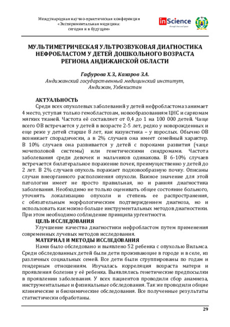 Мультиметрическая ультрозвуковая диагностика нефробластом у детей дошкольного возраста региона Андижанской области
Экспериментальная медицина: сегодня и в будущемСреди всех опухолевых заболеваний у детей нефробластома занимает 4 место, уступая только гемобластозам, новообразованиям ЦНС и саркомам мягких тканей. Частота её составляет от 0,4 до 1 на 100 000 детей. Чаще всего ОВ встречается у детей в возрасте 2-5 лет, редко у новорожденных и еще реже у детей старше 8 лет, как казуистика - у взрослых. Обычно ОВ возникает спорадически, а в 2% случаев она имеет семейный характер. В 10% случаев она развивается у детей с пороками развития (чаще мочеполовой системы) или генетическими синдромами. Частота заболевания среди девочек и мальчиков одинакова. В 6-10% случаев встречается билатеральное поражение почек, преимущественно у детей до 2 лет. В 2% случаев опухоль поражает подковообразную почку. Описаны случаи внеорганного расположения опухоли. Важное значение для этой патологии имеет не просто правильная, но и ранняя диагностика заболевания. Необходимо не только оценивать общее состояние больного, уточнять локализацию опухоли и степень ее распространения, с обязательным морфологическим подтверждением диагноза, но и использовать как можно больше инструментальных методов диагностики. При этом необходимо соблюдение принципа ургентности.
-
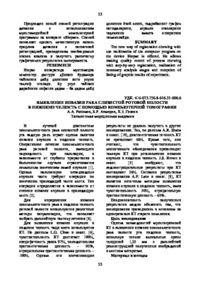 Detection of invasion of cancer of the oral mucosa into the lower jaw using computed tomographyThere is a significant role in detection of tumour invasion to mandible in diagnostics of oral cavity cancer. Modem imaging modalities has a great value m evaluation of invasion to mandible. In this article discussed possibilities of multidetectior computed tomography in evaluating of mandibular invasion in cancer of oral mucosa.
Stomatologiya -
 Nephroblastoma is one of the most often tumor in children. In concern of Wilm’s tumor it is necessary to provide different analysis and examinations in order to prove diagnosis and clarify the level of dissemination in body. After take answer to question we can start to plan of optimal treatment's tactic. Treatment pathomorphosis is one of the important factor of prognosis. The results of nonadjuvant therapy were analyzed by the depending of pathomorphosis. It gives possibility in each case to clarify the level of tumor progression, adequately chose treatment method and correctly assess prognosis.