Library search
Search Results
-
 JUSTIFICATION OF THE STUDY TO DEVELOP A SYSTEM OF PREDICTING OUTCOMES OF DENTAL IMPLANTSIn this article, thanks to the development of new implant systems and methods of reconstructive operations for atrophy of the alveolar bone tissue of the jaws, it is possible to use the method of dental implantation to replace dental defects of any localization with orthopedic structures. Prosthetics on implants helps to achieve the main goal - complete restoration of masticatory function in patients with partial or complete absence of teeth, improving the quality of life of the patient both in physiological and socio- psychological aspects.
Journal of oral medicine and craniofacial research -
In this article, thanks to the development of new implant systems and methods of reconstructive operations for atrophy of the alveolar bone tissue of the jaws, it is possible to use the method of dental implantation to replace dental defects of any localization with orthopedic structures. Prosthetics on implants helps to achieve the main goal - complete restoration of masticatory function in patients with partial or complete absence of teeth, improving the quality of life of the patient both in physiological and sociopsychological aspects.
-
 Improvement of diagnosis and treatment of patients with combined trauma of facial bone
Catalog of abstractsTopicality and demand of the subject of dissertation. In the world lat days chanchcd structures of trauma, increase the number of heavy combined traumas, which resulting in more heavy nature of simultaneous injuries of three , four or more anatomical regions, which creates difficulties in determining of the order of care and surgical tactics in patients with combined traumas of the facial skeleton bones (CTFSB). The syndrome of mutual burdening injuries of various anatomical regions, variety, hcavity and speed of the development of pathological process did difficulty of diagnosis of the CTFSB. Complexity of the clinical picture, features of the progress of post-traumatic shock, the development of traumatic disease cause difficulties which arise in the course of examination of patients and put tasks to the experts to find new ways of developing diagnostic algorithms and early surgical treatment of the CTFSB.
Frequency of CTFSB ranges from 34,8 to 63,3%. Fractures of orbit has been observed with an extremely high frequency (98%) in CTFSB, injury of the orbit is accompanied by damage of the eyeball and its subsidiary bodies has been observed in 66 % of eases. Consequences of eye injuries arc becoming the leading cause of disability and in 50% of eases could cause permanent loss of vision. By reason of death combined trauma take the third part after coronary heart diseases. Frequency of disfiguring defects and deformities of face occurs in 12 and 57%, disability in CTFSB reaches up to 23%. CTFSB, combined with TBI, causes up to 60% of deaths.
The causes of unsufficient results is non-availability of a diagnostic algorithm, which includes the most informative research methods, determining the order of interaction and priority of work of doctors of various specialties in CTFSB.
In some eases, requires specified an indications, character, scope, sequence and timing of surgical interventions, depending of the objective assessment of heaviness of injuries to various anatomical regions, prognosis criteria, the nature and heaviness of life-threatening consequences of combined trauma. The research work earned out within the framework of the achievement of the set by the Decree of the President of Republic of Uzbekistan “About measures on the further deepening reform the health care system” November 28, 2011, № PD-1652, maintenance of high-quality medical aid to the population under modem requirements and standards.In this regard the need for the development of algorithms of diagnosis and early methods of surgical treatment of patients with CTFSB constitute one of the important criteria demand the theme of dissertation.
Purpose of research is improvement of the diagnostic tactics and therapeutic interventions in patients with acute combined injuries of the facial bones according to the severity and location of the injury.
Scientific novelty of disscrtational research consists in the following: revealed the structure and features provide consistent care to patients with combined injuries in Republic of Uzbekistan;
The sequence of diagnostic and therapeutic measures, depending on the patient's general condition with CTFSB first determined by using created CT program "ADIL
developed innovative methods for early reduction and fixation of bone fragments in CTFSB;
identified endogenous factors, affecting on the wound process, disclosed the mechanisms of post-traumatic complications in CTFSB;
proved, that at 2 - 3rd days after the injury occurs the depression of cell and humoral immunity in the blood. Increases the level of proinflammatory cytokines, reduced the level of anti-inflammatory cytokine (in 2,8 at patients with heavy commonl condition. Increased levels of pro - and reducing anti - inflammatory cytokines is a poor prognostic factor in the development of inflammatory complications (bone wound suppuration, osteomyelitis of the jaw bones, soft tissue abscess);
patients with CTFSB at 2 - 3rd days after the injury occurs the depression of the content of protein and micronutrients (calcium, potassium and phosphorus) in the blood, which is a prognostic factor of the development of complications;
a scheme was developed for integrated medical correction of endogenous factors affecting on the development of posttraumatic complications;
1. CTFSB in 100% of cases combined with TBI, in 27.7 % with injuries of skeleton and internal injuries. In the diagnosis and treatment of patients with CTFSB should participate resuscitator, maxillofacial surgeon, neurosurgeon, ophthalmologist, and otolaryngologist. Primary debridement of wounds, reduction and fixation of bone fragments in patients in compensated state should be done within 3 hours after injury, while at subcompensated state - during the first day, and at the decompensated state - within 3 days.
2. With the CT program "ADIL" can determine the overall condition of patients in a short time. The most informative diagnostic criteria arc the general condition of patients, level of consciousness, hemodynamic stability, shock index and temperature gradient. The severity of the general condition of patients is directly dependent on the localization of the fracture of the facial bones. Multiple fractures of the upper and middle areas of the face arc the most serious injury in patients.
3. Patients with CTFSB in compensated and subcompensated state emergency surgical aid and diagnostic procedures should be performed in full volume (maxillofacial surgery, traumatology, neurosurgery, surgery, ophthalmology and otorhinolaryngologist), including the reduction and fixation of bone fragments in the first day. To patients with CTFSB in state decompensated should be performed at least diagnostic procedures, limiting the amount of emergency surgery. Reduction and fixation of bone fragments should be done after the restoration of function of vital organs and systems.
4. The method of choice for the treatment of depressed large bone fragments of facial bones is a titanium distractor, the use of which gives a good clinical and functional outcome.
5. When depressed fracture of the zygomatic arch application of the developed device will allow us to produce reduction and fixation of bone fragments in the early stages (within one day) with a good cosmetic result.
6. At patients with CTFSB in posttraumatic period (7- 14th day.) there arc a deep depression of CD3, CD4 cell composition, humoral factors and secretory immune system, increased necrosis factor CD95, increasing the levels of proin-flammatory (IL-6 ) and a decrease - anti- inflammatory (IL -10) cytokines. On 9-10th day reduced total protein, calcium, potassium and phosphorus in the blood .
7. Reduction of cellular and humoral immunity, increased proinflammatory cytokine and tumor necrosis factor, reducing the anti-inflammatory cytokine , the protein concentration in the blood, calcium, potassium and phosphorus arc predictors of complications.
8. Application of complex drug therapy within the 1-3 days after the injury with the inclusion of immune ( immunomoduline, ribomunil ), enzyme ( Voben-zym ) drugs osteoplastic materials allows to correct the violation of homeostasis, also used to prevent complications. -
 Possibilities of mini-invasive diagnostic and treatment methods in closed abdominal injuries
Catalog of dissertations and abstractsThe aim of the study is to improve the results of diagnosis and surgical treatment of victims with closed abdominal injury by developing a new approach to ultrasound assessment of the amount of hemoperitoneum, expanding and specifying indications for laparoscopy, taking into account the volume of free fluid in the abdominal cavity.
The object of the study were 160 patients with closed abdominal injury with stable hemodynamics, was hospitalized in the surgical Department of the Republican specialized scientific and practical center for emergency medicine of the Samarkand branch (clinical departments of surgical diseases № 2 and surgery postgraduate faculty of Samarkand state Medical Institute) for the period from 2010 to 2019.
The scientific novelty of the study is as follows: a fundamentally new approach to ultrasound evaluation of discrete volumes of free fluid in the abdominal cavity is proposed, based on taking into account the thickness of the fluid layer and its prevalence in the abdominal cavity zones; The expediency of using the ultrasound indicator "free fluid in the abdominal cavity < or >500 ml" in choosing the tactics of surgical treatment of patients with closed abdominal injury is substantiated; an algorithm for choosing surgical tactics for the treatment of patients with closed abdominal trauma was developed based on an ultrasound assessment of the volume of free fluid in the abdominal cavity.
Implementation of research results. Based on the results of a scientific study to improve the diagnosis and surgical treatment of patients with closed abdominal trauma:methodological recommendations "The choice of tactics for surgical treatment of closed abdominal trauma based on ultrasound assessment of the nature and severity of the injury" have been developed (certificate of the Ministry of Health No. 8n-z/1282 dated November 15, 2022). The proposed recommendations made it possible to increase the effectiveness of the diagnosis of intra-abdominal injuries in patients with abdominal trauma;
The results of scientific research on improving the diagnosis and surgical treatment of patients with closed abdominal injury have been introduced into medical practice, including the clinical practice of the Republican Scientific Center for Emergency Medical Care and its Samarkand, Surkhandarya and Navoi branches (conclusion of the Ministry of Health No. 8 n-z/699 dated December 21, 2022). The introduction of the obtained results into clinical practice allowed to improve the quality of high-tech surgical care provided to patients with isolated and combined abdominal injuries, to reduce the frequency of postoperative complications from 11.9 to 3.1% (p=0.144).
The structure and volume of the dissertation. The dissertation consists of an introduction, 4 chapters, conclusions and a list of cited literature. The volume of the text material is 107 pages. -
Subjects of research: 815 patients with the various forms acute pancreatitis, 235 patients with primary erronous diagnosis of acute pancreatitis.
The purpose of research: improve the treatment results of the patients with acute pancreatitis by upgrading existing and developing new diagnostic methods in the treatment complex.
Methods of research: clinical, biochemical investigation.
The results achieved and their novelty. On the basis of the analysis of occurance frequency of various clinical symptoms, diagnostics system of the acute pancreatitis is offered, the criteria of the laboratory and ultrasonic diagnostics for given disease arc advanced, for a quantitative estimation of severity of a condition of the patients the ball scale is offered; the complex conservative therapy is advanced, the technique of realization of long intraarterial catheter therapy is optimized, the optimum indications to realization of retrograd and pcrcutanc endobiliar interventions in treatment of acute biliar pancreatitis arc determined, drainage and sanation of omental bag for open operative interventons is offered, the technique of treatment of the patients in postoperative period is advanced and the algorithm of medical measures is offered at acute pancreatitis.
The developed diagnostic and treatment complex has allowed to reduce frequency of diagnostic errors from 28,4% to 16,8%, to increase efficiency of conservative therapy from 89,8% up to 95,3%, long intraarterial catheter therapy -from 72,0% up to 82,8%, to reduce mortality from 7,4% to 3,4%.
The practical importance. The offered clinical system for diagnostics and the advanced criteria of laboratory diagnostics promote to increase reliability of diagnostics, the systematized ultrasonic criteria allows verify the form of acute pancreatitis. The offered scale of definition of severity of the acute pancreatitis allows quantitatively characterize a condition of the patients, dynamical changes of pathological process and efficiency of used of a complex of medical measures. The advanced complex conservative therapy, technique of realization long intraarterial catheter therapy, definition of the indications to performance retrograd and pcrcutanc endobiliar interventions, advanced draining method and sanation of omentum bag at pancrcanccrosis, combined treatment in postoperative period allow to improve results of treatment of the given category of the patients.
Degree of introduction and economic efficiency: the received results arc introduced into practical activity of surgical branches of second clinics TMA.
Area of application: emergency and abdominal surgery. -
 This case described by us is a striking example of complexity, concerning scrupulous diagnostics at tumors of a gcpatopankrcatoduodcnal zone and shows high percent of diagnostic mistakes. Result. The patient at height of the complicated course of oncological process of a pancreas was given highly skilled specialized help by performance of adequate oncological operation. The cancer of a head of a pancreas can be shown for the first time in the form of pro fuze stomach-intestinal bleeding that not always allows to establish the correct diagnosis and to build adequate tactics of maintaining patients. We hope that this clinical case will increase vigilance of surgeons of the general medical network that tumors of a gcpatopankrcatoduodcnal zone can be shown for the first time in the form of complication of the main disease, thereby will reduce percent of diagnostic mistakes and will improve quality, the given highly specialized help to oncological patients at height of complication of an onkoprocess.
 -
 Disadvantages and ways of improvement in diagnostics and surgical treatment of the diabetic foot syndromePatients with diabetes mellitus, often, there are changes in the vessels in the form of calcification of the middle shell of arteries and arterioles (arteriolosclerosis of Menkeberg, mediacalcinosis). The determination of the x-ray stage of calcification of the arteries, gives additional information on the state of the vessels of the foot. Destructive changes in blood vessels, soft tissues and bones, conducted surgical interventions are naturally related. The study of these patterns is possible with a timely, critical study of anamnestic, diagnostic data and therapeutic measures. In this regard, to date remain relevant issues related to the development of differentiated treatment, taking into account all possible diagnostic data and improving surgical methods for treating the diabetic foot syndrome.
Doctor's Herald -
 Shift to nouns and adjectives as alterations produced in the wordThis article is about some verbs of using phonetic stress rules, change of meanings in the sentences
Renaissance in the paradigm of innovations in education and technology in the 21st century -
 3D planning of dental implantation using navigation templates on the example of a clinical caseIntroduction: In the modem protocol of treatment of patients with partial or complete adentia, implantation surgery is used with the help of implants. One of the most common complications is an incorrectly selected angle of inclination of the implant, which leads to a number of complications in the form of incorrect load distribution during prosthetics and bone resorption. In this regard, the use of 3D templates and virtual planning of dental implantation plays a special role in implantation. Objectives: Justification of the use of navigation templates during planning, as a prevention of complications of implantological surgery. Materials and methods: The study of CBCT images, removal of the spines from the jaws with a cast mass, marking of anatomically important structures, assessing the compatibility of the optimal final position of the teeth, used (Scan Marker 3-Diemme, Cantu Italy) to determine the central occlusion. Top Scan, Open Technology, Rezatto, Brescia, Italy) to obtain images of the jaws and make a navigation template. Results: The use of a surgical template significantly reduces the problem of the surgical result already at the stages of planning the operation. Conclusion: Thus, it can be concluded that when using surgical templates, it is possible to avoid intraoperative and postoperative complications and achieve high aesthetic results in a short time.
Medicine and innovations -
 IN THE FIELD OF MODERN DENTISTRY WITH THE HELP OF COMPUTER MODELING CONDUCTIVE DENTAL IMPLANTATION POSITIONThe most widely used direction in the restoration of complete or partial defects of the dentition is the direction of surgical dentistry- dental implantology. But at the same time, along with this surgical practice, the number of complications resulting from it also increases. This article shows that research in this area is still insufficient, and research on new effective treatments is needed.
Journal of oral medicine and craniofacial research -
 MAIN INDICATORS OF THE SCIENTIFIC AND EDUCATIONAL ACTIVITY OF THE HIGHER EDUCATION INSTITUTIONThis article examines the determination of goals and dimensions in the effective operation of higher education institutions, as well as the participation of interested parties. The model of rating indicators and strategic goals used by the organization is highlighted. The article offers a template that can be used to create a university indicator system.
Modern Science and Research -
 Сравнение точности трех типов хирургических шаблонов имплантатов
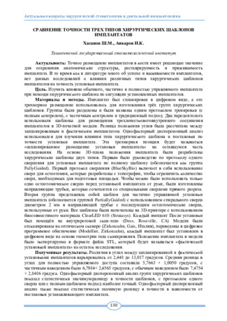
Topical issues of surgical dentistry and dental implantologyТочное размещение имплантатов в кости имеет решающее значение для сохранения анатомические структуры, реставрируемость и приживаемость имплантата. В то время как в литературе много об успехе и выживаемости имплантатов, нет данных исследований о влиянии различных типов хирургических шаблонов имплантатов на точность установки имплантата
-
 Клиническая значимость разработки и практического применения хирургического навигационного шаблона для мягкотканой трансплантации
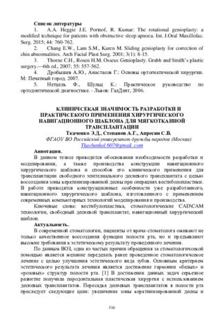
Actual problems of dentistry and maxillofacial surgery 4В данном тезисе приводится обоснования необходимости разработки и моделирования, а также производства конструкции навигационного хирургического шаблона и способов его клинического применения для трансплантации свободного эпителиального десневого трансплантата с целью воссоздания зоны кератинизированной десны при операциях вестиболопластики. В работе приводятся конструкционные особенности уже разработанного, навигационного хирургического шаблона, изготовленного с применением современных компьютерных технологий моделирования и производства.
-
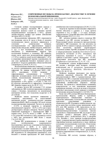
Согласно данным государственного доклада о состоянии здоровья населения РФ в 2003 г., пневмония является одной из основных причин внутрибольничной летальности (>10%), уровень диагностических ошибок при данном заболевании
достигает 40% -
 Clinical and radiological picture of septic pneumonia in childrenIn clinical practice, when managing patients with severe pneumonia, the doctor, when determining treatment tactics, has to solve difficult issues related to the primary or secondary (septic) nature of pneumonia. In this regard, the presence of clear diagnostic criteria for sepsis, pneumonic sepsis, and septic shock in patients with severe pneumonia is of great importance, since it largely determines the tactics of patient management. Severe respiratory failure and/or signs of severe sepsis or septic shock with a predicted risk of an adverse outcome requiring intensive therapy characterize severe pneumonia.
Journal of hepato-gastroenterological research -
 Особенности клинического течения атопического дерматита у детей раннего возраста
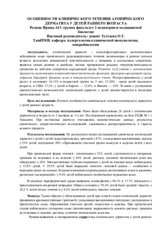
Prospects for the development of medicineАтопический дерматит - мультифакториальное воспалительное заболевание кожи хронического рецидивирующего течения, возникающее в раннем детском возрасте вследствие повышенной чувствительности к пищевым и контактным аллергенам, является самым ранним и наиболее частым проявлением реакции гиперчувствительности организма на действие аллергенов окружающей среды. Развитие АтД связано с комплексным процессом, включающим взаимодействия генетических факторов, факторов окружающей среды, дефектов барьерной функции кожи и иммунологического ответа. Распространенность у детей растет и составляет 10-15% в возрастной группе до 5 лет; 15-20% - среди детей школьного возраста, в США 17,2%, у детей в Европе — 15,6%, в Японии — 24%. В связи с улучшением диагностических возможностей, атопический дерматит у детей диагностируются в последнее время довольно часто.
-
 Ультразвуковые критерии в оценке венозного кровотока при циррозе печени у детейХронические вирусные гепатиты зачастую при отсутствии своевременной диагностики и лечения быстро формирует цирроз печени, приводящий к инвалидизации больного и летальному исходу. Одним их важнейших диагностических методов современной гепатологии является ультра-звуковое исследование с применением методик ультразвуковой допплерографии, имеющее неоспоримые преимущества: отсутствие лучевой нагрузки, высокая степень достоверности.
Journal problems of biology and medicine -
 The authors conducted a mass dental examination of 735 children (including 342 boys and 393 girls) in the period of milk and replacement bite at the age of 3 to 14 years. In 428 children (209 boys and 219 girls), there were detected abnormalities of PTSD. Of these. 23 children were diagnosed with an open bite with speech disorders, for which a complex of diagnostic and therapeutic measures was carried out. These children were recommended to wear “Infant” and T4K trainers. Was also made of a vestibular plate with the expanding screw; a vestibular arch and a lattice for the language. The use of the “Infant” trainer m complex orthodontic and speech therapy practice has given good effectiveness only in the milk bite. And in children over 5 years of age, positive effects are obtained from the use of the t4k trainer.
 -
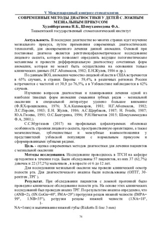 Современные методы диагностики у детей с ложным мезиальным прикусом
Actual problems of dentistry and maxillofacial surgery 5В последнее десятилетие во многих странах идет изучение мезиального прикуса, путем применения современных диагностических технологий, для своевременного лечения данной аномалии. Основой при постановке диагноза является рентгеноцефалометрическое исследование лицевого скелета, которое позволяет определить ведущие патогенетические механизмы и провести дифференциальную диагностику сочетанных форм аномалии, которая не может быть осуществлена на основании только клинических данных (Н.Г.Аболмасов, 1982; Е.Н.Жулев, 1986 и др.).
-
 Optimization of surgical treatment of chronic antero-medial dislocations of the radial head in children
Catalog of dissertations and abstractsThe aim of the research work is to improve the results of surgical treatment of chronic anterior-medial dislocations of the radial head in children based on the improvement of the method of surgical treatment.
The object of the study was 83 patients with chronic antero-medial dislocations of the head of the radius, treated in the department of the consequences of childhood injuries of the Samarkand branch of the RSSPMC of Traumatology and Orthopedics for the period 2017-2020. The scientific novelty of the research work is the following: it is proved by histological examination that, in case of injuries from up to 1 month ago, the anterior wall of the joint capsule is thin and elastic, which is easily stretched, and from 3 months ago, it thickens, scars and forms fibrous tissue; the possibility of using a fibrous-modified joint capsule for annular ligament plasty in the surgical treatment of chronic antero-medial dislocations of the radial head from 3 months ago was proved; the tactics of surgical treatment of chronic antero-medial dislocations of the head of the radius depending on the deformity of the bones of the forearm were determined; a direct relationship between the results of surgical treatment of chronic antero-medial dislocations of the head of the radius, depending on the duration of the injury, has been proven.
The introduction of research results. Based on the obtained scientific results on the optimization of surgical treatment of chronic antero-medial dislocations of the radial head in children:
based on the results of the development of a method for annular ligament plasty, a patent for an invention was obtained from the Intellectual Property Agency of the Russian Federation “A method for the surgical treatment of chronic anterior medial dislocation of the radial head in children by capsuloplasty according” (patent № 2749870 dated 06/17/2021). The results obtained made it possible to improve the results of surgical treatment, to reduce the period of penetration in the hospital and after the surgical rehabilitation period; based on the results of scientific research on the surgical treatment of chronic anterior medial dislocations of the radial head, the guidelines “Surgical treatment of chronic dislocations of the radial head in children” were approved (Conclusion of the Ministry of Health of the Republic of Uzbekistan 8 n-z / 81 dated February 21, 2022). The results obtained have improved the quality of early diagnosis and treatment of patients with chronic anterior medial dislocations of the radial head in children; based on the results of scientific research on the surgical treatment of chronic antero-medial dislocations of the head of the radius, the methodological recommendations “Conclusion of the Ministry of Health of the Republic of Uzbekistan 8 n-z / 289 of August 31, 2021” were approved. The results obtained have improved the quality of early diagnosis and treatment of patients with chronic anterior medial dislocations of the radial head in children;
Scientific results have been introduced into the practice of healthcare (Conclusion of the Ministry of Health 08-32955 of October 24, 2022), in particular, the Samarkand branch of the Republican Specialized Scientific and Practical Medical Center for Traumatology and Orthopedics, the Bukhara branch of the Republican Scientific Center for Emergency Medical Care, and the Samarkand Regional Children's Multidisciplinary Medical Center. The proposed method for the treatment of chronic anterior-medial dislocations of the radial head in children allowed to reduce the frequency of relapses, increase excellent and good results from 75.6% to 92.9%.
The structure and scope of the dissertation. The dissertation consists of an introduction, 5 chapters, conclusion, conclusions, practical recommendations, a list of references and applications. The volume of the dissertation is 109 pages. -
 Сравнительный анализ эффективности диагностических методов при опухолях носоглотки у детей и подростковЗлокачественные опухоли носоглотки у детей составляют 1-3% от всех злокачественных опухолей детского возраста и 10-12% от опухолей головы и шеи, а в структуре заболевания среди опухолевых заболеваний ЛОР-органов – составляют 25% [6, 9, 10].
Journal problems of biology and medicine -
 ROLE OF DIFFERENT ENDOSCOPIC STUDIES IN DIAGNOSTICS AND TREATMENT OF EROSIVE AND DYSPLASTIC CHANGES IN THE MUCOSA OF THE ESOPHAGUSThe development of modem medicine is reflected in all areas. This article also presents the results and the importance of using various endoscopic studies in the diagnosis and treatment of erosive and dysplastic pathologies of the esophageal mucosa, which is one of the urgent problems of medicine. Our research is based on the tactics of diagnosis and treatment of 2051 patients with gastroesophageal reflux disease complicated by various erosive pathologies of the esophageal mucosa, examined in the period 2019-2020 in the diagnostic departments of the Samarkand CMO and State Institution “RSSPMCS named after acad. V.Vakhidov".
Doctor's Herald -
 Clinical picture and diagnostic aspects of various tuberculosis in children and adolescents
Journal of Biomedicine and PracticeIn children’s tuberculosis inpatient departments of the PMCforPhandP and the Tashkent city tuberculosis hospital, 272 children and adolescents with various forms of tuberculosis were examined. Among children with various forms of tuberculosis, 28(10,3%) patients were diagnosed with complicated forms and 244 (89,7%)-uncomplicated forms of tuberculosis. When studying the risk factors for the disease, it was revealed that there were 225 (87,7%) children vaccinated with BCG, of which 58 (25,8%) children (76,8%), of whom anemia was found in 35 (12,9%) patients. In the structure of clinical forms in childrenand adolescents with tuberculosis, tuberculosis of the intrathoracic lymph nodes (57,0%) and primary tuberculosis complex (11,0%) predominate. Most of the children hada positivity to tuberculin (77,5±2,5%) and to the test for Diaskintest (69,5±2,7%).
-
 The basic principle of evidence-based medicine requires that in any infectious disease, topical diagnosis should be in the first place. However, due to the fact that a number of diseases are characterized by a variety of clinical manifestations, many forms of course (including mild or abortive), it is not always possible to identify the affected area. In addition, in many infectious diseases, other organs and systems are often involved in the process. This explains the fact that it is necessary to make more generalizing diagnoses. For example, the diagnosis of "urinary tract infection" would be more correct to consider as temporary, which in the course of diagnostic measures is transformed into a specific nosological form. Nevertheless, the diagnosis of "urinary tract infection" was included in the "International Classification of Diseases" (ICD-10, Geneva, 1995) as an independent nosological form. In children, this disease occurs quite often and ranks second after respiratory tract infections. Urinary tract infection (UTI) is more common in girls than in boys. The incidence rate increases with age in both, but boys have one early peak of morbidity — in the newborn period, and girls have two peaks: at the age of 3 to 6 years and at the beginning of sexual life.
 -
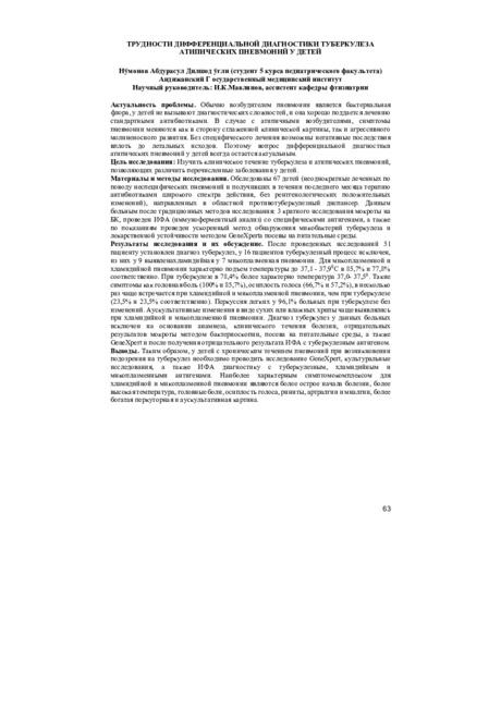 Трудности дифференциальной диагностики туберкулеза атипических пневмоний у детейОбычно возбудителем пневмонии является бактериальная флора, у детей не вызывают диагностических сложностей, и она хорошо поддается лечению стандартными антибиотиками. В случае с атипичными возбудителями, симптомы пневмонии меняются как в сторону сглаженной клинической картины, так и агрессивного молниеносного развития. Без специфического лечения возможны негативные последствия вплоть до летальных исходов. Поэтому вопрос дифференциальной диагностики атипических пневмоний у детей всегда остается актуальным.
Инновационные подходы к диагностике, лечению и профилактике туберкулеза и неспецефической респираторной патологии у взрослых и детей