Library search
Search Results
-
 Лечение и визуальный прогноз непрямой травматической оптической нейропатии при скулоорбитальном повреждении
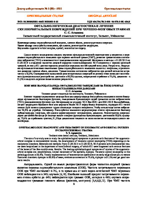
in LibraryВ структуре черепно-мозговой травмы краниофациальная составляет 6-7%, а от всех видов сочетанной черепно-мозговой травмы – 34%. Частота переломов орбиты при КФТ чрезвычайно высока – до 98%, что связано с особенностью строения глазницы. В 66% повреждения глазницы сопровождаются повреждениями глазного яблока и его вспомогательных органов. Повреждения зрительного нерва при черепно-мозговой травме встречаются в 0,5-5% наблюдений, при краниоорбитальных повреждениях – в 11,2%. Травматическая оптическая нейропатия, тяжелые травмы глаза в 50% случаев могут явиться причиной возникновения стойкой утраты зрения.
-
 OPHTHALMOLOGIC DIAGNOSTIC AND TREATMENT OF ZYGOMATIC AND ORBITAL INJURIES IN CRANIOCEREBRAL TRAUMAThe aim of our study was to study the ophthalmological symptoms in patients with fractures of the zygomatic-orbital complex in craniocerebral trauma, the development of traumatic optical neuropathy (TON) depending on hemodynamic disorders. Materials and methods: From 01.09.2015 to 01.09.2019, 3013 patients with craniocerebral trauma were hospitalized in the department of maxillofacial surgery, of which 821 were diagnosed with various fractures of the bones of the face middle zone. Results: The leading ophthalmological symptoms of injuries of the zygomatic-orbital complex are: enophthalmos, limited movement of the eyeballs, diplopia. Traumatic optical neuropathy in this pathology occurs in 20.9%. Timely implementation of reconstructive operations at an early stage allows you to restore functional disorders: dystopia in 89.8% of cases, strabismus correction in 73.6%. diplopia-in 91.5% and get good cosmetic results.
Doctor's Herald -
 Лечение и визуальный прогноз непрямой травматической оптической нейропатии при скулоорбитальном повреждении
in LibraryВ структуре черепно-мозговой травмы краниофациальная составляет 6-7%, а от всех видов сочетанной черепно-мозговой травмы – 34%. Частота переломов орбиты при КФТ чрезвычайно высока – до 98%, что связано с особенностью строения глазницы. В 66% повреждения глазницы сопровождаются повреждениями глазного яблока и его вспомогательных органов. Повреждения зрительного нерва при черепно-мозговой травме встречаются в 0,5-5% наблюдений, при краниоорбитальных повреждениях – в 11,2%. Травматическая оптическая нейропатия, тяжелые травмы глаза в 50% случаев могут явиться причиной возникновения стойкой утраты зрения. Последствия травм органа зрения, наличие выраженных косметических дефектов, а также функциональных расстройств нарушают качество жизни больного и влекут за собой стойкую утрату трудоспособности. Краниоорбитальные повреждения в 16-30% остаются не диагностированными даже после проведения компьютерной томографии пациентам с тяжелой черепно-мозговой травмы. В настоящее время остается открытым вопрос о раннем выявлении офтальмологической симптоматики краниофациальных повреждений в остром периоде черепно-мозговой травмы. Несвоевременное и неправильное их устранение может приводить к нарушениям функций органа зрения, тяжелым косметическим дефектам и гнойно-септическим осложнениям.
-
To evaluate the therapeutic efficacy of gliatilin for stabilizing visual functions after complex treatment of patients with progressive glaucoma-optical neuropathy with "normalized" pressure. Gliatilin is a nootropic drug, a central cholinomimetic with a primary effect on the central nervous system. The inclusion of gliatilin in the complex of therapeutic measures aimed at maintaining visual functions in patients with unstabilized glaucoma, aimed at various links in the pathogenesis ofglaucoma optical neuropathy, makes it possible to achieve stabilization of the process in 89% of patients with far-reaching unstabilized glaucoma for 6 months.
-
 Травматическая оптическая нейропатия и монокулярная слепота после трансназальное проникающее повреждение зрительного канала
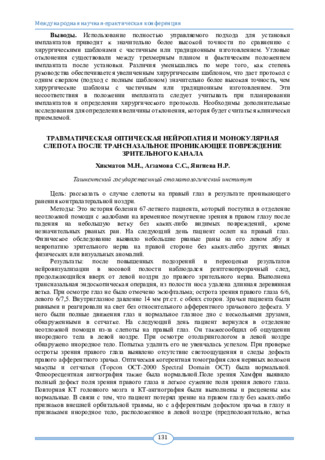
Topical issues of surgical dentistry and dental implantologyЦель: рассказать о случае слепоты на правый глаз в результате проникающего ранения контралатеральной ноздри.
-
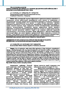 Assessment of influence of dialipon on the course of diabetic neuropathy at patients with sugar diabetes 2 typesAim of exploration: study about effect application of drug comprehensive therapy diabeticpolineropaty with way impact hemodynamic and functional condition nervous system. Exploration take in 54 sick people suffering from diabetic polineropaty in 2 group: in first group sick people get drug intravenously during 10 days, after that they pass tableted form; in second group patients take only traditional cure (angoprorector, resorption, vitamin therapy). Exploration revealed positive influence to the drug demonstration demolition neorapathy.Begin the second week of treatment, sick people mark reduction debility and sick people`s organism lower showings sensivitity and vegetative disorder. Take the tablet of prolong reach effect, at abolition of the drug therapy effect saved during 2-3 months. Thereby influence diabetic neropaty with using the neoprorector along the propose circuitry show substantial effect current method of treatment.
Journal problems of biology and medicine -
 Clinical and biochemical evaluation of the effektiveness of therapy for hereditary motosensory neuropathy in childrens
Journal of Biomedicine and PracticeThis article presents clinical, neurological and biochemical research methods in sick children with hereditary motorsensory neuropathy. It is known that some trace elements (Ca2 +, Mg2 +, P5 +) are characterized by high energy intensity and have a high penetrating ability into the bodies of neurons through ligand-mediated receptors and sites for the cooperation of these trace elements. Against the background of pathogenetic therapy and physiotherapeutic rehabilitation measures, it is possible to limit the severity of hereditary motor-sensory neuropathy, which is especially important in pediatric neurology.
-
 Clinical features, course and diagnosis of Guillain-Barre syndrome
Journal of Biomedicine and PracticeGuillain-Barre syndrome (GBS) is a very rare autoimmune disease that is associated with demyelination of the peripheral nervous system and progressive muscle weakness that occurs mainly in previously healthy people. The incidence of GBS is 1.1-1.8 cases per 100,000 per year, and the incidence increases with age. The clinical spectrum of GBS is heterogeneous and includes acute inflammatory demyelinating polyneuropathy (AIDP), acute motor axonal neuropathy (AMAN), acute motor and sensory axonal neuropathy (AMSAN), and Miller Fisher Syndrome (MFS). The disease is usually characterized by a rapid onset of symmetrical limb weakness, which progresses within a few days to 4 weeks and occurs in patients of any age. In developed countries, GBS has become the most common cause of acute flaccid paralysis. Despite improved recognition and treatment, GBS continues to be a serious disease.
-

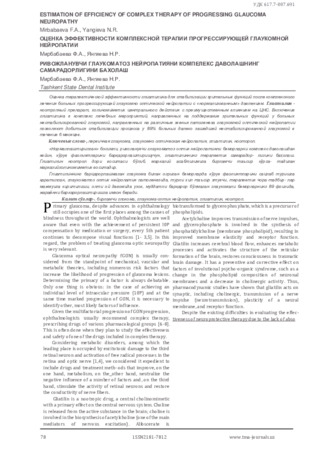
По данным ряда авторов и многих исследований, нестабильное течение глаукомы наблюдается даже при стойкой нормализации внутриглазного давления (ВГД), так как остаются факторы риска, не связанные с повышенным офтальмотонусом. Причина этого заключается в многофакторности глаукоматозной оптической нейропатии (ГОН) [3]. Наряду с общепризнанными механическими и сосудистыми факторами ГОН, значительная роль отводится метаболическим нарушениям, одно из ведущих мест в которых занимает активация свободнорадикального процесса в сетчатке и зрительном нерве. Избыточное усиление свободнора-дикального окисления в тканях глаза приводит к характерным изменениям — синдрому пероксидации, включающему повреждения мембран, инактивацию или трансформацию ферментов, нарушение процессов деления и дифференцировки клеток [1, 2, 4, 5].
-
 Clinical and functional evaluation of the effectiveness of endonasal electrophoresis in combination with electrical stimulation in the complex therapy of glaucomatous optic neuropathy
in LibraryClinical and functional assessment of the results of complex treatment of optic glaucoma neuropathy using tanakan in the form of endonasal electrophoresis in combination with electrical stimulation according to optical coherence tomography of the eye and ultrasound Doppler mapping. Material and methods: 43 patients (74 eyes) with glaucomatous optic neuropathy aged from 58 to 76 years were examined. Results: The used method effectively delays the development of optic nerve atrophy and, along with the improvement of visual functions, lengthens the positive effect of the main treatment, which was confirmed by a significant improvement in hemodynamic parameters ac-cording to the ultrasound Doppler study. Conclusions: The proposed method will improve the efficiency of treatment of patients with compensated open-angle glaucoma and improve the vision prognosis and the quality of rehabilitation measures.
-
Многочисленные исследования показали что, нестабилилизации течении глаукомы, даже при стойкой нормализации ВГД, так как остаются факторы риска не связанные с повышенным офтальмотонусом. Причина этого заключается в многофакторности глаукоматозной оптической нейропатии (ГОН). Наряду с общепризнанными механическими и сосудистыми факторами ГОН, значительная роль отводится метаболическим нарушениям, одно из ведущих мест в котором занимает активация свободно - радикального процесса в сетчатке и зрительном нерве. Избыточное усиление свободно - радикального окисления в тканях глаза приводит к характерным изменениям - индрому пероксидации, включающему повреждения мембран, инактивацию или трансформацию ферментов, нарушение процессов деления и дифференцировки клеток
-
 THE RESULTS OF THE USAGE OF ENDONASAL ELECTROPHORESIS IN COMBINATION WITH ELECTROSTIMULATION IN NEUROPROTECTIVE THERAPY OF THE GLAUCOMATOUS OPTIC NEUROPATHYGlaucoma is a chronic progressive optic neuropathy with characteristic morphologic changes in the head of optic nerve and progressive death of retinal ganglion fibers with narrowing of the visual field. Effective decrease of IOP is not able to serve as a guarantee of stabilization of glaucomatous process that continue to progress in part of patients. Thus, a search of a new direction of the drug therapy is needed because of the fact that hypotensive therapy is not completely effective. The most perspective of them is neuroprotection in combination with percutaneous electrostimulation that protect neurons of the retina and nerve fibers of optic nerve from different damage factors.
Journal of oral medicine and craniofacial research -
 Clinical And Functional Effectiveness Of Endonasal Electrophoresis In Combination With Electrostimulation In Complex Therapy Of Glaucomatous Optic Neuropathy
in LibraryThe aim of the study was the clinical and functional assessment of the complex treatment of glaucomatous optic neuropathy with the administration of the drug tanakan in the form of endonasal electrophoresis in combination with electrical stimulation according to IST and ultrasound Doppler mapping. We examined 43 (74 eyes) patients with GON aged from 58 to 76 years. The results of the study showed that this method effectively delays the development of optic nerve atrophy and, along with the improvement of visual functions, lengthens the positive effect of the main treatment, which was confirmed by a significant improvement in hemodynamic parameters according to the ultrasound Doppler study.
-
 Clinical and biochemical assessment of the use of antioxidant therapy in the treatment of glaucomatous optical neuropathy
in LibraryPurpose. To study the effectiveness of the use of the drug retinalamin in the complex treatment of patients with glaucomatous optical neuropathy (GON) and to compare the results with traditional means of treatment. Material and methods. A study of 70 patients (114 eyes), whose average age was 66.7+ 6.2 years old, suffered from POAG with compensated I0P. The diagnosis of the disease was made on the basis of anamnesis, clinical and instrumental studies. All patients studied were divided into 2 groups. Results. Treatment in both studied groups showed a positive effect on the improvement of biochemical parameters. So, the speed of products lipid peroxidation accumulation decreased from initial by 75.0% and 53.4%, for 77.2%
and 43.3%, for 73.8 and 60.2% according to the l-lll stages of POUG (P<0.001). Conclusion. The use of Retinalamin in the complex treatment of patients with glaucomatous optical neuropathy has an antioxidant effect, reduces the activity of PA2 and the level of MSM in patients with GON. -
 Abstract. According OCT and visometry data evaluation of the effectiveness of laser photocoagulation at central serous chorioretinopathy .Three patients with central chorioretinopathy have dynamic monitoring of the retina using OCT during medical treatment and after laser coagulation of leakage zones.The use of laser photocoagulation in patients with central serous chorioretinopathy in the absence of therapeutic treatment for 1-2 months has led to the rapid recovery of the architectonics of retina, and made possible to avoid irreversible changes in the neuroepithelium, improve the quality of vision.
 -
 Efficacy of traumatic optical neuropathy using color and magnetic stimulation method
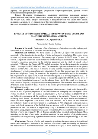
Topical issues of surgical dentistry and dental implantologyEvaluation of the effectiveness of simultaneous color and magnetic stimulation in the treatment of traumatic optic neuropathy.
-
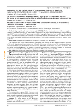 Progressive thinning of nerve fibers and retinal ganglion cells in traumatic optic neuropathy: 2 clinical cases
in LibraryThe aim of this work was an optical coherence tomography study to assess the state of the optic nerve in contusion of the eye. In 12 patients with eye contusion, an OCT study of the retina and optic nerve of the injured and fellow eye was performed. The layer of nerve fibers and ganglion cells of the retina was measured. Thus, broad-spectrum SS-OCT allowed us to obtain a wide-spectrum image of the macula and peripapillary areas, which is useful for monitoring various sequential developmental conditions in patients with TON as well as those with TON.
-
 DIAGNOSTICS AND TREATMENT OF OPHTHALMOLOGIC COMPLICATIONS IN ZYGOMATIC AND ORBITAL INJURIES.Purpose: The aim of the study was to study ophthalmic complications in patients with fractures of the zygomatic-orbital complex. Materials and methods: From 01.09.2015 to 01.09.2019, 3013 patients with craniocerebral trauma were hospitalized in the department of maxillofacial surgery, of which 821 were diagnosed with various fractures of the bones of the middle zone of the face. Results: The leading ophthalmological symptoms of injuries of the zygomatic-orbital complex are: cnophthalmos, limited movement of the eyeballs, diplopia. Traumatic optical neuropathy in this pathology occurs in 20.9%. Timely implementation of reconstructive operations at an early stage allows you to restore functional disorders: dystopia in 89.8% of cases, strabismus correction in 73.6%, diplopia-in 91.5% and get good cosmetic results
Medicine and innovations -
 Елка суягини диафизар синишларини жаррохлик услубларида даволашда келиб чиқадиган асоратлар.
Современная медицина глазами молодых ученыхЕлка суягини диафизар синишларини жаррохлик услубларида даволашда келиб чиқадиган асоратлар тахлил қилиб,уларни бартараф этиш чораларини ишлаб чиқиш.
-
 The Results of the Usage of Electro Stimulation in Neuroprotective Therapy of the Glaucomatous Optic Neuropathy
in LibraryGlaucoma is a chronic progressive optic neuropathy with characteristic morphologic changes in the head of optic nerve and progressive death of retinal ganglion fibers with narrowing of the visual field. Effective decrease of IOP is not able to serve as a guarantee of stabilization of glaucomatous process that continue to progress in part of patients. Thus, a search of a new direction of the drug therapy is needed because of the fact that hypotensive therapy is not completely effective. The most perspective of them is neuroprotection in combination with percutaneous electrostimulation that protect neurons of the retina and nerve fibers of optic nerve from different damage factors.
-
 Diagnostic and treatment complications of diabetes mellitus on evidence based medicineDiabetes mellitus (DM) is the result of a metabolic disorder of carbohydrates, proteins and fats of various etiologies and is manifested by chronic hyperglycemia. [1, 3, 5]. Complications of diabetes mellitus are divided into acute (hypoglycemia, ketoacidosis), which can occur suddenly at any period of the disease, and chronic (diseases of the cardiovascular system, kidney pathology, visual impairment, neuropathy), which develop several years after onset of the disease [10, 12].
Journal problems of biology and medicine -
The article is devoted to the problems of the development of Currently used antiretroviral therapy has made HIV infection a chronic controlled infection and has prolonged life and quality of life. However, any antiretroviral drug has side effects, although not on every patient and not always the same.
To assess the current state of the side effects problem of antiretroviral therapy in HIV - infected patients based on the analysis of literature data.
The following are data on the side effects of various groups of antiretroviral drugs used in the past and now, as well as some of their representatives. Antiretroviral drugs exhibit various side effects, in particular mitochondrial toxicity caused by nucleoside reverse transcriptase inhibitors (NRTIs), causing a wide range of side effects such as lactic acidosis, hepatic steatosis, myopathy, cardiomyopathy, peripheral neuropathy, pancreatitis, and possibly lipodystrophy syndrome. In general, integrase inhibitors are well tolerated and cause minimal drug interactions
The use of older antiretroviral drugs has been associated with a variety of side effects, which include lactic acidosis, hepatic steatosis, allergic reactions, myopathy, cardiomyopathy, peripheral neuropathy, pancreatitis and lipoatrophy.These side effects are less common with newer recommended ARVs. drugs. When treating, it is necessary to consider the likelihood of side effects and the possibility of their prevention.
-
 Клиническое значение факторов эндотелиальной дисфункции в диагностике пациентов с первичной открытоугольной глаукомой
Topical issues of surgical dentistry and dental implantologyГлаукома является частой причиной необратимой слепоты и слабовидения, занимая высокую ранговую позицию в перечне инвалидизирующих заболеваний органа зрения, значительно нарушающих качество жизни населения [1,2,4,7].В связи с возрастающей социальной значимостью заболевания повышение качества ранней диагностики и профилактики первичной глаукомы с медицинской стороны имеют особое значение в современном мире [3,5,8]
-
 Improving the prevention and treatment of glaucomatous optic neuropathy by predicting the risk of occurrence
in LibraryIn this article the findings of complex neuroprotective treatment of glaucomatous optic neuropathy (GON) in terms of prognostic criteria of the development of atrophic process in the optic nerve are given. Comparative analysis are carried out among patients who agreed to obtain given treatment and who preferred to reject from it. 54 (84 eyes) patients with GON aged 40 to 76 years old from them 31 (57,4%) were female, 23 (42,6%) – male with determined diagnosis glaucoma II and III stage and compensated intraocular pressure (21,3±3,2) were included in the group of examination. Duration of the examination made up 6 months. Effective criteria were reliable improvements of the vi-sual functions and positive structural dynamic according to tomographic findings, where thickness of thickness of the nerve fiber layer, ecscavatcia/disc ratio, and area of the neuroretinal rim.
-
The article presents the principles that are incorporated in the methods, as well as the methods themselves used to study the cornea: biomicroscopy, pachymetry, topographic keratotopography, optical coherence tomography, confocal microscopy and endothelial biomicroscopy. The use of traditional and modern high-tech methods of corneal examination allows for early and differential diagnosis in complex clinical cases, monitoring of treatment, as well as predicting the course and outcomes of corneal diseases