Library search
Search Results
-
Optimization Of Pelvic Prolaps Surgical Correction Using Its Own Tissues
The American Journal of Medical Sciences and Pharmaceutical ResearchPelvic organ prolapse (PTP) - pelvic floor and organ omission syndrome pelvic floor in isolation or in combination, which is extremely negatively affected by the quality of life of patients. According to world data, between 2.9 and 53% of women report some form of PTP. Up to 47% of pelvic organ prolapsed patients are women of working age. According to the Women Health Initiative Study, among 16,616 women of perimenopaus age, the incidence of uterine prolapsed was 14.2%, cystocele was 34.3%, and reconcile was 18.6%. In most cases, PTP is almost asymptomatic, which indicates its greater prevalence in the population.
-
BRIDGING DIVIDES: SWAP AND DOMINO ORGAN TRANSPLANTATION ACROSS SOCIOCULTURAL AND POLITICAL FRONTIERS
The American Journal of Interdisciplinary Innovations and ResearchSwap and domino organ transplantation represents a groundbreaking approach to address organ shortages by facilitating organ exchanges among incompatible donor-recipient pairs or utilizing organs from deceased donors to trigger multiple transplant chains. This paper explores the socio-cultural and political implications of swap and domino organ transplantation, transcending traditional boundaries and challenging existing norms in the field of organ donation and transplantation. Through a multidisciplinary lens, it examines the ethical considerations, legal frameworks, and societal attitudes surrounding swap and domino transplantation, highlighting both the opportunities and challenges inherent in this innovative approach. By fostering collaboration and cooperation across sociocultural and political divides, swap and domino transplantation offers a promising pathway to expand access to life-saving organ transplants and promote global health equity.
-
 Improvement of surgical aspects in providing help for sufficients wi thcombined pelvic and femoral bone damages
Catalog of dissertations and abstractsThe aim of the research work is to improve the results of treatment of patients with combined injuries of the pelvis and femur, by developing tactical and technical aspects based on the severity of the injury and the severity of the condition.
The object of the study was 130 patients with injuries of the pelvic and hip bones with concomitant trauma, treated at the Republican Scientific Center for Emergency Medical Aid and its Samarkand branch for the period 2016-2021 years.
The scientific novelty of the research work is the following: the structure and frequency of combined injuries of the pelvis and femur in the general structure of injuries, in the structure of injuries to the pelvis and femur separately were evaluated. the risk factors for the development of unsatisfactory results of treatment of concomitant injuries of the pelvis and hip, based on traditional clinical and diagnostic standards, have been determined; a direct relationship has been proven in the dynamics of the condition of the victims and the prognosis, taking into account the type and nature of segmental injuries; the device for external fixation for stable functional minimally invasive osteosynthesis has been improved and the possibility of expanding the indications for surgical treatment for combined injuries of the pelvis and hip in the early period of traumatic disease has been proved; the technical advantages of a complete set of an improved rod device for external fixation have been proved, the pelvic and femoral versions of which make it possible to use them for effective stabilization of the pelvis and hip separately during anti-shock measures, and for the final reposition of bone fragments; the direct dependence of treatment results on the proposed tactics of providing trauma care at an early hospital stage, depending on the type, nature, severity of pelvic and hip injuries, and the severity of the condition has been proved.
The introduction of research results. Based on the results of scientific research to improve the surgical aspects of providing assistance to victims with concomitant injuries of the pelvis and femur: based on the results of the development of a device for the treatment of fractures, a patent for an invention was obtained from the Intellectual Property Agency of the Russian Federation "Apparatus for the treatment of combined fractures of the pelvic and hip bones" (patent No. 2749897 dated 06/18/2020). The results obtained made it possible to improve the tactics of surgical treatment of patients, to shorten the period of hospitalization and the period of postoperative rehabilitation, to ensure the possibility of patients with minimal economic costs; on the basis of the results of scientific research on the diagnosis and treatment of concomitant injuries of the pelvic and femur bones, methodological recommendations were approved "Method for the treatment of victims with concomitant injuries of the pelvis and hip, depending on the severity" (Conclusion of the Ministry of Health of the Republic of Uzbekistan No. 8 n-z / 288 dated August 31, 2021 of the year). The results obtained made it possible to improve the quality of wound diagnosis and rehabilitation of patients with injuries of the pelvic and hip bones in concomitant injury; approved methodological recommendations "Tactics of rendering assistance to victims with combined injuries of the pelvis and hip, taking into account the severity of the condition" (Conclusion of the Ministry of Health of the Republic of Uzbekistan No. 8 n-z / 288 of August 31, 2020). The results obtained made it possible to improve the tactical and technical aspects in the treatment of injuries to the pelvic and hip bones, based on the severity of the injury and the severity of the patient's condition.
Scientific results have been introduced into the practical activities of healthcare, in particular, the Samarkand branch of the Republican Specialized Scientific and Practical Medical Center of Traumatology and Orthopedics, the Jizzakh Branch of the Republican Scientific Center for Emergency Medical Aid, the Samarkand branch of the Republican Scientific Center for Emergency Medical Aid (certificate of the Ministry of Health No. 08-09 / 18979 dated December 02, 2021). The proposed tactics for the treatment of combined injuries of the pelvis and femur made it possible to reduce the incidence of postoperative complications of excellent and good long-term functional results from 66.1% to 92.6%.
The structure and scope of the dissertation. The dissertation consists of an introduction, five chapters, conclusions, practical recommendations, a list of referencesand applications. The volume of the text material of the work is 111 pages.
-
 The role of magnetic resonance imaging in the comprehensive radial diagnosis of volumetric masses of the eye organ
Catalog of abstractsRelevance of the problem. The difficulties of diagnostics of orbital diseases are well known. Especially difficult is intraspecies differentiation among the multitude of tumour, pseudotumour, inflammatory, vascular, endocrine and other diseases occurring here, manifested by the symptom complex of unilateral exophthalmos [Beradze I.N., 1978; Brovkina A.F., 1993].
Malignant intraocular neoplasms are the main cause of death of patients with diseases of the organ of vision, with 45-48% of patients dying from metastases in the first 5 years after enucleation [Alekseeva I.B., 1990, Barkhash S.A.1978, Brovkina A.F..1991, 1997; Keizer R.W.. Viclvoyc G.L.,1986],
Retinoblastoma is the most frequent malignant neoplasm in children. According to different authors, the frequency of its occurrence is 1 case per 14000 - 35000 newborns. [Bobrova N.F. and Vit V.V., 1993; Brovkina A.F., 1997; Provenzale J.M., et al., 1995; Skulski M., et al., 1997; Weber A.L., Mafee M.F, 1992; Wilms G., et al., 1989]. The frequency of patients with the most malignant intraocular tumour in adults - uveal melanoma has recently reached 7-9 people per 1 million population [Brovkina A.F., 1997; Kotslyansky E.O., 1989; Yushko N.A., Peskova L.I., Kalenich L.A., 1989; Peyster R.G., Augsburger J..I., Shields J.A., 1988; Romani A.. Baldeschi L., ct al 1998; Scott I.U., 1998].
The fundamental difference in treatment tactics, depending on the stage of development, size and topography of the tumour, as well as the seriousness of the prognosis in retinoblastomas and melanomas sharply increase the requirements for the accuracy of their differential diagnosis. At the same time, the number of diagnostic errors in ocular tumours continues to be 10-30% even when complex clinical and instrumental examination is applied in specialised ophthalmological centres [Ternovoy S.K., Panfilova G.V., Rogozhin V.A., 1979; Friedman F.E., Malyuta G.D., Kodzov M.V., 1995; Song G.X., 1991].
Widely used in ophthalmological practice traditional diagnostic methods (ophthalmoscopy, gonioscopy, diaphanoscopy, fluorescence angiography, laboratory tests) are insufficient to obtain comprehensive information about the localisation, nature of growth and prevalence of volumetric pathological formations of the eye and orbit. This circumstance and not quite satisfactory results of surgical treatment are the causes of high mortality of patients [Muratova T.T., Nigmanova N.H., Kozlovskaya G.M.. 1989, Naches A.I., 1980; Cheremisin V.M., Trufanov G.E., Kholin A.V., 1991]. Untimely or erroneous recognition of pathological processes of the orbit leads to a sharp deterioration of visual functions, up to blindness, and in some cases to the death of the patient [Yuzhakov A.M., Travkin A.G., Kiseleva O.A., 1991]. All this determines the importance of timely and accurate diagnosis of diseases of the orbit, on the one hand, and the difficulty of such diagnosis - on the other [Gabunia R.I., Kolesnikova E.K., Tumanov L.B., 1982].
The fact that the orbit is closed from direct inspection and palpation by bone walls and the eyeball, indicates the advantage of radial diagnostics in comparison with other methods of examination. In the arsenal of clinicians there is a great variety of methods of clinical-radial diagnostics of orbital pathology, however, at present the information in the literature about their resolving capabilities and significance in comparative aspect is incomplete and not fully studied. The priority of using one or another instrumental investigation, their sequence and expedient combination have not been determined yet. This makes it difficult to choose the optimal standardised approach for diagnosis and adequate treatment [Cheremisin V.M., Trufanov G.E., 1993, Weber A.L., Sabates N.R., 1996; Wenig V.M., Mafee M.F., 1998].
Thus, the study of these and other questions, contributing to the improvement of diagnostics and treatment of patients with neoplasms of the eye and ocular cavity, should be recognised as urgent urgent.
Purpose of the study. Comparative evaluation of magnetic resonance tomography capabilities and development of algorithms for complex radial diagnostics of volumetric formations of the visual organ. To solve this goal we set the following tasks.
1. To study the normal picture of the magnetic resonance image of the visual organ in comparison with other methods of visualisation.
2. To find out the possibilities of magnetic resonance tomography, ultrasound and computed tomography in detection and evaluation of intraocular neoplasms.
3. To determine the role and place of magnetic resonance tomography in differential diagnostics of volumetric pathological formations of the eye cavity in comparison with other radial methods of research.
4. To determine the indications and to develop an algorithm for the complex application of radiography, ultrasound, computer and magnetic resonance tomography for diagnostics of volumetric formations of the eye organ.
Scientific novelty.
The present work is the first to give a detailed and detailed description of the complex clinical and radiation examination, with generalisation and standardisation of magnetic resonance, computer and ultrasound semiotics of volumetric pathological formations of the eye and eye cavity. The conducted clinical and instrumental investigations allowed to determine the diagnostic value and resolving capabilities of each of the applied methods. The ultrasound, CT and MRI signs of volumetric formations of the eye organ were studied, clarified and supplemented taking into account the use of low-field magnetic field and general-purpose ultrasound apparatus. The developed standardised diagnostic algorithm of examination of patients with this pathology is new, thanks to which the pre-oppositional diagnosis of tumour and other diseases of the visual organ is improved and the total radiation load on the patient is reduced.
Conclusions
1. MPT will provide an opportunity to study the weight of the soft tissue and anatomical components of the ocular cavity, up to the optic nerve sheath and perineural liquor space, the orbital apex and chiasmal-sellar region, as well as to assess the condition of adjacent structures of the brain and facial skull. The method is limited in the evaluation of changes in the bony walls of the orbital cavity.
2. MRI is inferior in detecting characteristic signs of retinoblastoma (presence of calcification). The sensitivity of MRI was 66.6%, while for ultrasound and CT these values were 96.1 and 100%, respectively. But when the tumour spreads rstrobulbarly outside the eyeball (at 3-4 stages) the informativeness of MRI increases significantly. In uveal melanoma the sensitivity and specificity of MRI reaches 100%.
3. Both MRI and CT have a high detection rate (98.1% and 95.8% respectively) of benign orbital tumours of both primary and secondary origin. However, MRI is the preferred method of investigation. MRI is especially informative when a cranioorbital tumour and pseudotumour are suspected. The sensitivity of the method is 90.9% and 91.6%, respectively
4. In some cases ultrasound can be used to differentiate between encapsulated and diffuse neoplasms, which facilitates the diagnosis. However, when the pathological process is localised near the orbital apex, the diagnostic value of ultrasound decreases. In such cases it is advisable to use MRI.
5. In detection of primary and secondary malignant tumours of the orbital cavity both MRI and CT are quite informative (sensitivity 97,2% and 95,4% respectively), but the most comprehensive information about the state of bone walls will be provided by CT. When the process spreads intracranially, the value of MRI increases significantly, especially with the use of contrast enhancement.
6. The developed algorithm of complex clinical and radiation examination of patients with the use of ultrasound, CT and MRI is the most effective in the diagnosis of volumetric pathological formations of the eye and eye cavity, allowing to reduce to an adequate minimum the total radiation load on the patient and diagnostic period, excluding duplication of research techniques and choosing the most informative in each case, which in turn allows to develop appropriate treatment tactics and reduce the level of disability of the patient. -
 COMORRIDITY OF ISCHEMIC DISEASES OF THE ORGAN OF VISION AND CHRONIC BRAIN ISCHEMIA IN ATHEROSCLEROSISThe purpose of this research was to study the features of the comorbid course of ischemic diseases of the organ of vision and chronic disorders of cerebral circulation in atherosclerosis. The material for this study was the results of complex examinations of 42 patients (84 eyes) with ischemic diseases of the organ of vision in combination with chronic brain ischemia. Important in the development chronic brain ischemia and progression of ischemic diseases of the organ of vision holds the consistency of collateral circulation. So. good consistency of collateral hemodynamics eliminates ischemia of brain and eyes tissue, and suffer less visual function.
Stomatologiya -
 Sravneniye effektivnosti enteral'nogo i parenteral'nogo pitaniya pri belkovo-energeticheskoy nedostatochnosti. 107 / 5 000 Результаты перевода Перевод Comparison of the effectiveness of enteral and parenteral nutrition in protein-energy malnutrition.
in LibraryParenteral nutrition (PN) is the most complex and technological variant of clinical nutrition, carried out by intravenous administration of nutrients into the body. It is intended for patients in whom it is impossible or insufficient to use other methods of nutrition and occupies the highest level in the hierarchy of clinical nutrition options, since it is considered the most difficult both in terms of technique and in terms of the variety of decision-making when prescribing it in clinical practice in the most difficult contingent of patients. . One of the main causes of death in patients with multiple organ failure is the development of an immune and inflammatory response. A number of studies have shown a decrease in the severity and incidence of septic complications in patients on enteral nutrition, after severe mechanical and thermal injuries, after major surgical interventions, it also allows you to maintain and maintain the barrier function of the intestine, which prevents the translocation of microflora. Based on the foregoing, it can be assumed that enteral nutrition will have a positive effect on the course of multiple organ failure.
-
To determine the hydrodynamic parameters of the uninjured fellow eye of children with combined injuries of the organ of vision. A prospective analysis of the hydrodynamic parameters of the fellow eye according to Friedenwald was carried out in 18 patients (18 eyes) aged 3 to 10 years 2–3 and 45–50 days after primary surgical treatment (PSD) of a penetrating wound of the cornea, who were hospitalized in the ophthalmological department of the clinic Tashkent Pediatric Medical Institute. Group I included 8 (44%) children with the following diagnosis: “Combined injury of the organ
of vision. Contusion of the eyeball severe. Complex penetrating wound of the cornea. Group II included 10 (56%) patients with complex penetrating wounds of the cornea. 2–3 days after PST of the wound, group I showed a statistically significant increase in Pt by 2.04±0.03 mm Hg. compared with the control group, while 1–2 days after the first measurement and 45–50 days after PST, the indicators decreased, on average, by 4.4±0.02 mm Hg. without the use of antihypertensive drugs. Changes in the hydrodynamics of the eye in children of group II were not statistically significant. The results of the examination of children revealed a transient increase in tonometric intraocular pressure in the paired uninjured eye 2–3 days after PST of a penetrating wound of the cornea with combined injuries of the organ of vision. -
 Features of the functional state of kidneys of newborn births in the breech presentation48 children who were bom in breech and head presentation are investigated revealed that risk factors at mothers of the given rise children in pelvic presentation are: early age, algodismenoriya and pathology of reproductive system. At pelvic presentation disorder of functional ability of kidneys that is connected with an ischemic nephropathy and with frustration of organ haemodynamics at pelvic presentation of a child is revealed.
Doctor's Herald -
 Exercise of a small pelvis with one-dimensional plastic the base of the base of the base the nearest resultsThe study analyzed the immediate results after a front pelvic exenteration with a one-stage pelvic floor osteoplasty in 28 patients with locally advanced cervical cancer (MR cervical cancer) with a bladder germination with stage T2b-4N0-1M0. The age of patients ranged from 35 to 60 years, the average age was 47.5 years. The reasons that have a significant impact on the development of postoperative complications, the advantages of pelvic floor plastic, which reduces the incidence of postoperative complications, are analyzed
Journal problems of biology and medicine -
 Clinical and epidemiological aspects of damage to the organ of vision in diabetes mellitus
in LibraryThe visual organ injuries in diabetes mellltus are an urgent problem-of ophthalmology to investigate. However, in our»Republic the studies of this kind are infriquent and do not use epidemiologic approach. Six hundred patients with diabetes mellltus of types I and II were examined, they being the residents of Tashkent and our examination revealed the visual organ changes. The eye fundus was injured in 83.68% of patients with dieletes. type I and in 84.53% of those with type II, the extent of the eye fundus injury being greater in IDDM (Insulin Dependent Diabetes Mellltus) than in IIDM ( Insulin Independent Diabetes Mellltus). The clinical analysis showed that the frequency of the lens clouding in patients with diabetes mellltus increased with the .age of the patients, duration of the disease and its severity. Me had developed the classification of the injuries of the eve fundus in patients with diatetes mellltus which-allows to choose the proper tactics of treatment and succession of specialists and measures at various stages of the treatment course. The integral assessment of the risk factors of diabetic retinopathy revealed that the risk factors in IDDM and IIDM are similar,but their influence on development of the disease is different. To improve prophylaxis of the retina injuries in patients with dia-betes mellltus-, we selected the following risk groups: "favourable prognosis", "attention", and " unfavourable prognosis". We also developed the criteria of time and frequency of examination of patients in these groups. The analysts of dynamics of morbidity rate for diabetes melli- tus by 2005 showed the further increase of this rate - in 1.4 times. Blindness in patients with I DOU is 5.65% and in those with IIDM - 11.11%. On the basis of the material described above, the neccessery conditions for organising the ophthalmo-diabetic service were formulated.
-
 CLINICAL AND ANAMNESTIC CHARACTERISTICS OF PATIENTS WITH CHANGES IN THE ORGAN OF VISION IN ATHEROSCLEROSISTo study the clinical and anamnestic characteristics of patients with changes in the organ of vision in atherosclerosis (AS). Material and methods: 156 patients with laboratory-clmically diagnosed atherosclerosis underwent examination and dynamic observation. All patients underwent a comprehensive examination, including the study of central visual acuity, kinetic and computed static perimetry, tonometry, gonioscopy. biomicroscopy, fundus ophthalmoscopy, ultrasound Doppler ultrasound of the vessels of the eye and the brachiocephalic trunk. Results: In patients with initial atherosclerosis in the absence of any subjective complaints and clinical manifestations of atherosclerotic vascular lesions, with a careful survey, episodes of short-term visual impairment after overeating, physical overstrain, visual impairment in one eye in bright light, periodic fog m front of the eye. discomfort, etc. dry eye. Patients with a clinical course of atherosclerosis, patients noted decreased vision, disappearance of the lower half of vision, upper half of vision, floating spots m front of the eye. pain in the eye. fog in front of the eye and loss of visual acuity, etc. Conclusions: Taking into account the presence of both modifiable in patients, and unmodifiable risk factors for the development of atherosclerosis, early and differential diagnosis of atherosclerosis should be carried out to prevent damage to target organs.
Stomatologiya -
Purpose. An analysis of complications in case of penetrating injuries of visual organ in children and methods of their elimination.
Material and methods. There were in the follow-up 36 patients (36 eyes) aged from 6 to 14 years with various ocular injuries: boys - 70% of cases, girls - 30% of cases. All children underwent ophthalmic and clinical examinations.
Results. The primary surgical treatment (PST) was performed in all patients at the place of residence in different regions of the Republic. The secondary surgical treatment (SST) was carried out according to the following indications: swelling cataract, filtration of corneal and scleral injuries, local endophthalmitis, iris incarceration, hypotension, secondary glaucoma, sutures failure, torpid uveitis, deformation of eyelids, abruption of lacrimal canaliculi, non-sutured scleral wound, suspect intraocular foreign body, the destruction of the eyeball, suspected intraocular foreign body, the destruction of the eyeball. Causes of PST complications were: the severity of injuries (47.2%), an unskilled first aid (41.7%), a late appeal of patient (47.2%).
Conclusion. As a SST result the inflammatory response was stopped and the eyes were maintained as an anatomical organ in 99.6% of cases, visual functions were preserved in 67.4% of cases. It is necessary to intensify educational work among the population, to improve the quality of emergency eye care and the adequate rehabilitation of patients.
-
 Usefulness of systemic inflammatory response syndrome as an independent Predictor of organ failure development in patients with acute pancreatitisSystemic inflammatory response syndrome is a protective response of the human organism aimed at eliminating the agent that caused the inflammatory process (infection, trauma, bums and tissue necrosis, etc.). The severity of the response depends on the amount of damage inflicted. This study conducted to assess the possibility of using systemic inflammatory response syndrome as an independent predictor in the development of complications of acute pancreatitis. For this, the following clinical and laboratory parameters were study: body temperature, pulse, respiratory rate and the count of leukocytes in the blood, which were obtain within the first 24 hours after hospitalization and before the development of organ failure. We investigated the relationship between the presence of systemic inflammatory response syndrome and organ failure.
Doctor's Herald -
 This article talks about the methodology of teaching the native language, which is founded on principles rooted in cognitive theory and interdisciplinary research. It emphasizes holistic language development, covering speech organ proficiency, linguistic comprehension, and expressive communication skills. The article discusses central tenets including the integration of linguistic components, fostering linguistic sensitivity, and assessing speech expressiveness. It highlights the importance of mastering written language before oral communication in instructional design.
 -
 The structure of traumatic lesions of the visual organ among the population of the Fergana Valley
Doctor's HeraldTrauma, particularly traumatic impairment of vision organ, is one of the most common percentage in the structure of diseases. Analysis of structure of traumatic impairments of vision organ was performed among the population of Fergana valley by the using archive materials of hospitals of Andijan. Fergana and Namangan regions for period 2006- 2009yy. Patients with the gender of Male, age of pre-school and school, active working age are the most sensitive for trauma of eyes. In-time diagnostics, primary medical and special ophtalmological aid are actual in the prevention of complication of traumatic impairments of vision organ
-
 Modern condition of the morbidity of the organ of vision in childrenAt present, a person is faced with growing actions on his body of various external, chemical and biological factors. The organ of vision, as part of the body, is also affected by these factors, including environmental ones. Mentioned factors affect not only the structure of the organ of vision, but also disrupt its activity, which is expressed by the development of various pathologies.
Journal problems of biology and medicine -
 For the purpose of the study, the clinical course of labor and the postpartum period was studied in 90 women who underwent massive obstetric postpartum hemorrhage. Group 1 - the main group, 15 women who underwent postpartum obstetric bleeding, who underwent amputation or hysterectomy with or without appendages. Group 2 - a comparative group, 45 women who underwent postpartum hemorrhage, which was stopped by conservative methods. Group 3 - control 30 women with normal childbirth and the postpartum period. Evaluation of the quality of life of women undergoing massive postpartum bleeding was performed using the Russian-language version of the MOS SF-36 common questionnaire. The most significant changes (more than 2 points) were found in respondents with removal of the organ when answering questions about the frequency of the nervous condition 2.07 ± 0.35 (without removing the organ - 1.41 ± 0.35); the absence of calm and pacification - 2.18 ± 0.36 (without removing the organ - 1.94 ± 0.33); lack of happiness - 2.2 ± 0.36 (without removing the organ - 1.54 ± 0.32).
 -
 An effective method of contraception for women with incomplete loss of the vaginal wallsNowadays the pelvic organ prolapse is the most common pathology among women of reproductive age. For these patients the actual is not only the correction of pelvic organ prolapse but also the search for the most effective contraceptive methods. Treatment of prolapse with simultaneous contraception for these patients is one of the topical problems of modem gynecology. Article suggests the method of trans-vaginal voluntary surgical contraception, produced simultaneously with surgical treatment of descent and prolapse of the vaginal walls. Were studied early and late results of surgery in women having at the time of surgical treatment of genital prolapse the simultaneous trans-vaginal tubal ligation.
Doctor's Herald -
 Reconstruction of the base bottom after the exercise of a small pancre with a locally distributed cancerThe study analyzed the results of treatment in a comparative aspect after a front pelvis exenteration with a single-stage pelvic floor plastic surgery in 88 patients with locally advanced cervical cancer (MR cervical cancer) with stage T2b-4N0-1M0 disease. An assessment of the possibilities and advantages of plastic recon-struction of the pelvic floor has been made, which reduces the incidence of postoperative complications
Journal problems of biology and medicine -
 ASSESSMENT OF IMMUNE STATUS AND EFFICIENCY OF IMMUNE CORRECTION OF INFLAMMATORY DISEASES OF THE PELVIC ORGANS IN WOMENThe aim of the study was to review and analyze the literature on the definition and condition of the immune system and the effectiveness of immune corrective measures. It was found that in acute and chronic IDPO (inflammatory diseases of the pelvic organs) the development of secondary immunodeficiency was noted, expressed by a decrease in the indicators of cellular and humoral immunity, multidirectional changes in pro- and antiinflammatory cytokines in patients. Almost all authors were unanimous in the opinion that immune corrective therapy should be used for inflammatory diseases of the pelvic organs, while offering various immune correctors.
Doctor's Herald -
 Our experience in the prevention and treatment of multiple organ failure in severely burnedThe article analyzes the results of the prevention and treatment of multiple organ failure in severely burned. The object of the study were 330 victims with deep burns, aged 18 to 74 years. 320 (96.97%) patients had IIIB-IV deep burns over 10% of the body surface. Studies have shown that complex against shock infusion- transfusion therapy with inotropic and organ-protective support, depending on impaired function of vital or- gans and early active surgical tactics facilitates the course of burn disease, reduces the number of multiple or- gan failure and mortality in severely burned
Journal problems of biology and medicine -
Parenteral nutrition (PN) is the most complex and technological variant of clinical nutrition, carried out by intravenous administration of nutrients into the body. It is intended for patients in whom it is impossible or insufficient to use other methods of nutrition and occupies the highest level in the hierarchy of clinical nutrition options, since it is considered the most difficult both in terms of technique and in terms of the variety of decision-making when prescribing it in clinical practice in the most difficult contingent of patients. . One of the main causes of death in patients with multiple organ failure is the development of an immune and inflammatory response. A number of studies have shown a decrease in the severity and incidence of septic complications in patients on enteral nutrition, after severe mechanical and thermal injuries, after major surgical interventions, it also allows you to maintain and maintain the barrier function of the intestine, which prevents the translocation of microflora. Based on the foregoing, it can be assumed that enteral nutrition will have a positive effect on the course of multiple organ failure.
-
In the treatment of patients with acute surgical diseases, even in the presence of multiple organ disorders and sepsis, prolonged artificial lung ventilation (ALV) through a tracheostomy is often required.
-
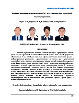 Dynamics of microcirculatory changes in the retina in patients after renal transplantationVisual disorders are the main factor determining the quality of life of patients with various somatic pathologies, a large part of which is represented by kidney diseases. The most famous ophthalmic manifestations of uremia are albuminuric retinopathy with eye hemorrhage, bilateral retinal detachment, typical cotton-like foci, and a star shape in the macula. The goal is to assess the state of the organ of vision in patients with end-stage chronic renal failure after kidney transplantation. As a result of studies in patients after 6-8 months, against the background of a decrease in normalization of blood pressure, regression of angiopathy and retinopathy, restoration of nerve conduction, and improvement of visual functions were noted. In patients with end-stage CRF, kidney transplantation is the most effective and only radical method of treating the body, which leads to a pronounced improvement in microcirculation, hemo-and hydrodynamics of the eye, improvement of the functions of the organ of vision, and the condition of the fundus.
Medicine and innovations -
 Ntensive treatment of multiple organ failure in severely burnedMultiple organ failure complicates the course of the burn disease and increases the likelihood of death in patients with severe thermal trauma. The analysis of 210 victims (mean age 25.5±5.4 years) with deep burns of more than 25% of the body surface and a burn shock of I-II-III degree (deep burn area of 20-40% bw) from 2010 to 2017 years. The structure of polyorganic insufficiency, frequency (cardiovascular, respiratory, renal, hepatic insufficiency, dysfunction of the central nervous system, gastrointestinal tract) and methods of its cor-rection are studied.
Journal problems of biology and medicine