Library search
Search Results
-
 The effect of treatment of diabetic nephropathy by “vessel due f”(sulodexide) on the risk of intraocular hemorrhages in patients with proliferative diabetic retinopathyDiabetic nephropathy and diabetic retinopathy as diabetes melhtus late complications are the main causes of disability and mortality in this category of patients. There is an evident pathogenic relationship between diabetic affection of the retiana and kidneys. Moreover, some methods of therapy targeting to normalization of renal function, may influence diabetic retinopathy course. The study is dealing with investigation of effect of standart treatment of diabetic nephropathy with “Vessel Due F” (sulodexide) to the risk of intraocular (pre-retinal and vitreal) hemorrhages in patients with proliferative retinopatia.
Doctor's Herald -
 Evaluation of the effectiveness of the biopeptide preparation corticin in the non-proliferative and preproliferative stages of diabetic retinoblastoma
in LibraryPurpose of work w as the cliniko-functional assessment of efficiency of a preparation “CORTEXIN” in the complex treatment of a diabetic retinopathy at non proliferative and preproliferative stages. Under supervision there were 30 patients (60 eyes) aged from 40 till 81 years old, with a diabetic retinopathy at nonproliferative and preproliferative stages who were divided into 2 groups. The first group (main) - 15 people (30 eyes) in addition to traditional treatment took “CORTEXIN” preparation of 10 mg on 0,5ml parabulbarly 1 time within 10 days. The second group (control) — 15 people (30 eyes) underwent traditional therapy. There were 12 males (40%) and 18 females (60%). The efficacy of treatment was studied by methods of visiometry, ophthalmoscopy and perimetry. The examinations have been performed before and 1, 3 and 6 months after treatment. During the carried out treatment in the main group of patients the improvement of visual acuity, field of vision and condition of retina w ith keeping the stable effect till 6 months was fixed. And ali this allows authors to recommend “CORTEXIN” application in the complex treatment of diabetic retinopathy at nonproliferative and preproliferative stages.
-
 METHODS OF THE FUNCTIONAL STATE OF THE KIDNEYS IN PATIENTS WITH DIABETES MELLITUSThis review article presents the results of studies in recent years to assess the structural and functional state of the kidneys in patients with diabetes mellitus. The data on pathogenesis and diagnostic markers in patients with diabetic nephropathy arc presented. Diabetic nephropathy is a common microvascular complication in diabetes mellitus (DM); the incidence of CKD ranges from 20 to 40% in patients with diabetes, inadequate glycemic control is considered the main risk factor for the development and progression of diabetic kidney damage. Diabetic nephropathy significantly reduces the quality of life in patients with diabetes mellitus. Early diagnosis of DN makes it possible to identify and prevent the development of renal complications in diabetes mellitus. Good nutrition, healthy lifestyle, symptomatic treatment and glycemic control are the main components of the treatment of DN.
Medicine and innovations -
Одно из самых тяжелых офтальмологических проявлений сахарного диабета (СД) - диабетическая ретинопатия (ДР), частота которой, по данным разных авторов, колеблется от 20 до 90% и неуклонно нарастает в последние годы. Среди причин необратимой потери зрения и инвалидности у лиц, в основном трудоспособного возраста, ДР занимает одно из первых мест. Так, 12% случаев слепоты во всех возрастных группах и 20% случаев слепоты, появившейся в возрасте от 45 до 74 !дет, обусловлено диабетическим поражением сетчатки (Brechncr R. J., 1992). Кроме того, слепота, связанная с ДР, обнаруживается у 0,6% больных СД, болеющих менее 6 лет, а при продолжительности заболевания более 15 лет она в 10 раз выше (Кацнельсон Л. А.. 1991).
-
Исследования последних лег, особенно эпидемиологического характера, тех или иных заболеваний показывают, что факторы влияющие на возникновение и развитие заболеваний, их частоту, структуру и распространенность, имеют комплексный характер.
-
The late complications of the DM are one of the basic reasons of premature physical inability and lethality of the DM patients, which puts essential harm to the health of the population and the economy as a whole (2). Proceeding from it in the foreground of the problem of prophylactics of the given DM complications is put forward.
-
 Assessment of efficiency of complex treatment of diabetic makulopatiya with use of the peptide bioregulator (retinalamin) and lazerkoagulyationResults of the laser treatment in patients with diabetic maculopathy complicated by macular edema have showed that the combined using lasercoagulation and peptid bioregulator Retinalamin contributed the resolu-tion of macular edema and allow to enhance of the visual functions. At application of traditional conservative therapy at DM only at 11,4% visual acuity, in 6 months after treatment doubtfully improved, visual acuity was authentically below initial. As a result of the combined treatment of DM by means of laser photocoagu-lation and Retinalamin's preparation visual acuity improved, with 0,140 to 0,333, changes of the area of rela-tive and absolute scotomas at DM, during treatment wasn't noted, the reached effect remains more than 6 months that speaks about stabilization of function of a retina.
Journal problems of biology and medicine -
Диабетическая ретинопатия (ДР) является одним из наиболее тяжелых осложнений сахарного диабета (СД), представляя одну из ведущих причин инвалидизации больных (М.И.Балаболкин, 1994). Заболевание нередко продолжает прогрессировать, несмотря на проводимое лечение, приводя пациента к слепоте (Ф.Е. Шадричев и др., 1998). Известно, что успех лечения и профилактики развития ДР определяется своевременным проведением терапевтических мероприятий, направленных на коррекцию обменных нарушений, гемореологических факторов с включением средств местного воздействия на сетчатку (Van der Pijl J.W. et all, 1998). Вместе с тем. несмотря на большое количество исследований, проблема лекарственного лечения ДР остается открытой и нуждается в дальнейшем изучении.
-
 Duration of diabetes is a major risk factor associated with the development of diabetic retinopathy. The severity of hyperglycemia is the key alterable risk factor associated with the development of diabetic retinopathy.The scientific article presents the results of the study of the risk factors for the development of the disease and the hroblems in the diagnosis of diabetes retinopathy
 -
 Диабетическая ретинопатия (ДР) - специфичное позднее сосудистое осложнение сахарного диабета, является основной причиной слепоты среди лип трудоспособного возраста. В экономически развитых странах она составляет 80-70% от всей инвалидности по зрению обусловленной сахарным диабелом
 -
Diabetic retinopathy (DR) is considered a microcirculatory disease of the retina. However, new data are emerging that suggest that retinal neurodegeneration is an early event in the pathogenesis of DR, which may precede the development of microcirculatory disorders that occur in DR, as well asparticipate in them. Therefore, studying the underlying mechanisms that lead to neurodegeneration will be essential for identifying new therapeutic targets in the early stages of DR. Elevated glutamate levels, oxidative stress, overexpression of the renin-angiotensin system, and activation of glycation end-product receptors play important roles in diabetic-induced retinal neurodegeneration. Finally, the balance between neurotoxic and neuroprotective factors is critical in determining the survival of retinal neurons. In this review, we focus on neurotrophic factors already synthesized by the retina under physiological conditions, and we also discuss current neuroprotective strategies and future directions for the treatment of DR
-
 The prevalence of diabetic retinopathy according to the optic center of "A.A.Yusupov" The city of samarkandSummary. In the ophthalmic center" A.A.Yusupov ” we examined 18880 patients, 384 (2%) of whom were diagnosed as diabetic retinopathy (DR).Proliferative stage of DR was found in 48.6 % of patients. On average, the rapid development of disease observed in age from 60-65 years. At this age the number of patients with proliferative stage prevailed. The findings suggest the need for and feasibility of population screening programs that may help identify the DR in the early stages of its development.
Doctor's Herald -
 Диабетическая ретинопатия - позднее неспецифическое сосудистое осложнение сахарного диабета, в равной степени характерное как для инсулин зависимого, так и для инсулин независимого диабета. Па возникновение и прогрессирование диабетической ретинопатии могут влиять разнообразные факторы. Наиболее существенными факторами, непосредственно влияющими на развитие диабетической ретинопатии, является длительность заболевания сахарным диабетом и гипергликемия
 -
Диабетическая ретинопатия (ДР) представляет собой одну из ведущих причин потери зрения, инвалидизации больных с сахарным диабетом (СД). Проблема патогенеза поражения сосудов глазного дна при диабете и поиск новых подходов в терапии этого страдания остается самой актуальной в диабетологии [1, 2, 3]. В литературе опубликовано большое количество исследований по лекарственному лечению ДР. Есть указания на положительное влияние ангиопротекторов, дезагрегантов, ангиодилятаТоров, антиоксидантов, витаминов, препаратов, нормализующих гемостаз, белковый и жировой обмены, имуннокоррегирующих средств. Стабилизирующее действие на течение ДР оказали ингаляции гепарина и внутрисосудистое лазерное облучение крови. Для рассасывания кровоизлияния успешно используются фибринолитики, особенно, рекомбинантная урокиназа человека в сочетании с ингаляциями гепарина [1, 2,4, 6, 7, 8].
-
 Значение прогностических тестов в оценке клубочковых и канальцевых нарушений при сахарном диабете II типа
Prospects for the development of medicineОдним из поздних осложнений сахарного диабета, приводящих к смертельному исходу, является диабетическая нефропатия. Имеющиеся исследования функционального состояния почек при диабетической нефропатии в основном посвящены оценке фильтрационной способности. В начальных стадиях диабетической нефропатии важно установить не только фильтрационную способность клубочков, но и функции канальцевого аппарата почек, которые могут быть ранним признаком диабетической нефропатии. В качестве маркеров поражения канальцевого эпителия могут быть использованы показатели экскреции молекулы повреждения почек (KIM-1), липокалин-2, цистатин С, гликозаминглаканы, экскреция канальцевых ферментов
-
 Особенности хирургического лечения катаракты методом тоннельной экстракции катаракты (тэк) у больных сахарным диабетом (сд)Количество больных сахарным диабетом (СД) во всем мире неуклонно растет и в настоящее время насчитывается около 160 млн. больных СД, а к 2025 году, по прогнозу Всемирной организации здравоохранения, это число достигнет 380 млн. человек. Диабет занимает первое место среди причин слепоты у пациентов в возрастной группе 30-60 лет [2], среди диабетических причин 70% приходится на долю диабетической ретинопатии (ДР) и 30% на другие глазные заболевания (диабетическая катаракта, вторичная глаукома и др.). У пациентов с СД своевременная хирургия катаракты крайне необходима для оценки состояния глазного дна и своевременного проведения лазерной коагуляции сетчатки, с целью предотвращения прогрессирования ДР и, тем самым, предупреждения развития конечной стадии ДР - отслойки сетчатки, рубеозной глаукомы и, в конечном счете, полной потери зрения. Любые хирургические вмешательства у пациентов с СД сопряжены с большим риском послеоперационных осложнений, возникающих на фоне декомпенсации диабета либо развития тяжелых гипогликемических состояний. Ответ организма на оперативное вмешательство проявляется активизацией симпато-адреналовой системы, что может вызвать резкую гипергликемию, липолиз, кетогенез и протеолиз, усугубляющиеся голоданием перед и во время операции. Воздержание от еды в пред- и послеоперационном периоде представляет особую опасность для пациентов с СД в плане развития гипогликемических состояний.
Doctor's Herald -
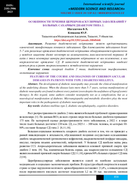 FEATURES OF THE COURSE AND DIAGNOSIS OF CEREBROVASCULAR DISEASES IN PATIENTS WITH TYPE 2 DIABETES MELLITUS.The development of diabetic neuropathy may precede the clinical manifestation of the underlying disease. When the disease lasts more than 5–7 years, various manifestations of diabetic neuropathy are found in almost every patient (even despite the usefulness of hypoglycemic therapy). In this regard, some authors consider neuropathy not as a complication, but as a neurological manifestation of diabetes. Microangiopathy and metabolic disorders play the most important roles in the pathogenesis of diabetic neuropathy.
Modern Science and Research -
 The role of magnetic resonance imaging in the comprehensive radial diagnosis of volumetric masses of the eye organ
Catalog of abstractsRelevance of the problem. The difficulties of diagnostics of orbital diseases are well known. Especially difficult is intraspecies differentiation among the multitude of tumour, pseudotumour, inflammatory, vascular, endocrine and other diseases occurring here, manifested by the symptom complex of unilateral exophthalmos [Beradze I.N., 1978; Brovkina A.F., 1993].
Malignant intraocular neoplasms are the main cause of death of patients with diseases of the organ of vision, with 45-48% of patients dying from metastases in the first 5 years after enucleation [Alekseeva I.B., 1990, Barkhash S.A.1978, Brovkina A.F..1991, 1997; Keizer R.W.. Viclvoyc G.L.,1986],
Retinoblastoma is the most frequent malignant neoplasm in children. According to different authors, the frequency of its occurrence is 1 case per 14000 - 35000 newborns. [Bobrova N.F. and Vit V.V., 1993; Brovkina A.F., 1997; Provenzale J.M., et al., 1995; Skulski M., et al., 1997; Weber A.L., Mafee M.F, 1992; Wilms G., et al., 1989]. The frequency of patients with the most malignant intraocular tumour in adults - uveal melanoma has recently reached 7-9 people per 1 million population [Brovkina A.F., 1997; Kotslyansky E.O., 1989; Yushko N.A., Peskova L.I., Kalenich L.A., 1989; Peyster R.G., Augsburger J..I., Shields J.A., 1988; Romani A.. Baldeschi L., ct al 1998; Scott I.U., 1998].
The fundamental difference in treatment tactics, depending on the stage of development, size and topography of the tumour, as well as the seriousness of the prognosis in retinoblastomas and melanomas sharply increase the requirements for the accuracy of their differential diagnosis. At the same time, the number of diagnostic errors in ocular tumours continues to be 10-30% even when complex clinical and instrumental examination is applied in specialised ophthalmological centres [Ternovoy S.K., Panfilova G.V., Rogozhin V.A., 1979; Friedman F.E., Malyuta G.D., Kodzov M.V., 1995; Song G.X., 1991].
Widely used in ophthalmological practice traditional diagnostic methods (ophthalmoscopy, gonioscopy, diaphanoscopy, fluorescence angiography, laboratory tests) are insufficient to obtain comprehensive information about the localisation, nature of growth and prevalence of volumetric pathological formations of the eye and orbit. This circumstance and not quite satisfactory results of surgical treatment are the causes of high mortality of patients [Muratova T.T., Nigmanova N.H., Kozlovskaya G.M.. 1989, Naches A.I., 1980; Cheremisin V.M., Trufanov G.E., Kholin A.V., 1991]. Untimely or erroneous recognition of pathological processes of the orbit leads to a sharp deterioration of visual functions, up to blindness, and in some cases to the death of the patient [Yuzhakov A.M., Travkin A.G., Kiseleva O.A., 1991]. All this determines the importance of timely and accurate diagnosis of diseases of the orbit, on the one hand, and the difficulty of such diagnosis - on the other [Gabunia R.I., Kolesnikova E.K., Tumanov L.B., 1982].
The fact that the orbit is closed from direct inspection and palpation by bone walls and the eyeball, indicates the advantage of radial diagnostics in comparison with other methods of examination. In the arsenal of clinicians there is a great variety of methods of clinical-radial diagnostics of orbital pathology, however, at present the information in the literature about their resolving capabilities and significance in comparative aspect is incomplete and not fully studied. The priority of using one or another instrumental investigation, their sequence and expedient combination have not been determined yet. This makes it difficult to choose the optimal standardised approach for diagnosis and adequate treatment [Cheremisin V.M., Trufanov G.E., 1993, Weber A.L., Sabates N.R., 1996; Wenig V.M., Mafee M.F., 1998].
Thus, the study of these and other questions, contributing to the improvement of diagnostics and treatment of patients with neoplasms of the eye and ocular cavity, should be recognised as urgent urgent.
Purpose of the study. Comparative evaluation of magnetic resonance tomography capabilities and development of algorithms for complex radial diagnostics of volumetric formations of the visual organ. To solve this goal we set the following tasks.
1. To study the normal picture of the magnetic resonance image of the visual organ in comparison with other methods of visualisation.
2. To find out the possibilities of magnetic resonance tomography, ultrasound and computed tomography in detection and evaluation of intraocular neoplasms.
3. To determine the role and place of magnetic resonance tomography in differential diagnostics of volumetric pathological formations of the eye cavity in comparison with other radial methods of research.
4. To determine the indications and to develop an algorithm for the complex application of radiography, ultrasound, computer and magnetic resonance tomography for diagnostics of volumetric formations of the eye organ.
Scientific novelty.
The present work is the first to give a detailed and detailed description of the complex clinical and radiation examination, with generalisation and standardisation of magnetic resonance, computer and ultrasound semiotics of volumetric pathological formations of the eye and eye cavity. The conducted clinical and instrumental investigations allowed to determine the diagnostic value and resolving capabilities of each of the applied methods. The ultrasound, CT and MRI signs of volumetric formations of the eye organ were studied, clarified and supplemented taking into account the use of low-field magnetic field and general-purpose ultrasound apparatus. The developed standardised diagnostic algorithm of examination of patients with this pathology is new, thanks to which the pre-oppositional diagnosis of tumour and other diseases of the visual organ is improved and the total radiation load on the patient is reduced.
Conclusions
1. MPT will provide an opportunity to study the weight of the soft tissue and anatomical components of the ocular cavity, up to the optic nerve sheath and perineural liquor space, the orbital apex and chiasmal-sellar region, as well as to assess the condition of adjacent structures of the brain and facial skull. The method is limited in the evaluation of changes in the bony walls of the orbital cavity.
2. MRI is inferior in detecting characteristic signs of retinoblastoma (presence of calcification). The sensitivity of MRI was 66.6%, while for ultrasound and CT these values were 96.1 and 100%, respectively. But when the tumour spreads rstrobulbarly outside the eyeball (at 3-4 stages) the informativeness of MRI increases significantly. In uveal melanoma the sensitivity and specificity of MRI reaches 100%.
3. Both MRI and CT have a high detection rate (98.1% and 95.8% respectively) of benign orbital tumours of both primary and secondary origin. However, MRI is the preferred method of investigation. MRI is especially informative when a cranioorbital tumour and pseudotumour are suspected. The sensitivity of the method is 90.9% and 91.6%, respectively
4. In some cases ultrasound can be used to differentiate between encapsulated and diffuse neoplasms, which facilitates the diagnosis. However, when the pathological process is localised near the orbital apex, the diagnostic value of ultrasound decreases. In such cases it is advisable to use MRI.
5. In detection of primary and secondary malignant tumours of the orbital cavity both MRI and CT are quite informative (sensitivity 97,2% and 95,4% respectively), but the most comprehensive information about the state of bone walls will be provided by CT. When the process spreads intracranially, the value of MRI increases significantly, especially with the use of contrast enhancement.
6. The developed algorithm of complex clinical and radiation examination of patients with the use of ultrasound, CT and MRI is the most effective in the diagnosis of volumetric pathological formations of the eye and eye cavity, allowing to reduce to an adequate minimum the total radiation load on the patient and diagnostic period, excluding duplication of research techniques and choosing the most informative in each case, which in turn allows to develop appropriate treatment tactics and reduce the level of disability of the patient. -
 Comparative analysis of the prevalence of malignant neoplasms among children of Andijan region and the republic of KarakalpakstanPopulation based analysis of cancer prevalence have done among children of Andijan region and Republic of Karakalpakstan in Uzbekistan. We used pediatric cancer cases registered at regional oncology dispancers. Cancer cases among children aged 0-17 consist from 0,38% to 0,55% from all oncology cases in Andijan region and in the republic of Karakalpakstan from 1,1% to 1,2%. In 2013 the annual incidence rate of pediatric cancer was 35/100 000 in Andijan region and 40 in the republic of Karakalpakstan. In both regions the most prevalent cancers were Hodjkin’s lymphoma, cancer of neurology systemand leucosis with higher distribution among male representatives. Retinoblastoma identified only among Andijan region children.
Doctor's Herald -
 It has taken in researches and analysis of patients with diabetes mellitus (DM), subjective clinical and neurological examinations, electromyography (EMG). The study has showed asymmetrical violation of simi-lar nerves, reducing the amplitude of potential action (PA) and motor response (M), the velocity of propaga-tion of excitation (VPE) in the field of compression. Based on this algorithm differential diagnosis was carried out with defeat, which showed the need for theevaluation of sensory symptoms, evaluation of EMG amplitude M-answers from two sides. The study showed that the benefit of tunneling damage in patients with diabetic polyneuropathy (DPN) and the exclusion of cervical radiculopathy, and plexopathy will testify, pronounced asymmetry of similar violations of nerves on both sides, with the exclusion of radiculopathy, and plexopathy.The study showed that the benefit of tunneling damage in patients with diabetic polyneuropathy (DPN) will testify pronounced asymmetry of similar violations of nerves on both sides, with the exclusion of radiculopathy, and plexopathy.
 -
 RISK FACTORS FOR THE DEVELOPMENT OF DIABETIC NEPHROPATHY IN PATIENTS WITH TYPE 1 DIABETEThe article presents data from a study that included 150 patients with type 1 diabetes mcllitus (type 1 diabetes) aged 6 to 18 years, who were registered at the Bukhara regional dispensary for 2018 - 2019. The study made it possible to study the clinical and epidemiological indicators of children and adolescents with type 1 diabetes in the city of Bukhara. We were able to identify priority risk factors for the development of diabetic nephropathy, which included age, onset of sexual development, anthropometry feature (tall), female sex. Adverse metabolic factors that save the development of diabetic nephropathy in children and adolescents with type 1 diabetes include high hyperglycemia at the onset of the disease. Arterial hypertension, dyslipidemia, development of diabetic retinopathy arc not among the main risk factors for diabetic nephropathy in children and adolescents.
Doctor's Herald -
 Перинатальные факторы риска у новорожденных с диабетической фетопатией.
Prospects for the development of medicineПроблема перинатальной патологии, обусловленной сахарным диабетом (СД) у матерей, остается одной из актуальнейших в акушерстве, неонтологии и педиатрии.
-
 Evaluating the effectiveness of the drug "nucleo cmp forte" in treatment of diabetic polyneuropathyStudy included 49 patients with type 2 diabetes (18 men and 31 women) patients were divided into two groups. Patients core (n = 28) and a comparison group (n = 21). Patients of the group, along with the complex therapy received glucose-lowering therapy, drugs with alpha-lipoic acid, В vitamins, as well as in the treatment of complex was added to the drug-Nucleo CMP Forte. Patients comparison group received all of the above drugs, except Nucleo CMP Forte. Results In general, the effectiveness of combined therapy with the addition of the preparation of CMP Nucleo fort in all cases were satisfactory, since improvement in NDS NSS and ranged from 40% to 80%, averaging 66.5%. Patients reported less pain on feet, especially at night. Also noted a decrease in feelings of injection on the side. Also noted a positive trend in terms of carbohydrate metabolism. Conclusion The tests showed that the addition of the complex treatment of diabetic neuropathy drug Nucleo CMP Forte positive impact on the state of the peripheral nervous system, as indicated by a significant decrease in performance NSS and NDS by 66 and 43%, respectively.
Doctor's Herald -
 One of frequent complications of diabetes mellitus are trophic and it is purulent - necrotic defeats of feet, as a result of angiopatiya, polyneuropathy, integrated under the name "diabetic foot". The syndrome of diabetic foot meets at 30-80% of patients. At syndrome of "diabetic foot" there is center of infectious defeat, ulcer defect, destructions of deep fabrics of feet which lead to formation of purulent necrotic phlegmon and gangrene of extremities which demand operational treatment. In purulent department of the Samarkand city medical association observation over 67 patients with syndrome of "diabetic foot" is made. Into the present in practice we have entered double-stage operational treatment. At the first stage diabetic phlegmon is opened long rasslablyayushimi cuts with necretomy. At the second stage radical operation is made. Thanks to use of active surgical tactics by us the percent of proximal amputation has decreased from 19% to 4,5%.
 -
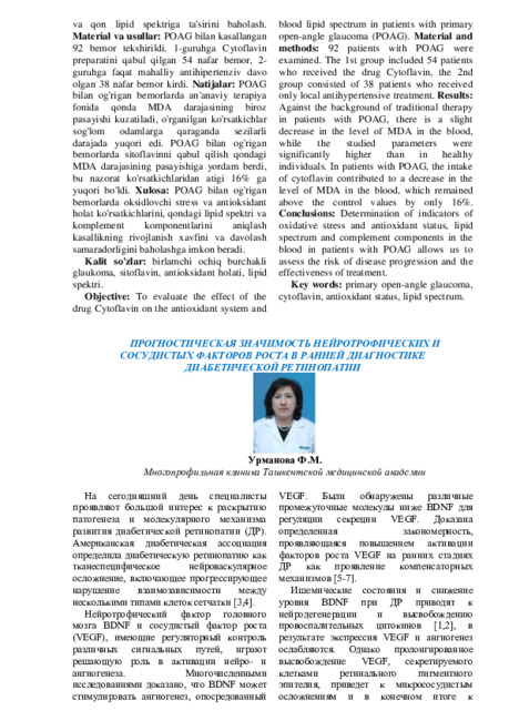 Prognostic value of neurotrophic and vascular growth factors in the early diagnosis of diabetic retinopathy
StomatologiyaDiabetik retinopatiya,mikrosirkulyatsiya, qon tomir o'sish omili VEGF, miyadan olingan neyrotrofik omil BDNF, DR rivojlanishi uchun biomarkerlar.